Review
. 2018 Mar;78(3):221-237.
doi: 10.1002/dneu.22546. Epub 2017 Oct 24.

The role of mitochondria in axon development and regeneration

Affiliations
Review

The role of mitochondria in axon development and regeneration

George M Smith et al. Dev Neurobiol. 2018 Mar.

Abstract

Mitochondria are dynamic organelles that undergo transport, fission, and fusion. The three main functions of mitochondria are to generate ATP, buffer cytosolic calcium, and generate reactive oxygen species. A large body of evidence indicates that mitochondria are either primary targets for neurological disease states and nervous system injury, or are major contributors to the ensuing pathologies. However, the roles of mitochondria in the development and regeneration of axons have just begun to be elucidated. Advances in the understanding of the functional roles of mitochondria in neurons had been largely impeded by insufficient knowledge regarding the molecular mechanisms that regulate mitochondrial transport, stalling, fission/fusion, and a paucity of approaches to image and analyze mitochondria in living axons at the level of the single mitochondrion. However, technical advances in the imaging and analysis of mitochondria in living neurons and significant insights into the mechanisms that regulate mitochondrial dynamics have allowed the field to advance. Mitochondria have now been attributed important roles in the mechanism of axon extension, regeneration, and axon branching. The availability of new experimental tools is expected to rapidly increase our understanding of the functions of axonal mitochondria during both development and later regenerative attempts. © 2017 Wiley Periodicals, Inc. Develop Neurobiol 78: 221-237, 2018.

Keywords: growth cone; mitochondrion; organelle.


Conflict of interest statement

The authors do not have any conflicts of interests to disclose.

Figures
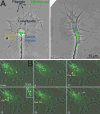
Figure 1
Figure 1
Growth cone morphology and mitochondria. (A) The two panels show examples of the growth cones of cultured chicken sensory neurons (phase contrast and Mitotracker Green labeled mitochondria). The approximate location of the central (C) domain and transition zone of the growth cone are denoted by the lines of orange and blue dots, respectively. In these examples, the C-domain and transition zone are approximated for didactic presentation using these phase contrast images, as direct determination of the extent of these two components of the growth cones requires imaging of the cytoskeleton. The peripheral (P) domain of the growth cone extends distal to the C-domain and consists of lamellipodia and filopodia. The mitochondria reside in the C-domain and are not usually observed to penetrate the P-domain. (B) Examples of the dynamics of mitochondria in growth cones. Phase contrast and Mitotracker Green labeled mitochondria are shown in these panels for a time lapse imaging sequence. Three mitochondria labeled a-c are used to show the behaviors of mitochondria in growth cones and referred to as Ma, Mb, and Mc (M for mitochondria). Ma can be noted to undergo changes in morphology from bent (0–6 sec) to a more linear appearance as it eventually undergoes “retrograde” transport away from the leading edge of the growth cone (purple arrow at 30 sec). Mb undergoes fission between 12–24 sec and the more proximal emergent mitochondria then undergoes retrograde transport (purple arrow at 24 sec). The inset at 18 sec shows a 3× magnified view of Mb (region bracketed by white lines) and the yellow arrow in the inset points to the site of fission. Mc undergoes “anterograde” transport from its initial position at the base of the growth cone toward the leading edge (red arrow).
Figure 2
Figure 2
Overview of the mechanism of the axonal transport and stalling of mitochondria. (A) Schematic summary of the types of mitochondrial behaviors in axons. Mitochondria can undergo runs in both the anterograde and retrograde directions, remained stalled in place with no detectable movement or exhibit dynamic pausing wherein the mitochondrion undergoes “wobbling” between short movements in opposite directions. (B) Machinery of anterograde transport. The mitochondrial outer membrane protein Miro scaffolds Milton/TRACK proteins to the mitochondrial surface. Milton/TRACK in turn engage the kinesin motor proteins responsible for the anterograde microtubule + end directed transport. (C) Machinery of retrograde transport. Similar to its role in anterograde transport, the Miro-Milton/TRAK complex binds the retrograde motor protein complex consisting of dynain and dynactin. (D) Machinery underlying microtubule based stalling of mitochondria. The mitochondrial outer membrane protein syntaphilin directly links mitochondria to microtubules through a microtubule binding domain. LC8 promotes the interaction of syntaphilin with microtubules. (E) Localized signals that locally generate accumulations of actin filaments trap and stall en passant mitochondria in a filament dependent manner. Although not directly addressed, myosin V and VI likely mediate this interaction (see main text).
Figure 3
Figure 3
Overview of the mechanisms of mitochondria fission and fusion. (A) Fission. The Drp1 GTPase associates with the outer membrane of mitochondria through a variety of adaptors (e.g., mitochondrial fission factor, MiD49/51; Lee and Yoon, 2016). Drp1 dynamically redistributes along the mitochondrial surface resulting in localized accumulations that predict the future site of fission. Drp1 oligomers form and ring around the mitochondrion that upon constriction fissions the mitochondrion. An example of fission along the axon of a cultured embryonic chicken sensory neuron expressing mitochondrially targeted DsRed. The yellow arrow denotes the site of fission. At 3 sec a constriction along the mitochondrion is noted which by 6 sec has resulted in fission. (B) Fusion. Fusion is initiate by the close apposition of mitochondrial outer membranes resulting in the engagement of mitofusins from the two contacting surfaces and accumulation and the site of contact. Mitofusins promote the fusion of the outer membranes. Following outer membrane fusion, contact of the inner membranes of the two mitochondria results in engagement of Opa1 proteins that drive the fusion of the inner membrane. As in panel B an example of fusion is shown below the schematic. The blue arrow denotes that the mitochondrion on the right moves toward the one on the left between 0–3 sec. The ends of the two mitochondria then spatially overlap and undergo apparent full fusion by 42 sec. (The time lapse sequences of mitochondrial fission/fusion in the axons of chicken sensory axons were provided by L. Armijo-Wingart in the laboratory of G. Gallo).
Figure 4
Figure 4
Overview of the role of stalled mitochondria in axon branching. Stalled mitochondria locally regulate actin filament and filopodial dynamics through ATP production. Signals that promote or inhibit branching increase and decrease mitochondrial respiration, respectively. Mitochondrial respiration also promotes mitochondria-associated intra-axonal translation of actin regulatory proteins involved in branching. Both the mitochondria-dependent regulation of actin dynamics and intra-axonal translation are in part regulated by modulation of mitochondrial respiration by signals that control axon branching.

Similar articles

Cited by

References

    1. Arismendi-Morillo G, Hoa NT, Ge L, Jadus MR. Mitochondrial network in glioma's invadopodia displays an activated state both in situ and in vitro: potential functional implications. Ultrastruct Pathol. 2012;36:409–14. - PubMed
    1. Armijo-Weingart L, Gallo G. It takes a village to raise a branch: Cellular mechanisms of the initiation of axon collateral branches. Mol Cell Neurosci. 2016;S1044–7431(16):30209–3. - PMC - PubMed
    1. Ashrafi G, Schlehe JS, LaVoie MJ, Schwarz TL. Mitophagy of damaged mitochondria occurs locally in distal neuronal axons and requires PINK1 and Parkin. J Cell Biol. 2014;206:655–70. - PMC - PubMed
    1. Aschrafi A, Kar AN, Gale JR, Elkahloun AG, Vargas JN, Sales N, Wilson G, Tompkins M, Gioio AE, Kaplan BB. A heterogeneous population of nuclear-encoded mitochondrial mRNAs is present in the axons of primary sympathetic neurons. Mitochondrion. 2016;30:18–23. - PMC - PubMed
    1. Berthet A, Margolis EB, Zhang J, Hsieh I, Zhang J, Hnasko TS, Ahmad J, Edwards RH, Sesaki H, Huang EJ, Nakamura K. Loss of mitochondrial fission depletes axonal mitochondria in midbrain dopamine neurons. J Neurosci. 2014;34:14304–17. - PMC - PubMed

Publication types

LinkOut - more resources