. 2018 Jan:61:225-237.
doi: 10.1016/j.neurobiolaging.2017.09.001. Epub 2017 Sep 15.

Atypical PKC, PKCλ/ι, activates β-secretase and increases Aβ1-40/42 and phospho-tau in mouse brain and isolated neuronal cells, and may link hyperinsulinemia and other aPKC activators to development of pathological and memory abnormalities in Alzheimer's disease

Affiliations

Atypical PKC, PKCλ/ι, activates β-secretase and increases Aβ1-40/42 and phospho-tau in mouse brain and isolated neuronal cells, and may link hyperinsulinemia and other aPKC activators to development of pathological and memory abnormalities in Alzheimer's disease

Mini P Sajan et al. Neurobiol Aging. 2018 Jan.

Abstract

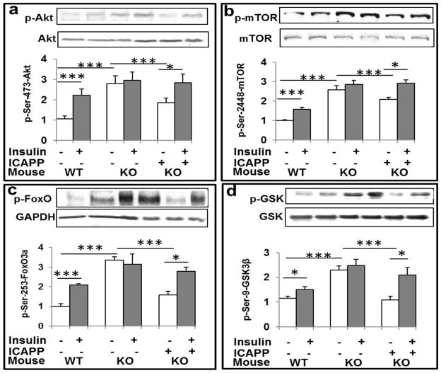
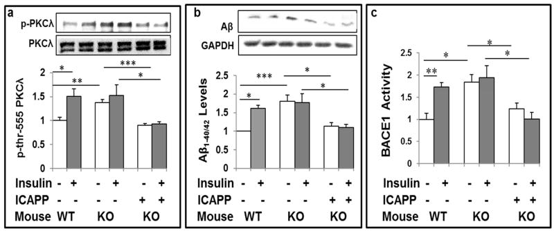
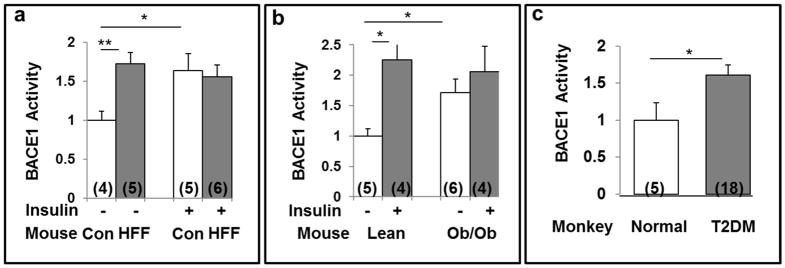
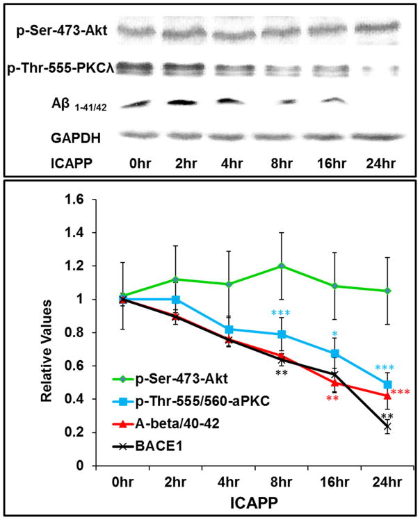
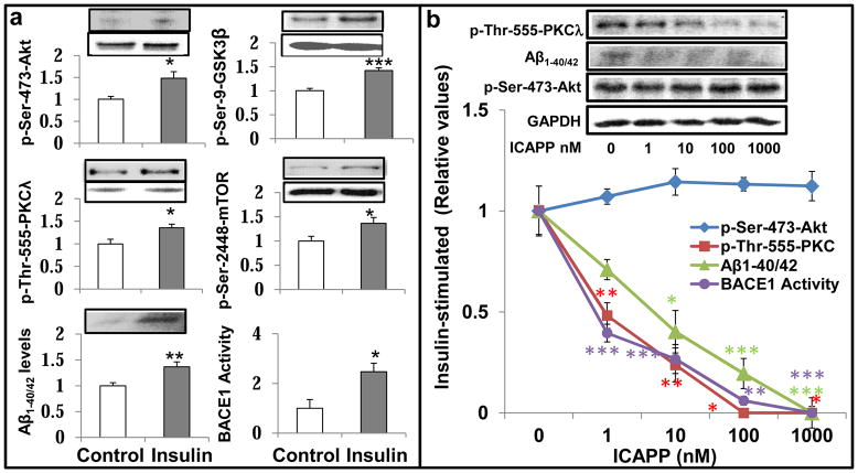
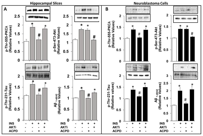
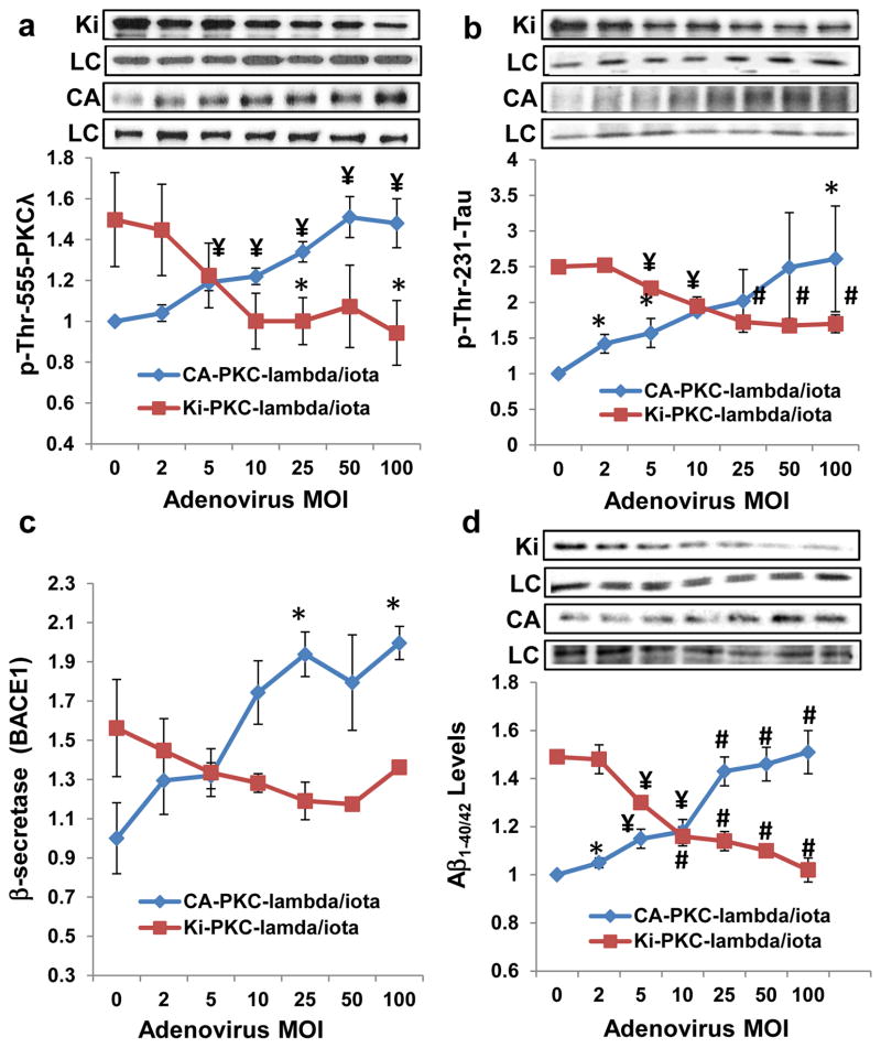
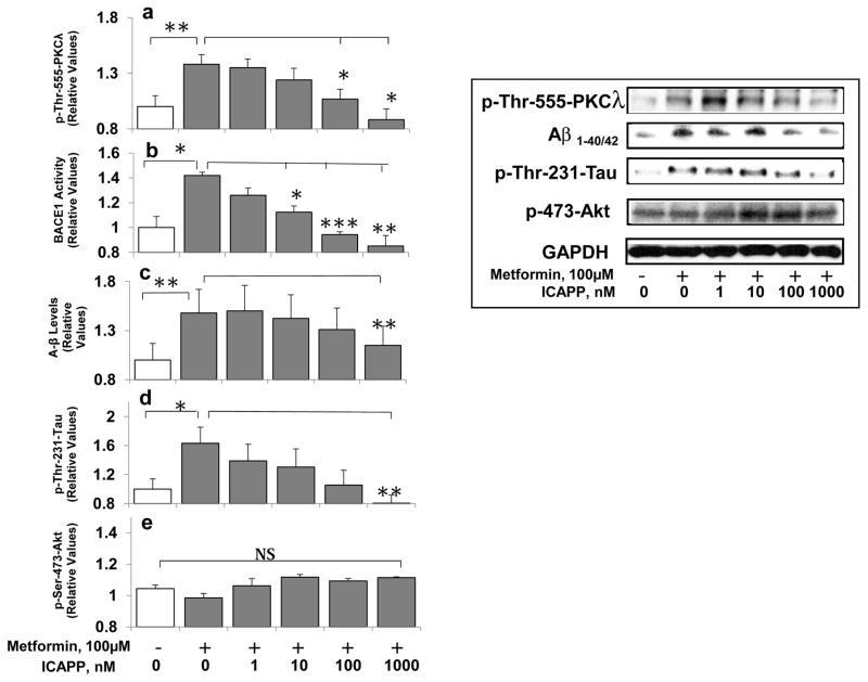
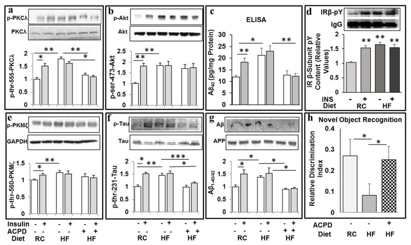
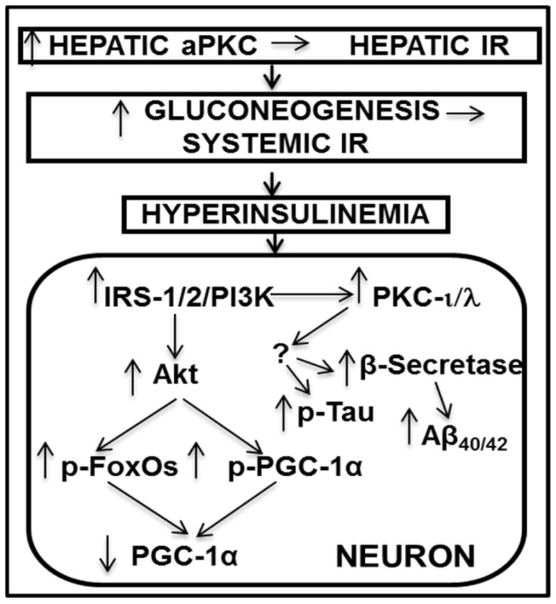
Hyperinsulinemia activates brain Akt and PKC-λ/ι and increases Aβ1-40/42 and phospho-tau in insulin-resistant animals. Here, we examined underlying mechanisms in mice, neuronal cells, and mouse hippocampal slices. Like Aβ1-40/42, β-secretase activity was increased in insulin-resistant mice and monkeys. In insulin-resistant mice, inhibition of hepatic PKC-λ/ι sufficient to correct hepatic abnormalities and hyperinsulinemia simultaneously reversed increases in Akt, atypical protein kinase C (aPKC), β-secretase, and Aβ1-40/42, and restored acute Akt activation. However, 2 aPKC inhibitors additionally blocked insulin's ability to activate brain PKC-λ/ι and thereby increase β-secretase and Aβ1-40/42. Furthermore, direct blockade of brain aPKC simultaneously corrected an impairment in novel object recognition in high-fat-fed insulin-resistant mice. In neuronal cells and/or mouse hippocampal slices, PKC-ι/λ activation by insulin, metformin, or expression of constitutive PKC-ι provoked increases in β-secretase, Aβ1-40/42, and phospho-thr-231-tau that were blocked by various PKC-λ/ι inhibitors, but not by an Akt inhibitor. PKC-λ/ι provokes increases in brain β-secretase, Aβ1-40/42, and phospho-thr-231-tau. Excessive signaling via PKC-λ/ι may link hyperinsulinemia and other PKC-λ/ι activators to pathological and functional abnormalities in Alzheimer's disease.

Keywords: Akt; Alzheimer's; Atypical PKC; Aβ; Beta-secretase; Insulin; Metformin; PKC-iota/lambda; PKM-zeta; Phospho-tau.


Conflict of interest statement

Conflicts

The authors report no conflicts of interest

There are no actual or potential conflicts of interest amongst the authors.

Figures

Figure 1
Figure 1
Hepatic aPKC inhibitor ICAPP reverses hyperinsulinemia-induced increases in basal/resting Akt activity (a) and phosphorylation of Akt substrates, ser-2448-mTOR (b), ser-253-FoxO3a (c), and ser-9-GSK3β (d) in brains of insulin-resistant Het-MλKO (KO) mice. Where indicated, PKC-λ/ι inhibitor, ICAPP (0.4mg/kg body weight), was administered subcutaneously once daily for 8 days, and insulin (1U/kg body weight) (shaded bars) was administered intraperitoneally 15-min before killing. Other features of these Het-MλKO (KO) and littermate wild type (WT) mice were reported previously (Sajan et al., 2012b; Sajan et al., 2016). Note that, by inhibiting hepatic aPKC and aPKC-dependent increases in hepatic gluconeogenic enzymes, ICAPP corrects hyperinsulinemia in Het-MλKO mice (Sajan et al., 2012b), which in turn reduces hyperinsulinemia-dependent increases in resting/basal brain Akt activity and Akt substrate phosphorylation ((Sajan et al., 2016); accordingly, acute effects of exogenous insulin treatment on these Akt-dependent parameters were restored by ICAPP treatment. Representative Western blots of indicated proteins are shown; loading controls are shown below phospho-proteins. Relative bar values are mean ± SEM of 6 mice. Asterisks: *, P<0.05; **, P<0.01; ***, P<0.001 (ANOVA).
Figure 2
Figure 2
aPKC inhibitor ICAPP inhibits hyperinsulinemia-stimulated increases in basal PKC-λ/ι activity (a), Aβ1-40/42 peptide production (b), and β-secretase (BACE1) activity (c) in brains of Het-MλKO (KO) mice, and subsequent stimulatory effects of acute insulin treatment on these parameters. PKC-λ/ι inhibitor, ICAPP (0.4mg/kg body weight), was administered subcutaneously once daily for 8 days to Het-MλKO (KO) mice, and insulin (1U/kg body weight) (shaded bars) was administered intraperitoneally 15-min before killing. Other features of these mice are described in Fig 1 or were previously reported [8,9]. Representative Western blots of indicated proteins are shown; loading controls levels, which were not significantly altered, are not shown in lower panels. Relative bar values are mean ± SEM of 6 mice. Asterisks: *, P<0.05; **, P<0.01; ***, P<0.001 (ANOVA).
Figure 3
Figure 3
Activation of brain β-secretase activity (BACE1) by insulin (1U/kg body weight) administered intraperitoneally 15-min before killing (shaded bars) in control chow-fed (a) and lean (ob+) control (Con) mice (b), and activation of brain β-secretase (BACE1) by hyperinsulinemia in high fat-fed (HFF) mice (a), ob/ob mice (b), and obese/type 2 diabetic (T2DM) monkeys (c). Increases in fasting serum or plasma insulin levels in obese/diabetic versus lean/non-diabetic mice were 4.9 for HFF mice, 4.2 for ob/ob mice, and 2.2 for obese/T2D monkeys (actual data are given in Sajan et al., 2014, Sajan et al., 2015, and Sajan et al., 2016., respectively). Other features of these mice, including alterations in brain Aβ1-40/42, were reported previously [8,9]. Relative bar values are mean ± SEM of (N) mice or monkeys. Asterisks: *, P<0.05; **, P<0.01; ***, P<0.001 (ANOVA).
Figure 4
Figure 4
ICAPP provokes time-related decreases in insulin-stimulated aPKC activity (blue), β-secretase (BACE1) activity (black) and Aβ1-40/42 peptide production (red), but spares insulin-stimulated Akt activation (green) in brains of normal mice. ICAPP (1.5mg/kg body weight) was administered subcutaneously as a single dose at zero time, and, at indicated times, insulin (1U/kg body weight) was administered intraperitoneally15-min before killing. Results in insulin-stimulated samples were compared to results in vehicle-injected control mice (see Figs 1–3 for comparison of control and insulin-stimulated values). Relative insulin-stimulated values plotted here are mean ± SEM of 3–4 mice. Asterisks: *, P<0.05; **, P<0.01; ***, P<0.001 (ANOVA). Representative blots of insulin-stimulated parameters are shown at top, Constant values of p-ser-273-Akt and glyceraldehydes-3-phosphate dehydrogenase (GAPDH) serve as loading controls.
Figure 5
Figure 5
Effects of insulin and ICAPP on aPKC and Akt activities, phosphorylation of Akt substrates (GSK3β and mTOR), β-secretase activity (BACE1) and Aβ1-40/42 peptide levels in LA1-5s human neuroblastoma cells. Effects of 24- hour treatment ± 200nM insulin are shown at left (a), and dose-related effects of ICAPP on insulin-stimulated parameters are shown at right (b). Loading controls, which did not differ significantly; in panel a are shown below phospho proteins; and in panel b, unchanged glyceraldehyde phosphate dehydrogenase (GAPDH) served as a loading control. Relative bar values in panel a are mean ± SEM of 6 determinations. Values in panel b are means ± SEM of 2–4 determinations. Asterisks: *, P<0.05; **, P<0.01; ***, P<0.001 (ANOVA). Note that the incubation time of 24 hours was arbitrarily chosen in anticipation of conducting studies requiring time for expression studies as in Figure 7, wherein cells were incubated for 48 hours to allow time for virally-mediated expression of mutated enzymes.
Figure 6
Figure 6
Effects of aPKC versus Akt inhibition on insulin-stimulated increases in Aβ1-40/42 and p-thr-231-tau in LA1-5s human neuroblastoma neuronal cells (A) and hippocampal slices (B). Neuroblastoma neuronal cells were treated for 24 hours without or with 200nM insulin, 1μM ACPD and 10μM Akti, as indicated. [Note that the dose of 200nM insulin was arbitrarily chosen to be certain that any losses of insulin owing to internalization and/or degradation by insulin-degrading enzyme would not limit maximal effects of insulin; the incubation time of 24 hours was arbitrarily chosen to allow attainment of maximal effects of a variety of agents, including insulin, metformin, metabolites, etc., on tissue levels of activated PKC-λ and increases in PKC-λ-dependent parameters.] Hippocampal slices were incubated for 1 hour without or with 100nM insulin, 1μM ACPD and 10μM Akti. Loading controls, which did not differ significantly in are shown below phospho proteins. The loading control for Aβ was 120kDa APP. Values are Mean ± SEM of 4 determinations. *, P<0.05 vs, unstimulated control mean value (clear bars); #, P<0.05, vs. insulin-stimulated mean value in absence of inhibitor treatment.
Figure 7
Figure 7
Adenovirus (Adv) encoding constitutively active (CA) PKC-λ/ι (in blue) dose-relatedly phosphorylates/activates total aPKC (a) and increases phospho-tau levels (b), β-secretase activity (BACE1) (c) and Ab 1-40/42 levels (d) in LA1-5s human neuroblastoma cells. Adv encoding kinase-inactive (KI) PKC-λ/ι (in red) dose-relatedly blocks effects of 200nM insulin on these parameters. Cells were incubated for 48 hours with (red) and without (blue) 200nM insulin and indicated Adv multiplicity of infection (MOI), the total level of which kept constant at 100 MOI for all samples by adding non-coding Adv. Note that the explanation for inhibitory effects of kinase-inactive (KI) PKC-λ [mutagenized in the ATP-binding site of the catalytic domain (Kotani et al, 1998) on endogenous wild type (WT) PKC-λ during insulin action is that KI-PKC-λ competes with WT-PKC-λ for either the PKC-λ-activating lipid, PIP3, or for the substrate-recognition motif (including the auto(trans)phosphorylation site at thr-555), or both. Shown here are Mean ± SEM (N=4) values relative to the mean initial basal or insulin-stimulated level of indicated protein/peptide. Symbols: *, P<0.05; ¥, P<0.01; #, P<0.001 (ANOVA). Abbreviations: KI, kinase-inactive PKC-λ; LC, loading control, glyceraldehyde phosphate dehydrogenase; and CA, constitutively-active PKC-λ.
Figure 8
Figure 8
Metformin activates aPKC (a), and increases β-secretase activity (BACE1) (b), A-beta (Aβ1-40/42) levels (c), and phospho-thr-231-tau levels (d), but not Akt activity (e) in LA1-5s human neuroblastoma cells, and PKC-λ/ι inhibitor, ICAPP, dose-relatedly blocks metformin-induced increases in aPKC activity, β-secretase activity (BACE1), and levels of Aβ1-40/42 and phospho-thr-231-tau. As in Fig 6, cells were incubated for 24 hours with metformin and ICAPP. Shown here are Mean ± SEM (N=4) values relative to the mean initial basal or insulin-stimulated level of indicated immunoreactive protein/peptide. Asterisks: *, P<0.05; **, P<0.01; ***, P<0.001 (ANOVA). Abbreviation: GAPDH, glyceraldehyde phosphate dehydrogenase, was used for loading controls.
Figure 9
Figure 9
Treatment of high-fat-fed mice with aPKC inhibitor, ACPD, reduces high fat diet-induced and acute insulin-stimulated increases in 70kDa PKC-ι/λ activity (a), Aβ1-40 levels (as per ELISA) (c), 4kDa Aβ1-40/42 levels (as per Western) (g), and phospho-thr-231-tau (f) levels, and simultaneously reverses/prevents a memory impairment in novel object recognition (NOR) (h), without affecting high fat (HF) diet-induced and acute insulin-stimulated increases in Akt activity (b) or PKM-ζ activity (e). Effects of hyperinsulinemia in HFF mice, and acute insulin treatment in regular chow (RC) fed and HFF mice, on insulin receptor (IR) activity (as per total pY content of immunoprecipitated IR β-subunit) are shown in panel (d). Mice were fed regular chow (RC) or a high fat (HF) diet, and treated ± ACPD (20mg/kg body weight, Monday, Wednesday and Friday of each week) over 10 weeks. Novel Object Recognition (NOR) testing was conducted at week 9. At 10 weeks, mice were treated acutely ± insulin (1U/kg body weight) or vehicle 15 min before killing. Values are mean ± SEM (N=4). Asterisks: *, P<0.05; **, P<0.01; ***, P<0.001 (ANOVA).
Figure 10
Figure 10
Schematic of pathogenesis of neuronal signaling abnormalities in insulin-resistant states that lead to production of factors that may abet development of Alzheimer’s disease. In this scheme, diet-induced increases in hepatic aPKC activity lead to impaired Akt activation by insulin, i.e., hepatic insulin resistance (IR), increases in hepatic gluconeogenesis, systemic IR, and hyperinsulinemia, which persistently hyperactivates brain Akt and aPKC. Increases in brain Akt activity lead to phosphorylation and thus diminished activities of all FoxOs (1/3a/4/6), and decreased activity and expression of PGC-1α (all needed for neuronal function and integrity). Increases in brain aPKC activity, either directly or indirectly, provoke increases in β-secretase activity, and levels of Aβ1-40/42 and phospho-thr- 231-tau, and thus abet plaque and tangle development.

References

    1. Antunes M, Biala G. The novel object recognition memory: neurobiology, test procedure and its modification. Cogn Process. 2012;13:93–110. - PMC - PubMed
    1. Barini E, Antico O, Zhao Y, Asta F, Tucci V, Catelani T, Marotta R, Xu H, Gasparini L. Metformin promotes tau aggregation and exacerbates abnormal behavior in a mouse model of tauopathy. Molec Neurodegen. 2016;11:16–36. - PMC - PubMed
    1. Barron AM, Rosario ER, Pike CJ. Sex-specific effects of high fat diet on indices of metabolic syndrome in 3xTg-AD mice: Implications for Alzheimer’s disease. LPOS ONE. 2013;8:1–8. - PMC - PubMed
    1. Chen Y, Zhou K, Wang R, Liu Y, Kwak Y-D, Ma T, Thompson RC, Zhao Y, Smith L, Gasparini L, Luo Z, Xu H, Liao F-F. Antidiabetic drug metformin (Glucophage) increases biogenesis of Alzheimer’s amyloid peptides via upregulating BACE1 transcription. Proc Natl Acad Sci USA. 2009;106:3907–3912. - PMC - PubMed
    1. Craft S. Insulin resistance and Alzheimer’s disease pathogenesis: mechanisms and implications for treatment. Curr Alzheimer Res. 2007;4:147–152. - PubMed

Publication types