. 2018 Jan 3;26(1):256-268.
doi: 10.1016/j.ymthe.2017.09.014. Epub 2017 Sep 19.

Safety and Long-Term Efficacy of AAV4 Gene Therapy in Patients with RPE65 Leber Congenital Amaurosis

Affiliations

Safety and Long-Term Efficacy of AAV4 Gene Therapy in Patients with RPE65 Leber Congenital Amaurosis

Guylène Le Meur et al. Mol Ther. .

Abstract

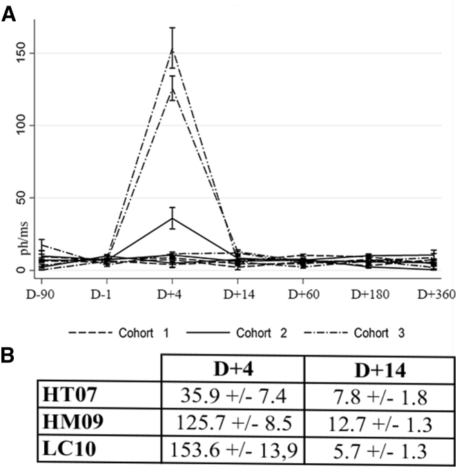
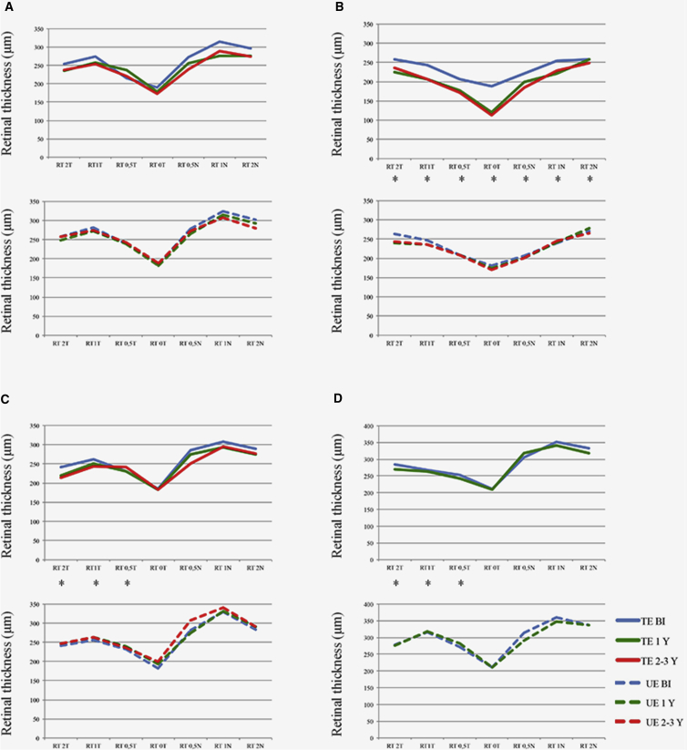
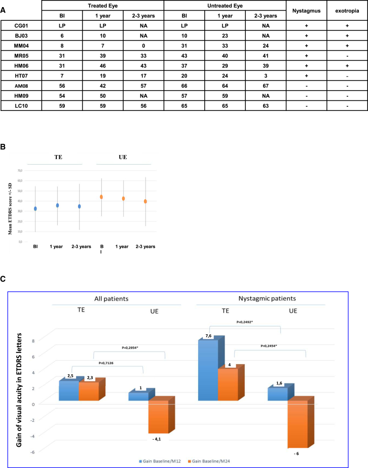
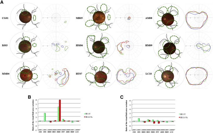
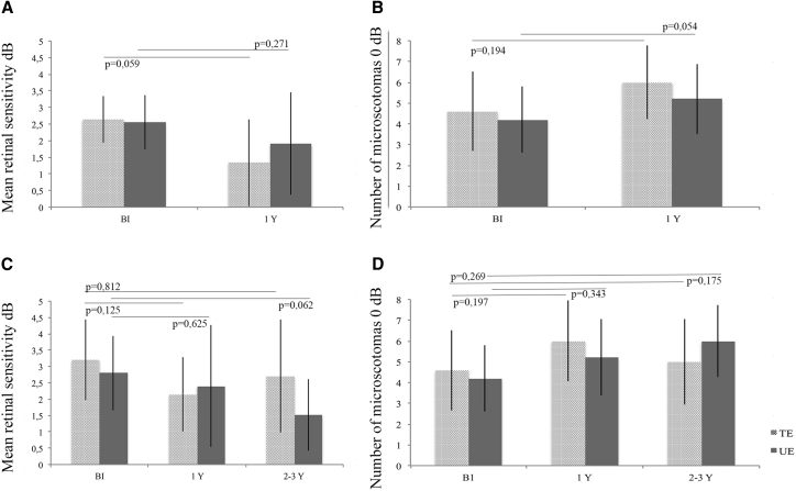
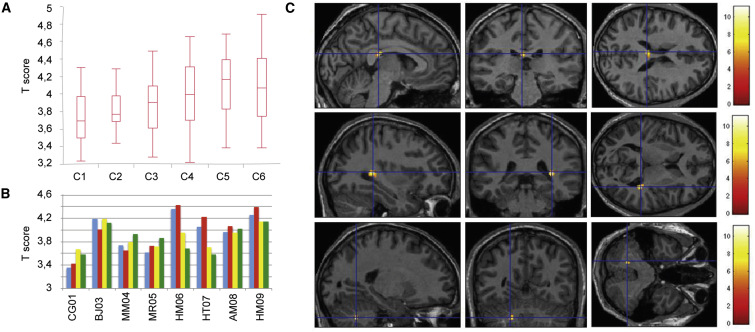
The aim of this study was the evaluation of the safety and efficacy of unilateral subretinal injection of the adeno-associated vector (AAV) serotypes 2 and 4 (AAV2/4) RPE65-RPE65 vector in patients with Leber congenital amaurosis (LCA) associated with RPE65 gene deficiency. We evaluated ocular and general tolerance and visual function up to 1 year after vector administration in the most severely affected eye in nine patients with retinal degeneration associated with mutations in the RPE65 gene. Patients received either low (1.22 × 10¹⁰ to 2 × 10¹⁰ vector genomes [vg]) or high (between 3.27 × 10¹⁰ and 4.8 × 10¹⁰ vg) vector doses. An ancillary study, in which six of the original nine patients participated, extended the follow-up period to 2-3.5 years. All patients showed good ophthalmological and general tolerance to the rAAV2/4-RPE65-RPE65 vector. We observed a trend toward improved visual acuity in patients with nystagmus, stabilization and improvement of the visual field, and cortical activation along visual pathways during fMRI analysis. OCT analysis after vector administration revealed no retinal thinning, except in cases of macular detachment. Our findings show that the rAAV2/4.RPE65.RPE65 vector was well tolerated in nine patients with RPE65-associated LCA. Efficacy parameters varied between patients during follow-up.

Keywords: AAV vector; RPE65; clinical trial; gene therapy; inherited retinal dystrophies.


Figures

Figure 1
Figure 1
Evaluation of Post-injection Inflammation (A) Laser flare meter analysis of treated eyes (Ph/ms) at D−90, D−1, D+4, D+14, D+60, D+180, and D+360. (B) Laser flare meter values (Ph/ms) for the three patients exhibiting inflammation.
Figure 2
Figure 2
SD-OCT Evaluation of Changes in Total Retinal Thickness according to Eccentricity of the Fovea SD-OCT evaluation of changes in total retinal thickness according to eccentricity of the fovea was measured in treated eyes (upper graphs, solid line) and untreated eyes (lower graphs, dotted line) at baseline (blue line), 1 year post-treatment (green line), and 2–3 years post-treatment (red line). (A) Mean retinal thickness in all patients. (B) Patient who experienced foveal detachment during surgery (MM04). (C) Patient with the bleb close to the fovea (HM06). (D) Patient with the bleb close to the fovea (HM09). BI, before injection; N, nasal; T, temporal; TE, treated eye; UE, untreated eye; Y, year, Asterisks (*) indicate subretinal injection site.
Figure 3
Figure 3
Visual Acuity Measured in Treated Eyes and Untreated Eyes at 1 (Blue) and 2–3 Years (Orange) Post-injection (A) ETDRS visual acuity of injected and uninjected eyes before injection, 1 year post-injection, and at the final examination. (B) Mean visual acuity in the six patients who underwent long-term follow-up. (C) Changes in mean visual acuity after the surgery as measured in the untreated and treated eyes in the six patients who underwent long-term follow-up. Data are shown for all patients (left) and for nystagmic patients only (right). BI, before injection; LP, light perception; TE, treated eye; UE, untreated eye.
Figure 4
Figure 4
Visual Field Analysis during Follow-up (A) For each patient a composite retinal photograph is shown (left): the area exposed to the vector is indicated with a green line. The Goldmann visual field is shown on the right: blue lines delineate the V4 surface before injection, green lines indicate the V4 surface 1 year post-injection, and red lines indicate the V4 surface at the final follow-up examination. (B) Variation in the visual field surface area of the treated eye: 1 year post-treatment versus values obtained at final follow-up examination. (C) Variation in the visual field surface area in untreated eye: 1 year post-treatment versus values obtained at final follow-up examination. TE, treated eye; UT, untreated eye; Y, year.
Figure 5
Figure 5
Microperimetry Data for All Patients at 1 Year Post-injection and for Patients Who Underwent Long-Term Follow-up (A) Mean retinal sensitivity at 1 year post-injection. (B) Number of microscotomas at 1 year post-injection. (C) Mean retinal sensitivity in patients who underwent long-term follow up. (D) Number of microscotomas in patients who underwent long-term follow-up. Treated and untreated eyes are represented by light gray and dark gray bars, respectively. BI, before injection; Y, year.
Figure 6
Figure 6
Functional MRI Analysis (A) Activity in the primary visual cortex as a function of the intensity of light intensity of stimulation. T score is based on the stimulation conditions (C1, lowest luminance; C6, highest luminance), as illustrated in the boxplot. (B) Primary visual cortex activity in individual patients. The mean T score per patient is shown. Blue, treated eye before treatment; red, treated eye after treatment; yellow, untreated eye before treatment; green, untreated eye after treatment. (C) Increase in activity after treatment. Statistical parametric map showing the voxels with a significant activation (p < 0.001 uncorrected, cluster correction at p < 0.05, random effect; n = 8) after versus before treatment, all stimulation conditions together, projected onto a mean MRI template. Color bar indicates t scores.

References

    1. Leber T. Ueber Retinitis pigmentosa und angeborene Amaurose. Graefes Arch. Clin. Exp. Ophthalmol. 1869;15:1–25.
    1. Kaplan J. Leber congenital amaurosis: from darkness to spotlight. Ophthalmic Genet. 2008;29:92–98. - PubMed
    1. Chacon-Camacho O.F., Zenteno J.C. Review and update on the molecular basis of Leber congenital amaurosis. World J. Clin. Cases. 2015;3:112–124. - PMC - PubMed
    1. Hanein S., Perrault I., Gerber S., Tanguy G., Rozet J.-M., Kaplan J. Leber congenital amaurosis: survey of the genetic heterogeneity, refinement of the clinical definition and phenotype-genotype correlations as a strategy for molecular diagnosis. Clinical and molecular survey in LCA. Adv. Exp. Med. Biol. 2006;572:15–20. - PubMed
    1. Mata N.L., Moghrabi W.N., Lee J.S., Bui T.V., Radu R.A., Horwitz J., Travis G.H. Rpe65 is a retinyl ester binding protein that presents insoluble substrate to the isomerase in retinal pigment epithelial cells. J. Biol. Chem. 2004;279:635–643. - PubMed

Publication types

MeSH terms

Substances