. 2017 Nov 16;171(5):1110-1124.e18.
doi: 10.1016/j.cell.2017.09.039. Epub 2017 Oct 12.

The DNA Inflammasome in Human Myeloid Cells Is Initiated by a STING-Cell Death Program Upstream of NLRP3

Affiliations

The DNA Inflammasome in Human Myeloid Cells Is Initiated by a STING-Cell Death Program Upstream of NLRP3

Moritz M Gaidt et al. Cell. .

Abstract

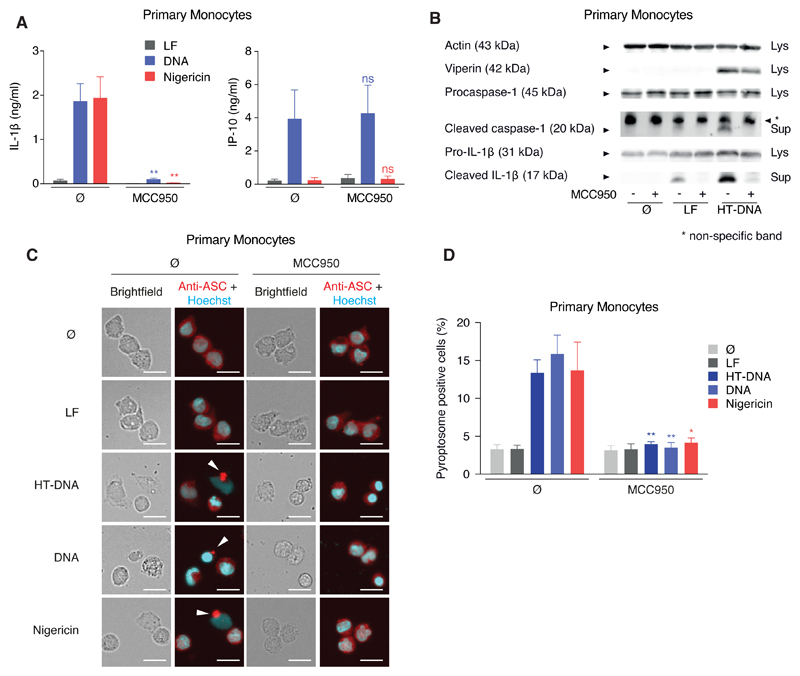
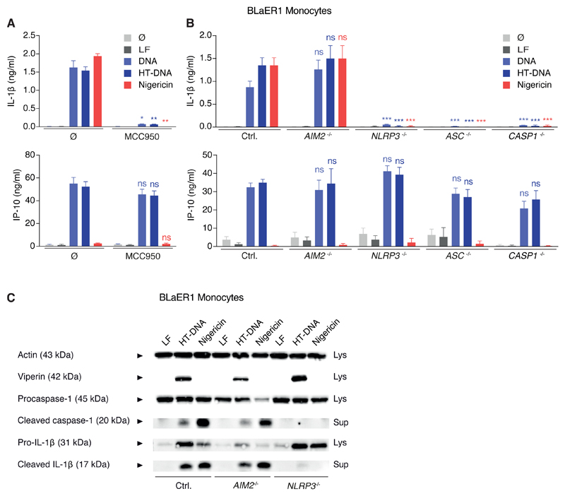
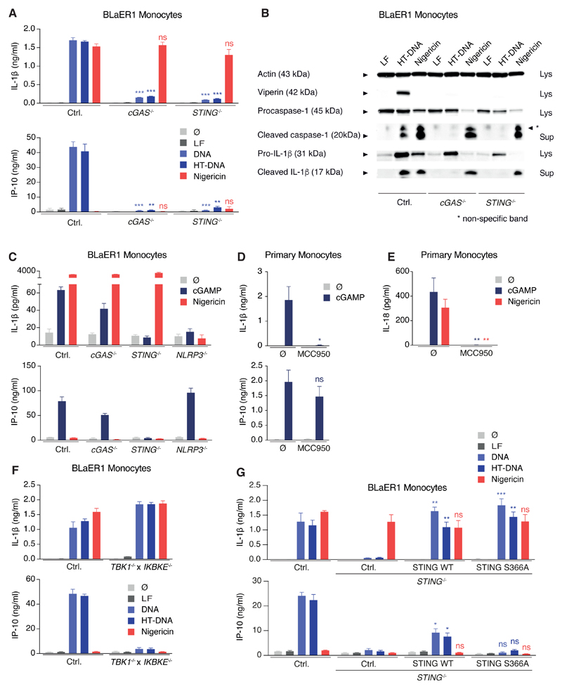
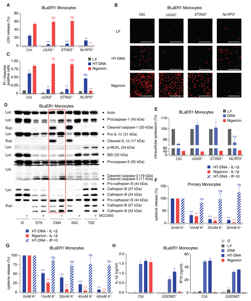
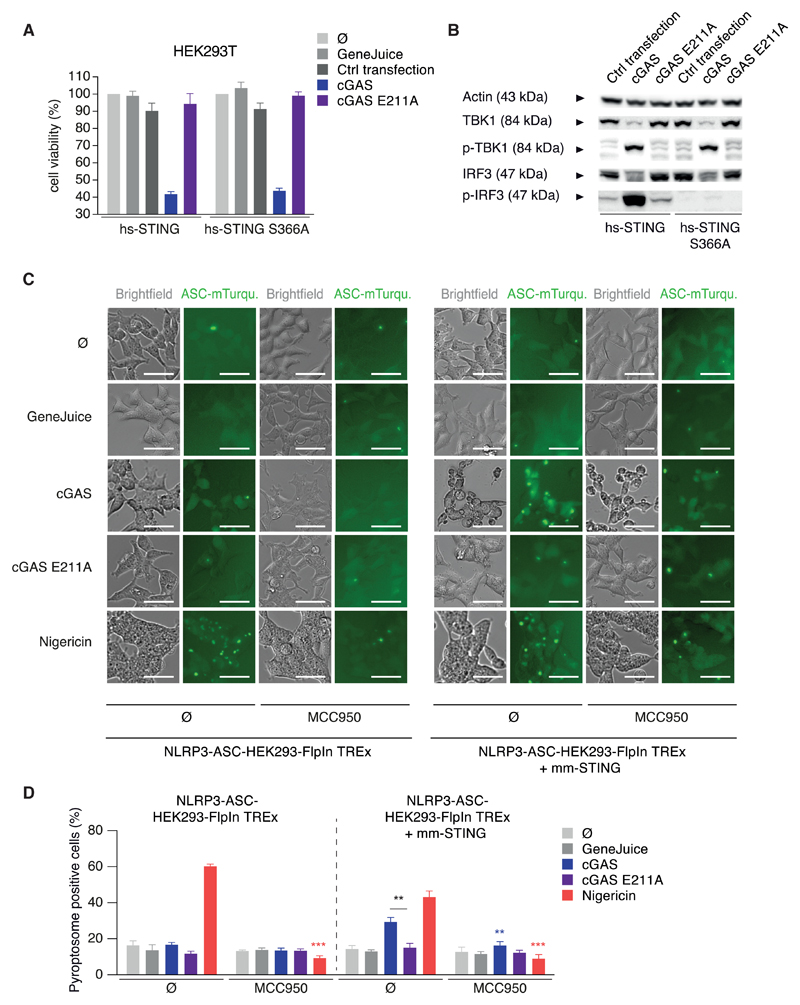
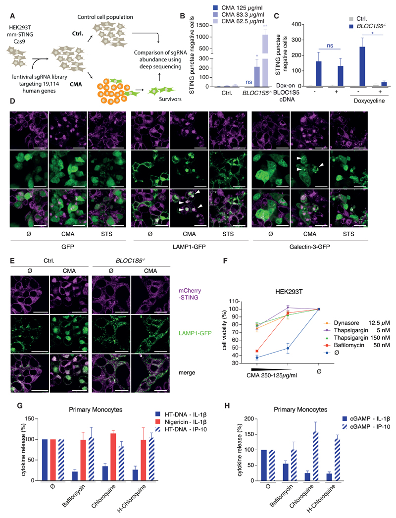
Detection of cytosolic DNA constitutes a central event in the context of numerous infectious and sterile inflammatory conditions. Recent studies have uncovered a bipartite mode of cytosolic DNA recognition, in which the cGAS-STING axis triggers antiviral immunity, whereas AIM2 triggers inflammasome activation. Here, we show that AIM2 is dispensable for DNA-mediated inflammasome activation in human myeloid cells. Instead, detection of cytosolic DNA by the cGAS-STING axis induces a cell death program initiating potassium efflux upstream of NLRP3. Forward genetics identified regulators of lysosomal trafficking to modulate this cell death program, and subsequent studies revealed that activated STING traffics to the lysosome, where it triggers membrane permeabilization and thus lysosomal cell death (LCD). Importantly, the cGAS-STING-NLRP3 pathway constitutes the default inflammasome response during viral and bacterial infections in human myeloid cells. We conclude that targeting the cGAS-STING-LCD-NLRP3 pathway will ameliorate pathology in inflammatory conditions that are associated with cytosolic DNA sensing.

Keywords: AIM2; Caspase-1; DNA; IL-1β; NLRP3; STING; cGAS; inflammasome; lysosomal cell death.


Figures

Figure 1
Figure 1. Cytosolic DNA Recognition Leads to NLRP3-Dependent Classical Inflammasome Activation in Human Monocytes
(A and B) Pam3CSK4-primed primary human monocytes were treated with or without MCC950 and stimulated with lipofected DNA, HT-DNA, or lipofection reagent alone (LF) for 8 hr. If indicated, nigericin was added for the last 2 hr of the experiment. Cytokines were quantified by ELISA or samples were analyzed by immunoblotting. Data are depicted as mean + SEM of seven donors (A) or one representative immunoblot of three (B). **p < 0.01; ns, not significant. (C and D) Primary human monocytes were stimulated as in (A) and pyroptosome formation was analyzed. Representative images (C) and quantifications from four donors are shown as mean + SEM (D). Scale bars denote 15 µm. **p < 0.01, *p < 0.05. See also Figure S1.
Figure 2
Figure 2. BLaER1 Monocytes Faithfully Recapitulate DNA-Mediated NLRP3-Inflammasome Activation Independently of AIM2
(A) LPS-primed BLaER1 monocytes were stimulated as indicated. Data are depicted as mean + SEM of three experiments. **p < 0.01, *p < 0.05, ns, not significant. LF, Lipofectamine. (B and C) BLaER1 monocytes of indicated genotype were stimulated as in (A). Data are shown as mean + SEM of three experiments (B) or one representative immunoblot from four experiments (C). ***p < 0.001; ns, not significant. See also Figure S2.
Figure 3
Figure 3. cGAS/STING Signaling Activates NLRP3 Independently of Type I Interferon Induction
(A and B) LPS-primed BLaER1 monocytes of indicated genotype were stimulated with lipofected DNA for 8 hr. Nigericin was added for the last 2 hr. (A) Cytokine release is depicted as mean + SEM from three experiments. (B) One immunoblot from two experiments is shown. ***p < 0.001, **p < 0.01; ns, not significant. (C) LPS-primed BLaER1 monocytes were stimulated with 3 µg cGAMP for 16 hr. If indicated, nigericin was added for the last 2 hr of the experiment. Data are depicted as mean + SEM of 3–5 experiments. (D and E) Pam3CSK4-primed primary human monocytes with or without MCC950 were stimulated with 3 µg cGAMP for 16 hr. Cytokine release from four donors (D) or seven donors (E) is depicted as mean + SEM. **p < 0.01, *p < 0.05; ns, not significant. (F) LPS-primed BLaER1 monocytes were stimulated as in (A). Cytokine release is depicted as mean + SEM from three experiments. (G) WT or the S366A mutant of hs-STING were stably expressed in STING−/− BLaER1 monocytes and indicated cells stimulated as in (A). Data are shown as mean + SEM of three independent experiments. **p < 0.01, *p < 0.05; ns, not significant. See also Figure S3.
Figure 4
Figure 4. STING Activation Leads to a Lytic Cell Death that Drives NLRP3 Activation by Inducing K+ Efflux
(A–C) LPS-primed BLaER1 monocytes were stimulated with lipofected DNA for 8 hr or with nigericin (last 2 hr). Cell death was quantified by LDH-release or by microscopy for PI-positive cells. Data are depicted as mean + SEM from three experiments (A), representative micrographs from four experiments (B), or quantifications from four experiments as mean + SEM (C). ***p < 0.001, **p < 0.01; ns, not significant. Scale bars denote 100 µm. (D) LPS-primed BLaER1 mm-STING monocytes were stimulated with staurosporine (STS), CMA, nigericin (NIG) or hsTNF, birinapant, Z-VAD-FMK (TBZ) with or without MCC950. One immunoblot of three experiments is shown. (E) LPS-primed BLaER1 monocytes were stimulated as in (A) and intracellular K+ content quantified by ICP-MS. Data are shown as mean + SEM from three experiments. *p < 0.05. (F) Pam3CSK4-primed primary monocytes were stimulated as indicated in presence of increasing concentrations of extracellular K+. Data are shown as mean + SEM of four donors. (G) LPS-primed BLaER1 monocytes were stimulated as in (A) with increasing concentrations of extracellular K+. Cytokine release was quantified from three experiments and is depicted as mean + SEM. (H) LPS-primed BLaER1 monocytes were stimulated as in (A). Data are depicted as mean + SEM of three experiments. See also Figure S3.
Figure 5
Figure 5. The cGAS-STING-NLRP3 Inflammasome Can Be Reconstituted in HEK293 Cells
(A and B) Indicated HEK293T cells were transfected with mm-cGAS or the inactive E211A mutant. Cell viability was quantified by CTB assay and is depicted as mean + SEM of three experiments (A). One immunoblot of two is shown (B). (C and D) Indicated HEK293 NLRP3 ASC cells were transfected with cGAS or the E211A mutant or stimulated as indicated with or without MCC950. Representative micrographs are shown (C) and were quantified from three experiments (D) and data are depicted as mean + SEM. **p < 0.001, **p < 0.01. Scale bars indicate 50 µm. See also Figure S4.
Figure 6
Figure 6. A Genome-wide sgRNA-Screen Identifies BLOC1 as a Modulator of STING-Mediated Cell Death Guiding Its Characterization as Lysosomal Cell Death
(A) A schematic overview of the screening procedure is shown. (B) HEK293T mm-STING cells of indicated genotype were stimulated with CMA for 16 hr. STING punctae negative cells per visual field from four experiments are depicted as mean + SEM showing one representative clone of four. (C) HEK293T mm-STING cells of indicated genotype that were transduced with a dox-on BLOC1S5 cDNA or an empty vector were stimulated with doxycycline for 24 hr and subsequently with 83.3 µg/mL CMA for 16 hr. STING punctae negative cells from three experiment are depicted as mean + SEM. **p < 0.01, *p < 0.05; ns, not significant. (D) HEK293T mm-STING cells were transfected with GFP, LAMP1-GFP, or Galectin-3-GFP expression constructs. 24 hr later, cells were stimulated with CMA or staurosporine (STS) for 8 hr. Confocal micrographs from one experiment out of three are depicted. Scale bars indicate 25 µm. (E) HEK293T mm-STING cells of indicated genotype that were transduced with a LAMP1-GFP reporter were stimulated with 62.5 µg/mL CMA for 16 hr. Representative confocal micrographs from one experiment of two are depicted. Scale bars indicate 25 µm. (F) HEK293T mm-STING cells were stimulated with indicated inhibitors and CMA for 16 hr. CTB assay and is depicted as mean + SEM of three experiments. (G and H) Pam3CSK4-primed primary monocytes with or without indicated inhibitors were stimulated with HT-DNA for 8 hr (G) or with 3 µg cGAMP (H) for 16 hr. Nigericin was added for the last 2 hr. Normalized cytokine release from 4–7 donors is depicted as mean + SEM. See also Figures S5 and S6.
Figure 7
Figure 7. The cGAS-STING-NLRP3 Inflammasome Is Involved in Bacterial and Viral Sensing and Constitutes the Main DNA-Sensing Inflammasome in Human Myeloid Cells
(A and B) LPS-primed BLaER1 monocytes were stimulated with different MOIs of the respective strain of F. novicida (A) or with different MOIs of MVA (B) for 8 hr. Data are depicted as mean + SEM of three experiments. (C) Indicated LPS-primed BLaER1 monocytes were stimulated for 8 hr. Data are depicted as mean + SEM of three independent experiments. (D and E) Mononuclear cells from human bone marrow were primed with Pam3CSK4 for 2 hr and stimulated with lipofected DNA or nigericin for 8 hr or with 3 µg cGAMP for 16 hr in absence or presence of MCC950. Cytokine secretion from three (D) or six (E) donors is depicted as mean + SEM. **p < 0.01, *p < 0.05. (F) Human BMDM were primed with Pam3CSK4 for 2 hr and stimulated with lipofected DNA for 8 hr in absence or presence of MCC950. Nigericin was added for the last 2 hr. Cytokine secretion from 4 donors is depicted as mean + SEM. IL-1β responses were normalized to nigericin-mediated IL-1β secretion. (G) A schematic view of the cGAS-STING-LCD-NLRP3 signaling cascade is shown. See also Figure S7.

References

    1. Abe T, Barber GN. Cytosolic-DNA-mediated, STING-dependent proinflammatory gene induction necessitates canonical NF-kappaB activation through TBK1. J Virol. 2014;88:5328–5341. - PMC - PubMed
    1. Ablasser A, Schmid-Burgk JL, Hemmerling I, Horvath GL, Schmidt T, Latz E, Hornung V. Cell intrinsic immunity spreads to bystander cells via the intercellular transfer of cGAMP. Nature. 2013;503:530–534. - PMC - PubMed
    1. Aits S, Jaattela M. Lysosomal cell death at a glance. J Cell Sci. 2013;126:1905–1912. - PubMed
    1. Baum R, Sharma S, Carpenter S, Li QZ, Busto P, Fitzgerald KA, Marshak-Rothstein A, Gravallese EM. Cutting edge: AIM2 and endosomal TLRs differentially regulate arthritis and autoantibody production in DNase II-deficient mice. J Immunol. 2015;194:873–877. - PMC - PubMed
    1. Broz P, Dixit VM. Inflammasomes: mechanism of assembly, regulation and signalling. Nat Rev Immunol. 2016;16:407–420. - PubMed