. 2017 Oct 19;68(2):398-413.e6.
doi: 10.1016/j.molcel.2017.09.016. Epub 2017 Oct 12.

The Elongation Factor Spt6 Maintains ESC Pluripotency by Controlling Super-Enhancers and Counteracting Polycomb Proteins

Affiliations

The Elongation Factor Spt6 Maintains ESC Pluripotency by Controlling Super-Enhancers and Counteracting Polycomb Proteins

A Hongjun Wang et al. Mol Cell. .

Abstract

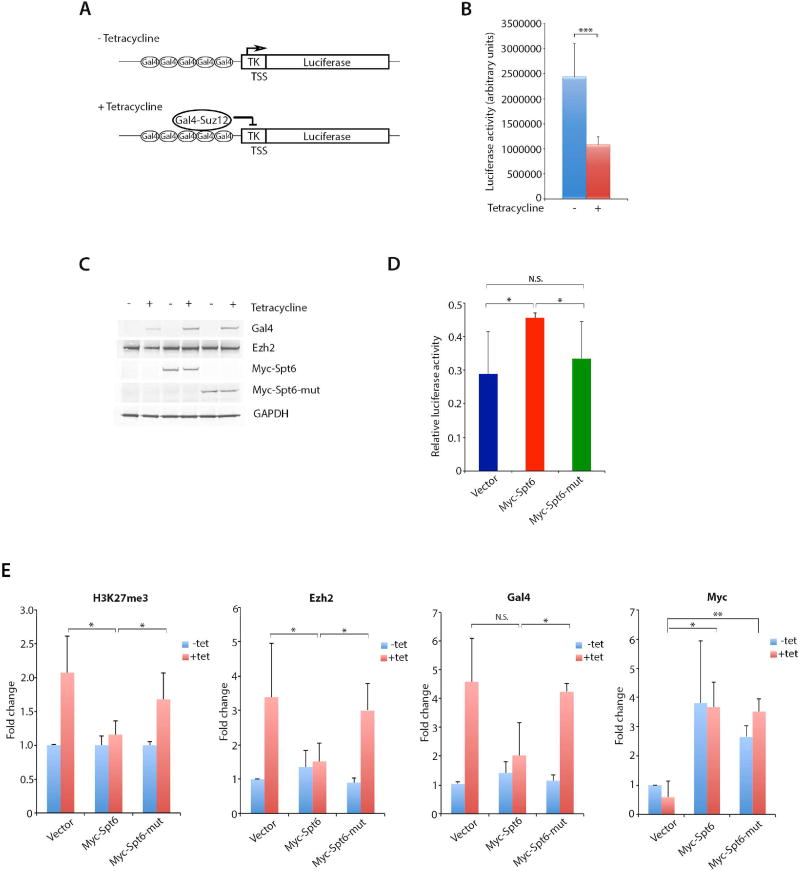
Spt6 coordinates nucleosome dis- and re-assembly, transcriptional elongation, and mRNA processing. Here, we report that depleting Spt6 in embryonic stem cells (ESCs) reduced expression of pluripotency factors, increased expression of cell-lineage-affiliated developmental regulators, and induced cell morphological and biochemical changes indicative of ESC differentiation. Selective downregulation of pluripotency factors upon Spt6 depletion may be mechanistically explained by its enrichment at ESC super-enhancers, where Spt6 controls histone H3K27 acetylation and methylation and super-enhancer RNA transcription. In ESCs, Spt6 interacted with the PRC2 core subunit Suz12 and prevented H3K27me3 accumulation at ESC super-enhancers and associated promoters. Biochemical as well as functional experiments revealed that Spt6 could compete for binding of the PRC2 methyltransferase Ezh2 to Suz12 and reduce PRC2 chromatin engagement. Thus, in addition to serving as a histone chaperone and transcription elongation factor, Spt6 counteracts repression by opposing H3K27me3 deposition at critical genomic regulatory regions.

Keywords: Spt6; embryonic stem cells; polycomb proteins; super-enhancers.


Figures

Figure 1
Figure 1. Genome-Wide Distribution of Spt6 in ESCs Negatively Correlates with PRC2 and H3K27me3
(A) Genome-wide distribution of Spt6 binding in ESCs. (B) Venn diagrams of genomic regions co-occupied by Spt6 and H3K4me3, H3K4me1, H3K27ac, or H3K27me3. (C) Bar graph depicting percentages of Spt6 peaks at TSS H3K4me3+/H3K27ac+, TSS H3K4me3+/H3K27me3+, H3K4me1+/H3K27ac+, H3K4me1+/H3K27me3+, and TSS (H3K4me3/H3K27me3+). (D) Spt6, H3K4me3, H3K4me1, H3K27ac, and H3K27me3 ChIP-seq profiles at pluripotency genes (Pou5f1, Nanog, Sox2) and cell lineage-affiliated genes (Gata2, Myod1). (E) Averaged normalized tag densities of H3K27me3, Suz12, or Ezh2 ChIP-seq signals at regions with different Spt6 enrichment signal (low Spt6: lowest quartile; medium low and medium high Spt6: middle quartile; high Spt6; upper quartile) at transcriptional start sites (TSS). (F) Averaged normalized tag densities of H3K27me3 at KDM6A, KDM6A+, KDM6A/high Spt6, or KDM6A/low Spt6 TSS. (G) Spt6 and H3K27me3 ChIP-seq and RNA-seq profiles at pluripotency genes (Zfp42 and Klf2) and cell lineage gene Pitx2.
Figure 2
Figure 2. Spt6 Maintains Expression of Pluripotency Genes in ESC s
(A) Relative expression measured by qPCR of Nanog, Sox2, Pou5f1, Esrrb, Klf2, Tcl1, and Prdm14 transcripts from ESCs transfected with either control or Spt6 siRNA. Data are presented as mean ± SD (standard deviation) (n=3) (**) P<0.01. (B) ESCs were transfected with either control or Spt6 siRNA and cell extracts were immunoblotted with Spt6, Oct4, Nanog, and Gapdh antibodies. (C) Scatter plot of the RNA-Seq datasets from ESCs transfected with either control or Spt6 siRNA. Up- and down-regulated transcripts are in red and blue, respectively. (D) GO-biological processes associated with genes with ≥ 1.5-fold decreased (upper panel) or increased (lower panel) expression from ESCs transfected with either control or Spt6 siRNA. (E) Heatmaps representing the log2 expression values for genes associated with pluripotency, mesoderm, ectoderm, and trophoectoderm obtained by RNA-Seq of ESCs transfected with either control or Spt6 siRNA. (F) Phase contrast microscopy of ESCs transfected with either control or Spt6 siRNA (top panel), and immunohistochemistry staining with the pluripotency marker alkaline phosphatase (AP) (lower panel).
Figure 3
Figure 3. Spt6 is Enriched at ESC Super-Enhancers
(A) Distribution of Med1, H3K27ac, and Spt6 normalized ChIP-seq signals across a subset of 8,794 ESC enhancers. Med1, H3K27ac, and Spt6 are enriched at a subset of enhancers (super-enhancers). (B) Metagenes of Spt6 ChIP-seq density (reads per million reads per base pair) across the 231 super-enhancers (SE) and the 8,563 typical enhancers (TE). (C) Box plot of Spt6 ChIP-seq density at SE and TE regions. (D) Spt6, Med1, Oct4, Sox2, and Nanog occupancies for super-enhancer regions of Nanog and Pou5f1 (Oct4) loci. (E) Heatmap representing the log2 expression values of SE-associated genes in ESCs transfected with either control or Spt6 siRNA.
Figure 4
Figure 4. H3K27ac is Reduced at Super-Enhancers and Associated Promoters of Spt6-Depleted ESCs
(A) Cell extracts of control or Spt6 siRNA ESCs were immunoblotted with Spt6, H3K27ac, and unmodified histone H3 antibodies. (B) Genome-wide H3K27ac coverage in control and Spt6 siRNA ESCs. (C) Average ChIP-seq read density of H3K27ac at typical enhancers in control and Spt6 siRNA ESCs. (D) Average ChIP-seq read density of H3K27ac at super-enhancers in control and Spt6 siRNA ESCs. (E) Violin plots representing H3K27ac read coverage at typical enhancers in control and Spt6 siRNA ESCs. (F) Violin plots representing H3K27ac read coverage at super-enhancers in control and Spt6 siRNA ESCs. (G) Violin plots representing H3K27ac read coverage at promoters associated with typical enhancers in control and Spt6 siRNA ESCs. (H) Violin plots representing H3K27ac read coverage at promoters associated with super-enhancers in control and Spt6 siRNA ESCs. (I) H3K27ac ChIP-seq profiles at the Fgfr3 locus in control and Spt6 siRNA ESCs. (J) H3K27ac ChIP-seq profiles at the Klf2 locus in control and Spt6 siRNA ESCs.
Figure 5
Figure 5. H3K27me3 is Increased at Super-Enhancers and Associated Promoters of Spt6-Depleted ESCs
(A) Violin plots representing H3K27me3 read coverage at typical enhancers in control and Spt6 siRNA ESCs. (B) Violin plots representing H3K27me3 read coverage at super-enhancers in control and Spt6 siRNA ESCs. (C) Violin plots representing H3K27me3 read coverage at promoters associated with typical enhancers in control and Spt6 siRNA ESCs. (D) Violin plots representing H3K27me3 read coverage at promoters associated with super-enhancers in control and Spt6 siRNA ESCs. (E) H3K27me3 ChIP-seq profiles at the Fgfr3 locus in control and Spt6 siRNA ESCs. (F) H3K27ac ChIP-seq profiles at the Klf2 locus in control and Spt6 siRNA ESCs. (G) RT-qPCR for Nanog, Tmem 131, and Pcf15 enhancer RNA transcripts in ESCs transfected with either control or Spt6 siRNA. Data are presented as mean ± SD (n=3) (*) P < 0.05, (**) P < 0.01.
Figure 6
Figure 6. Spt6 Directly Interacts with Suz12 and Competes for Ezh2 binding
(A) FLAG-Spt6 and HA-Ezh2, HA-Suz12 and HA-Eed constructs were transfected in 293T cells. Extracts from transfected cells were immunoprecipitated with FLAG resin (M2) and immunoblotted with either HA or FLAG antibodies. IgG indicates a band corresponding to immunoglobulin heavy chains. (B) ESC nuclear extracts were immunoprecipitated with IgG, Spt6 or Suz12 and immunoblotted with Spt6 or Suz12 antibodies. (C) Baculovirus–expressed Spt6, Ezh2, Suz12 and Eed proteins were incubated and immunoprecipitated with a Spt6 antibody. Immunocomplexes were subjected to immunoblotting with Spt6, Ezh2, Eed or Suz12 antibodies. IgG indicates a band corresponding to immunoglobulin heavy chains. (D) FLAG-tagged Spt6 full-length (amino acids 1–1726) and deletion mutants and HA-Suz12 constructs were transfected in 293T cells. Extracts from transfected cells were immunoprecipitated with FLAG resin (M2) and immunoblotted with either HA or FLAG antibodies. (E) Schematic representation of Spt6 constructs transfected in experiments described in (D). YqgFc indicates a potential resolvase/ribonuclease domain; S1 an RNA-binding domain; and SH2 a Src-Homology 2 domain (Sun et al., 2010). (F) Baculovirus-expressed and purified Spt6 protein and bacterially-expressed and purified GST-Suz12 polypeptides were incubated and immunoprecipitated with a Spt6 antibody. Immunocomplexes were subjected to immunoblotting with either Spt6 or GST antibodies (top panel). Coomassie staining of input GST control and GST-Suz12 polypeptides (lower panel). (G) Schematic representation of the GST-Suz12 constructs employed in experiments described in (F). VEFS indicate a Vrn2-Emf2-Fis2-Su(z)12 domain and ZnF_CH2 a zinc finger domain (Birve et al., 2001). (H) GST-Suz12 (amino acids 406–545) (corresponding to construct 4 in G), Ezh2 and different amounts of Spt6 proteins were incubated and processed for GST pull-down assay. The beads-bound complexes were subjected to immunoblotting with Spt6, Ezh2 or GST antibodies. Decreasing amounts of Ezh2 (arrow) are retrieved by Suz12 immunoprecipitation in the presence of increasing amounts of Spt6. (I) Cell extracts from 293T cells transfected with Ezh2, Eed, Suz12 and different amounts of Spt6 expression vectors were incubated and immunoprecipitated with a Suz12 antibody. Immunocomplexes were subjected to immunoblotting with Spt6, Ezh2, Eed and Suz12 antibodies. Decreasing amounts of Ezh2 (arrow) are retrieved by Suz12 immunoprecipitation in the presence of increasing amounts of Spt6.
Figure 7
Figure 7. Spt6 Counteracts PRC2 Chromatin Recruitment and Transcriptional Repression
(A) Schematic representation of the 293 Gal4-Suz12/Gal4-TK-Luc reporter system. (B) Luciferase activity in 293 cells stably transfected with Gal4-TK-luciferase reporter and tetracycline-regulated Gal4-Suz12 expression vector. The values of luciferase activity are the mean ± SD (standard deviation) of three independent biological replicates. (***) P<0.005. (C) Cell extracts from 293 Gal4-Suz12/Gal4-TK-Luc cells grown in the absence (−) or presence (+) of tetracycline for 24h after being transfected with control, full length or C-terminal amino acids 1299–1726 Myc-tagged Spt6 vectors were immunoblotted with Gal4, Ezh2, Myc, and GAPDH antibodies. (D) Luciferase activity in 293 Gal4-Suz12/Gal4TK-Luc cells transiently transfected with control (vector), full length or C-terminal amino acids 1299–1726 Myc-tagged Spt6 vectors. The values of luciferase activity are the mean ± SD (n=3). (*) P<0.05 when compared control and Spt6 vectors with tetracycline induction. N.S., not statistically significant. (E) 293 Gal4-Suz12/Gal4TK-Luc cells grown in the absence (−) or presence (+) of tetracycline were transfected with control, full length or C-terminal amino acids 1299–1726 Myc-tagged Spt6 vectors and processed for ChIP assay using the indicated antibodies. The immunoprecipitated DNA was quantified by qPCR. Data are presented as mean ± SD (n=3) (*) P < 0.05, (**) P < 0.01.

Comment in

References

    1. Ardehali MB, Yao J, Adelman K, Fuda NJ, Petesch SJ, Webb WW, Lis JT. Spt6 enhances the elongation rate of RNA polymerase II in vivo. The EMBO journal. 2009;28:1067–1077. - PMC - PubMed
    1. Azuara V, Perry P, Sauer S, Spivakov M, Jorgensen HF, John RM, Gouti M, Casanova M, Warnes G, Merkenschlager M, et al. Chromatin signatures of pluripotent cell lines. Nature cell biology. 2006;8:532–538. - PubMed
    1. Bannister AJ, Schneider R, Myers FA, Thorne AW, Crane-Robinson C, Kouzarides T. Spatial distribution of di- and tri-methyl lysine 36 of histone H3 at active genes. Journal of Biological Chemistry. 2005;280:17732–17736. - PubMed
    1. Belotserkovskaya R, Oh S, Bondarenko VA, Orphanides G, Studitsky VM, Reinberg D. FACT facilitates transcription-dependent nucleosome alteration. Science. 2003;301:1090–1093. - PubMed
    1. Bernstein BE, Mikkelsen TS, Xie X, Kamal M, Huebert DJ, Cuff J, Fry B, Meissner A, Wernig M, Plath K, et al. A bivalent chromatin structure marks key developmental genes in embryonic stem cells. Cell. 2006;125:315–326. - PubMed

MeSH terms