. 2017 Nov 2;21(5):665-678.e6.
doi: 10.1016/j.stem.2017.09.001. Epub 2017 Oct 12.

Melanocyte Stem Cell Activation and Translocation Initiate Cutaneous Melanoma in Response to UV Exposure

Affiliations

Melanocyte Stem Cell Activation and Translocation Initiate Cutaneous Melanoma in Response to UV Exposure

Hyeongsun Moon et al. Cell Stem Cell. .

Abstract

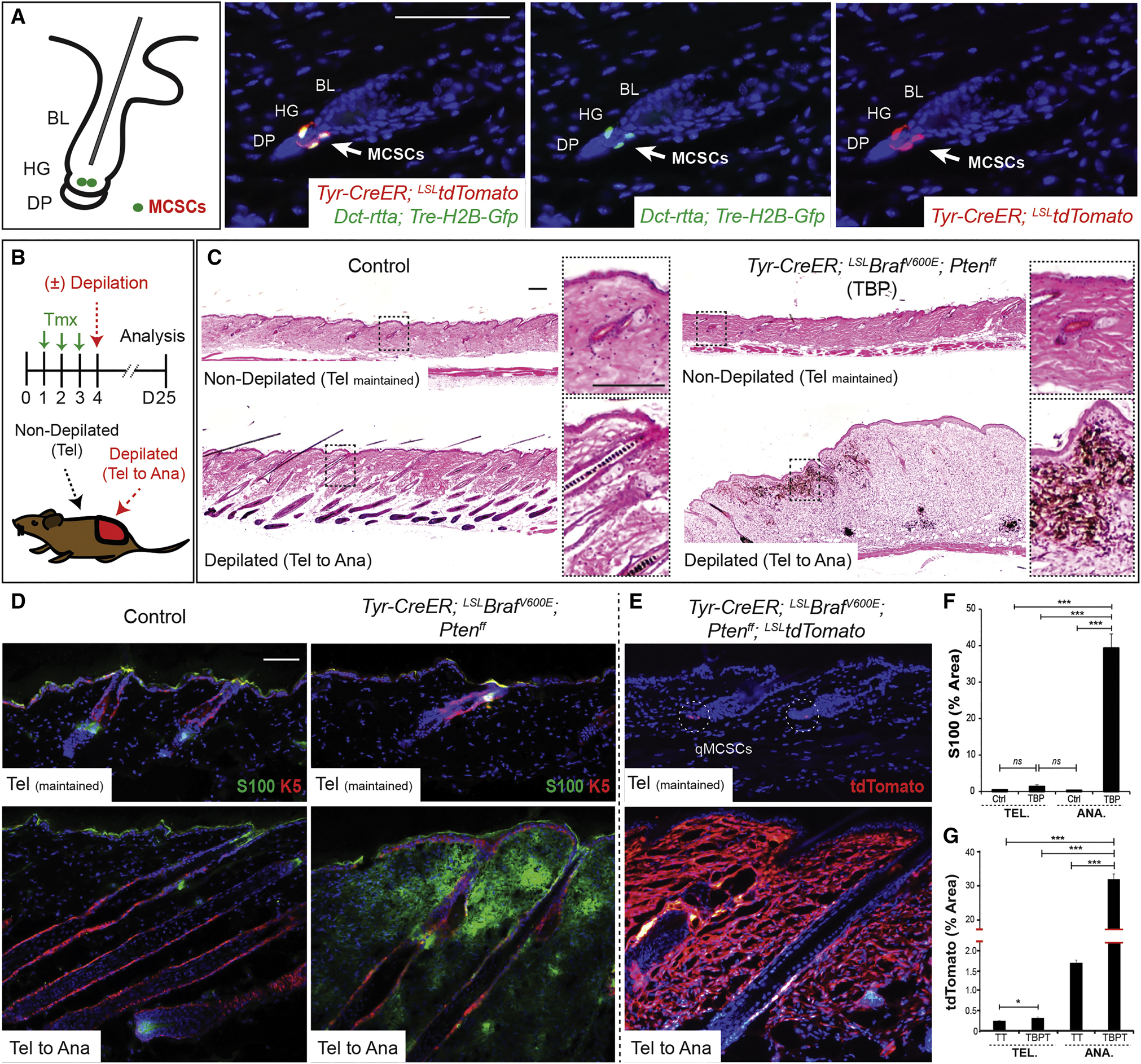
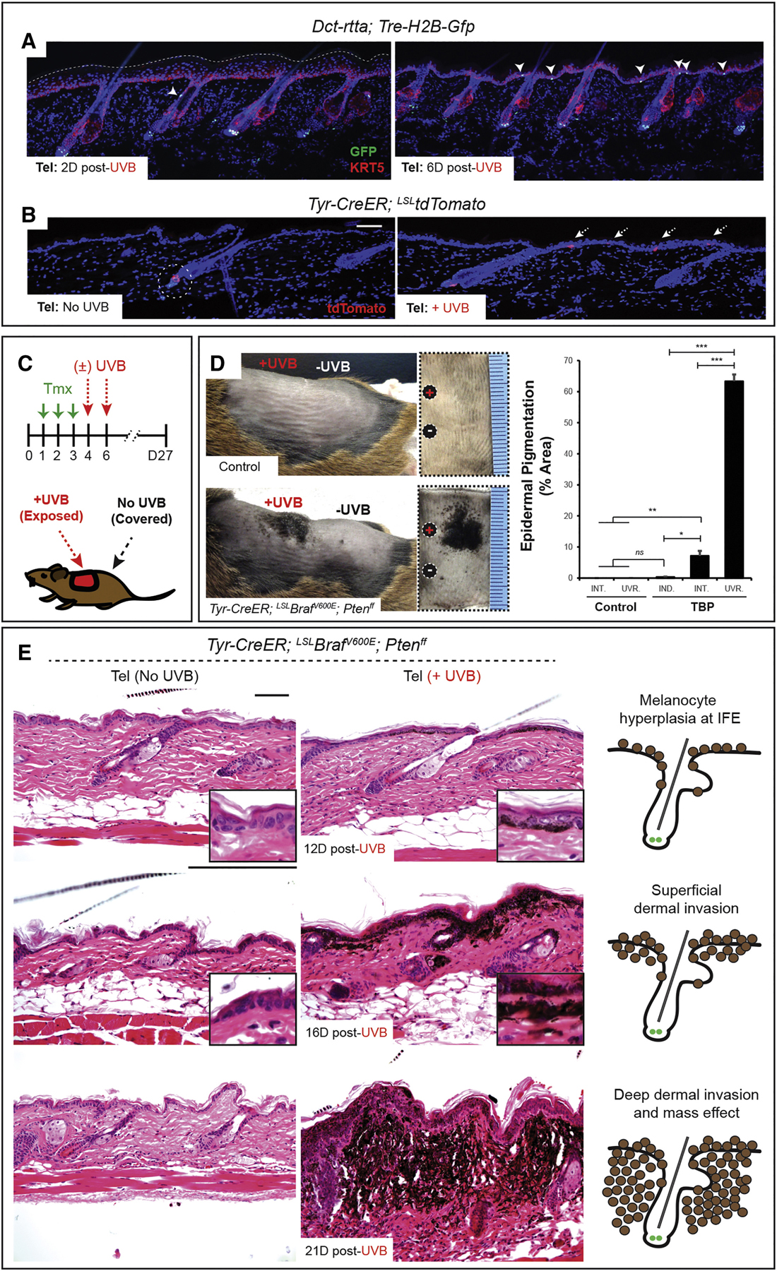
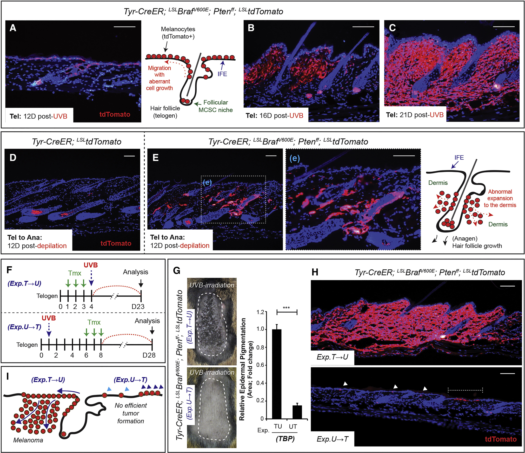
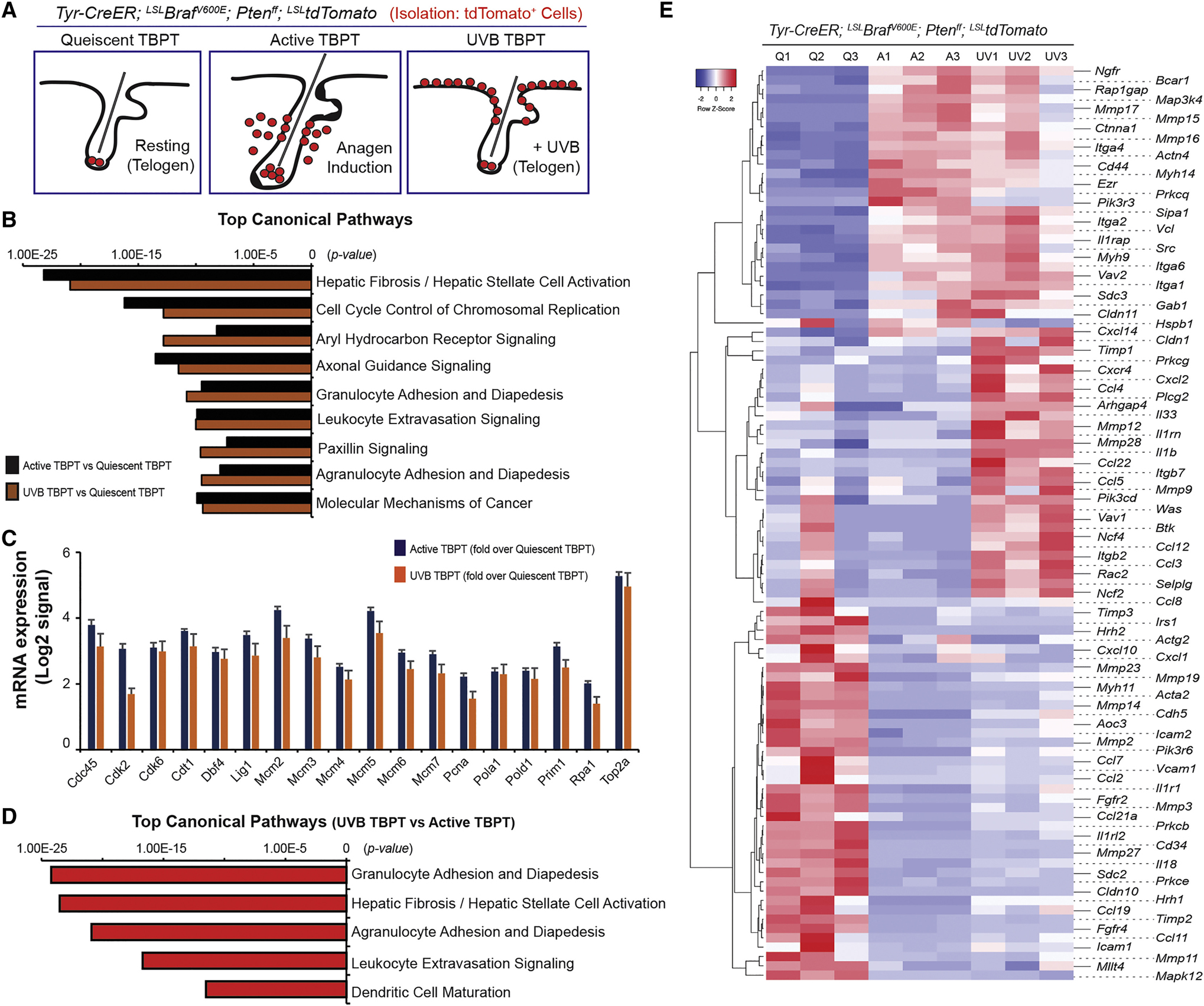
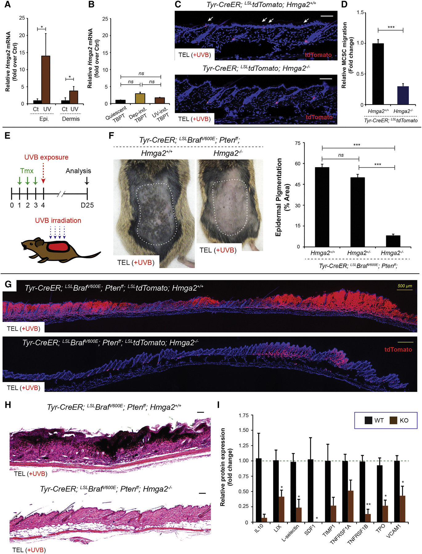
Melanoma is one of the deadliest cancers, yet the cells of origin and mechanisms of tumor initiation remain unclear. The majority of melanomas emerge from clear skin without a precursor lesion, but it is unknown whether these melanomas can arise from melanocyte stem cells (MCSCs). Here we employ mouse models to define the role of MCSCs as melanoma cells of origin, demonstrate that MCSC quiescence acts as a tumor suppressor, and identify the extrinsic environmental and molecular factors required for the critical early steps of melanoma initiation. Specifically, melanomas originate from melanoma-competent MCSCs upon stimulation by UVB, which induces MCSC activation and translocation via an inflammation-dependent process. Moreover, the chromatin-remodeling factor Hmga2 in the skin plays a critical role in UVB-mediated melanomagenesis. These findings delineate melanoma formation from melanoma-competent MCSCs following extrinsic stimuli, and they suggest that abrogation of Hmga2 function in the microenvironment can suppress MCSC-originating cutaneous melanomas.

Keywords: Hmga2; melanocyte stem cell; melanoma; ultraviolet radiation.


Figures

Figure 1.
Figure 1.. Melanocyte stem cells (MCSCs) can act as melanoma cells of origin and adult MCSC activation is required for melanoma initiation.
(A) Location of adult MCSCs, shown by co-expression of H2B-GFP and tdTomato in MCSCs at the hair germ (HG). BL, bulge; DP, dermal papillae. (B) Experimental scheme. Tmx, tamoxifen; Tel, telogen; Ana, anagen. (C-G) Histological phenotypes, and immunostaining for melanoma marker S100 and tdTomato lineage tracing in control, Tyr-CreER; LSL-tdTomato (TT) and TBP or TBP; LSL-tdTomato (TBPT) mice. Sequential photomicrographs were reconstructed for the panoramic images of histology (C). S100, n ≥ 60 fields, 8 mice; tdTomato, n ≥ 160 fields, 6 mice per group. IF counterstaining, DAPI. Scale bars, 100 μm. Data are represented as mean ± SEM. Also see Figures S1 and S2.
Figure 2.
Figure 2.. Ultraviolet radiation stimulates melanoma initiation by inducing the activation and translocation of quiescent melanoma-competent MCSCs.
(A-B) The lineage tracing alleles, H2B-GFP and tdTomato, show MCSC translocation during telogen upon ultraviolet-B (UVB) exposure. (C) Experimental scheme. Tmx, tamoxifen. (D) Macroscopic epidermal pigmentation in control and TBP mice with/without UVB exposure. Mean ± SEM. n = 10 UVB-irradiated control, n = 7 non-UVB TBP and n = 10 UVB-irradiated TBP mice. INT., non-UVB internal control; UVR., UVB-irradiated; IND., independent control skin. (E) Histological progression of melanoma in TBP mice with/without UVB irradiation. IF counterstaining, DAPI. Scale bars, 100 μm. Also see Figures S3 and S4.
Figure 3.
Figure 3.. Ultraviolet radiation efficiently induces MCSC-originating melanomas which arise in the epidermis and progress to the dermis.
(A-C) Lineage tracing allele tdTomato shows the progression of UVB-initiated melanoma from tumor-prone MCSCs during telogen. (D-E) Lineage tracing allele tdTomato shows normal melanocytogenesis and malignant melanocytic tumorigenesis in control (TT) and TBPT mice by MCSC activation through anagen induction. (F) Experimental scheme. T, Tmx, tamoxifen; U, UVB during telogen. (G) Macroscopic phenotypes with quantification of epidermal pigmentation from TBPT mice. Mean ± SEM. n = 6 / group. (H-I) Lineage tracing of tdTomato and the summary of microscopic phenotypes. Scale bars, 100 μm. IF counterstaining, DAPI.
Figure 4.
Figure 4.. Ultraviolet-B exposure initiates melanomagenesis through activation of quiescent melanoma-prone MCSCs.
(A) Experimental scheme and hypothesis showing a lack of efficient melanomagenesis during MCSC quiescence (Q) periods. Tmx, tamoxifen; Tel, telogen; Ana, anagen; Cat, catagen; A, active status of MCSCs. (B) Histological phenotypes with tdTomato lineage tracing. (C) Experimental scheme and hypothesis for the role of UVB exposure in melanomagenesis through MCSC activation during the quiescent period of telogen (circled A, UVB-mediated activation). (D) Melanomagenesis via UVB irradiation during late anagen, and demonstration of melanoma initiation by tdTomato lineage tracing. (E) Early UVB-induced melanocytic hyperplasia throughout the interfollicular epidermis (IFE) demonstrated by lineage tracing. (F) Histology and tdTomato lineage tracing demonstrating UVB-induced melanomagenesis from quiescent tumor-prone MCSCs during late anagen. (G) Summary of UVB effects in melanomagenesis originating from quiescent melanoma-competent MCSCs. Activation of MCSCs is required for melanomagenesis (1, 2 and 4), UVB can significantly activate quiescent melanoma-competent MCSCs in telogen (3) and anagen (5) to induce melanomagenesis in a manner similar to human tumor initiation and progression. IF counterstaining, DAPI. Scale bars, 100 μm.
Figure 5.
Figure 5.. Transcriptomic analysis demonstrates differentially expressed genes during early melanomagenesis.
(A) Cells were isolated for quiescent tumor-prone MCSCs (Quiescent TBPT), depilation-induced active melanocytes (Active TBPT), and UVB-induced active melanocytes (UVB TBPT) from TBPT mice. (B) Top canonical pathways were enriched for quiescent TBPT versus Active TBPT, and quiescent TBPT versus UVB-TBPT. (C) mRNA expression associated with cell cycle control of chromosomal replication measured by RNAseq. (D) Top canonical pathways were enriched for UVB-mediated versus depilation-induced active melanocytes from TBPT mice. (E) Heatmap of 98 genes differentially expressed related to leukocyte migration (adhesion and extravasation) between tumor-prone quiescent MCSCs (Q1-Q3), depilation-induced active (A1-A3) and UVB-mediated active early malignant melanocytes (UV1-UV3) from TBPT mice.
Figure 6.
Figure 6.. Melanoma formation from quiescent melanoma-competent MCSCs can be attenuated by suppression of inflammation and sunscreen application.
(A) MCSC translocation by topical TPA treatment shown by tdTomato lineage tracing. (B) Experimental scheme. (C) Macroscopic phenotypes in TBP mice with/without topical TPA treatment, and quantification of epidermal pigmentation. n = 10 TPA-treated control, n = 7 non-TPA TBP and n = 7 TPA-treated TBP mice. (D) Histological phenotypes of TPA-induced melanoma during quiescent MCSC, telogen period, and co-immunostaining for Sox10, a melanocyte stem cell/melanoma marker, with Ki67, a marker of proliferation. (E) Experimental scheme. Dex., dexamethasone. (F) Relative number of MCSC migration was quantified via tdTomato lineage tracing in TT mice, compared to the total number of hair follicles (≥ 500 hair follicles / each). Mean ± SEM, n = 10 / group. (G) Experimental scheme. (H) Macroscopic phenotypes with the quantification of epidermal pigmentation. n = 6 low-dose (80 mJ cm−2) UVB, n = 10 per each group (UVB only or UVB + Dex.). Data are represented as Mean ± SEM. Also see Figures S5 and S6.
Figure 7.
Figure 7.. Loss of Hmga2 function in the microenvironment significantly suppresses MCSC activation/translocation and melanomagenesis in quiescent melanoma-competent MCSCs.
(A) Relative Hmga2 mRNA expression between control and UVB-exposed epidermis or dermis was measured using ΔCt method. n = 5. (B) Relative mRNA expression of Hmga2 was measured by RNAseq using sorted quiescent tumor-prone MCSCs (Quiescent TBPT), and depilation-induced (Dep-ind. TBPT) or UVB-induced early melanoma cells (UV-ind. TBPT). n = 3. ns = not significant. (C) tdTomato lineage tracing demonstrates no significant MCSC migration (white arrows) by UVB irradiation in Hmga2−/− mice. (D) Relative MCSC migration between TT Hmga2+/+ and TT Hmga2−/−. n = 6. (E) Experimental scheme. Tmx, tamoxifen. (F) Suppressed UVB-induced melanomagenesis in Hmga2−/− mice. Macroscopic phenotypes and quantification of epidermal pigmentation show significantly suppressed UVB-induced melanomagenesis in Hmga2−/− mice. n = 10 TBP Hmga2+/+, n = 11 TBP Hmga2+/− and n = 11 TBP Hmga2−/−. (G-H) tdTomato lineage tracing and histological phenotypes in TBP Hmga2+/+ and TBP Hmga2−/−. Sequential photomicrographs were reconstructed for the panoramic images for tdTomato lineage tracing. Counter staining, DAPI. Scale bars, 100 μm and 500 μm (yellow bars). (I) Relative protein expression level for differentially expressed chemokines/cytokines in the skin post-UVB irradiation between Hmga2+/+ and Hmga2−/−. n = 4. Data are represented as Mean ± SEM. Also see Figure S7.

Comment in

References

    1. Anand A, and Chada K (2000). In vivo modulation of Hmgic reduces obesity. Nat Genet 24, 377–380. - PubMed
    1. Belmadani A, Jung H, Ren D, and Miller RJ (2009). The chemokine SDF-1/CXCL12 regulates the migration of melanocyte progenitors in mouse hair follicles. Differentiation 77, 395–411. - PMC - PubMed
    1. Bevona C, Goggins W, Quinn T, Fullerton J, and Tsao H (2003). Cutaneous melanomas associated with nevi. Arch Dermatol 139, 1620–4; discussion 1624. - PubMed
    1. Bosenberg M, Muthusamy V, Curley DP, Wang Z, Hobbs C, Nelson B, Nogueira C, Horner JW, Depinho R, and Chin L (2006). Characterization of melanocyte-specific inducible Cre recombinase transgenic mice. Genesis 44, 262–267. - PubMed
    1. Boumahdi S, Driessens G, Lapouge G, Rorive S, Nassar D, Le Mercier M, Delatte B, Caauwe A, Lenglez S, Nkusi E, et al. (2014). SOX2 controls tumour initiation and cancer stem-cell functions in squamous-cell carcinoma. Nature 511, 246–250. - PubMed

MeSH terms