Review
. 2017 Sep 20:11:1707-1718.
doi: 10.2147/OPTH.S130165. eCollection 2017.

Spotlight on reticular pseudodrusen

Affiliations
Review

Spotlight on reticular pseudodrusen

Alessandro Rabiolo et al. Clin Ophthalmol. .

Abstract

Age-related macular degeneration (AMD) is a leading cause of vision loss in patients >50 years old. The hallmark of the disease is represented by the accumulation of extracellular material between retinal pigment epithelium and the inner collagenous layer of Bruch's membrane, called drusen. Although identified almost 30 years ago, reticular pseudodrusen (RPD) have been recently recognized as a distinctive phenotype. Unlike drusen, they are located in the subretinal space. RPD are strongly associated with late AMD, especially geographic atrophy, type 2 and 3 choroidal neovascularization, which, in turn, are less common in typical AMD. RPD identification is not straightforward at fundus examination, and their identification should employ at least 2 different imaging modalities. In this narrative review, we embrace all aspects of RPD, including history, epidemiology, histology, imaging, functional test, natural history and therapy.

Keywords: age-related macular degeneration; choroidal neovascularization; geographic atrophy; reticular drusen; reticular macular degeneration; reticular macular disease; reticular pseudodrusen; subretinal drusenoid deposit.


Conflict of interest statement

Disclosure GQ is a consultant for Alimera Sciences (Alpharetta, Georgia, USA), Allergan Inc. (Irvine, California, USA), Bayer Schering-Pharma (Berlin, Germany), Heidelberg (Germany), Novartis (Basel, Switzerland), Sandoz (Berlin, Germany), Zeiss (Dublin, OH, USA). FB has the following disclosures: Allergan (s), Alimera (s), Bayer (s), Farmila-Thea (s), Schering Pharma (s), Sanofi-Aventis (s), Novagali (s), Pharma (s), Hoffmann-La Roche (s), Genentech (s) and Novartis (s). The other authors report no conflicts of interest in this work.

Figures

Figure 1
Figure 1
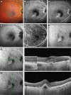
Multicolor imaging, FAF, IR and SD-OCT of a patient affected by RPD. Notes: (A) Multicolor imaging showing a yellowish–green reticular pattern and some RPD with a target appearance characterized by a more intense yellowish/greenish core surrounded by a decreased intensity. (B) Blue FAF showing RPD as hypoautofluorescent dots surrounded by a faint hyperautofluorescent halo. Several RPD have a isoautofluorescent core, conferring a target aspect. (C) IR showing RPD as small lesions clustered in a reticular pattern and a variable target aspect, with an isoreflective core surrounded by hyporeflective halo. (D) Combined IR and structural SD-OCT B-scan passing through the fovea showing RPD as discrete accumulation of hyperreflective material above the RPE in the subretinal space. Green arrow shows the position of the scan lines used to generate the cross-sectional SD-OCT images. Magnifications showing an example of four different stages of RPD: 1) diffuse accumulation of granular hyperreflective material between RPE and EZ (hash); 2) mounds of material bowing and distorting EZ profile (star); 3) conical accumulations with focal interruption of EZ (asterisk); 4) drusenoid material reabsorption and migration in inner retinal layers (triangle). Abbreviations: FAF, fundus autofluorescence; IR, infrared reflectivity; SD-OCT, spectral-domain optical coherence tomography; RPD, reticular pseudodrusen; RPE, retinal pigment epithelium; EZ, ellipsoid zone.
Figure 2
Figure 2
OCT-A of a patient affected by RPD. Notes: (A, B) Superficial and deep capillary plexuses (A and B, respectively), corresponding en-face OCT and B-scan with flow showing no significant alterations. (C, D) Choriocapillaris and choroidal segmentation (C and D, respectively) showing choriocapillaris and choroidal impairment of the vessel density (dashed triangles and circles, respectively). Measurement: 3×3 mm OCT-A. Abbreviations: OCT-A, optical coherence tomography angiography; RPD, reticular pseudodrusen; OCT, optical coherence tomography.
Figure 3
Figure 3
Multicolor imaging, FA, fundus autofluorescence, ICGA and structural SD-OCT of a patient affected by RPD and a type 1 CNV. Notes: (A, D) Multicolor imaging (A) and fundus autofluorescence (D) showing RPD, a large hemorrhage around the central lesion. FA (B, C) and ICGA (E, F) revealing the presence of a type 1 CNV with a typical hyperfluorescent plaque in the late phase of ICGA (F). (G, H) Combined ICGA and structural horizontal and vertical SD-OCT B-scans passing through the fovea revealing the presence of RPD and an active type 1 CNV. Green arrows show the position of the scan lines used to generate the cross-sectional SD-OCT images. Abbreviations: FA, fluorescein angiography; ICGA, indocyanine green angiography; SD-OCT, spectral-domain optical coherence tomography; RPD, reticular pseudodrusen; CNV, choroidal neovascularization.

References

    1. Lim LS, Mitchell P, Seddon JM, Holz FG, Wong TY. Age-related macular degeneration. Lancet. 2012;379(9827):1728–1738. - PubMed
    1. Mimoun G, Soubrane G, Coscas G. Les drusen maculaires. [Macular drusen] J Fr Ophtalmol. 1990;13(10):511–530. French. - PubMed
    1. Klein R, Davis MD, Magli YL, Segal P, Klein BE, Hubbard L. The Wisconsin age-related maculopathy grading system. Ophthalmology. 1991;98(7):1128–1134. - PubMed
    1. Arnold JJ, Sarks SH, Killingsworth MC, Sarks JP. Reticular pseudodrusen. A risk factor in age-related maculopathy. Retina. 1995;15(3):183–191. - PubMed
    1. Finger RP, Chong E, McGuinness MB, et al. Reticular Pseudodrusen and Their Association with Age-Related Macular Degeneration: The Melbourne Collaborative Cohort Study. Ophthalmology. 2016;123(3):599–608. - PubMed

LinkOut - more resources