

In vivo evaluation of cetuximab-conjugated poly(γ-glutamic acid)-docetaxel nanomedicines in EGFR-overexpressing gastric cancer xenografts
- PMID: 29033568
- PMCID: PMC5628680
- DOI: 10.2147/IJN.S143529
In vivo evaluation of cetuximab-conjugated poly(γ-glutamic acid)-docetaxel nanomedicines in EGFR-overexpressing gastric cancer xenografts
Erratum in
-
In vivo evaluation of cetuximab-conjugated poly(γ-glutamic acid)-docetaxel nanomedicines in EGFR-overexpressing gastric cancer xenografts [Corrigendum].Int J Nanomedicine. 2019 Jun 28;14:4681-4682. doi: 10.2147/IJN.S217605. eCollection 2019. Int J Nanomedicine. 2019. PMID: 31388299 Free PMC article. No abstract available.
Abstract
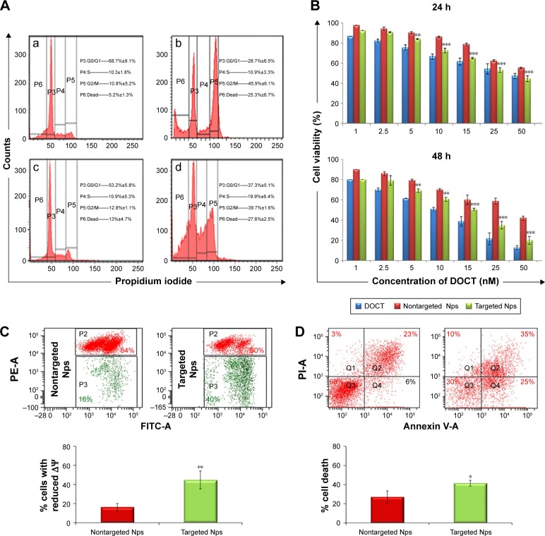
Epidermal growth factor receptor (EGFR), upregulated in gastric cancer patients, is an oncogene of interest in the development of targeted cancer nanomedicines. This study demonstrates in silico modeling of monoclonal antibody cetuximab (CET MAb)-conjugated docetaxel (DOCT)-loaded poly(γ-glutamic acid) (γ-PGA) nanoparticles (Nps) and evaluates the in vitro/in vivo effects on EGFR-overexpressing gastric cancer cells (MKN-28). Nontargeted DOCT-γ-PGA Nps (NT Nps: 110±40 nm) and targeted CET MAb-DOCT-γ-PGA Nps (T Nps: 200±20 nm) were prepared using ionic gelation followed by 1-Ethyl-3-(3-dimethyl aminopropyl)carbodiimide-N-Hydoxysuccinimide (EDC-NSH) chemistry. Increased uptake correlated with enhanced cytotoxicity induced by targeted Nps to EGFR +ve MKN-28 compared with nontargeted Nps as evident from MTT and flow cytometric assays. Nanoformulated DOCT showed a superior pharmacokinetic profile to that of free DOCT in Swiss albino mice, indicating the possibility of improved therapeutic effect in the disease model. Qualitative in vivo imaging showed early and enhanced tumor targeted accumulation of CET MAb-DOCT-γ-PGA Nps in EGFR +ve MKN-28-based gastric cancer xenograft, which exhibited efficient arrest of tumor growth compared with nontargeted Nps and free DOCT. Thus, actively targeted CET MAb-DOCT-γ-PGA Nps could be developed as a substitute to conventional nonspecific chemotherapy, and hence could become a feasible strategy for cancer therapy for EGFR-overexpressing gastric tumors.
Keywords: cetuximab; docetaxel; epidermal growth factor receptor; gastric cancer; poly(γ-glutamic acid) nanoparticles; targeted nanoparticles.
Conflict of interest statement
Disclosure The authors report no conflicts of interest in this work.
Figures


Similar articles
-
Actively targeted cetuximab conjugated gamma-poly(glutamic acid)-docetaxel nanomedicines for epidermal growth factor receptor over expressing colon cancer cells.J Biomed Nanotechnol. 2014 Aug;10(8):1416-28. doi: 10.1166/jbn.2014.1841. J Biomed Nanotechnol. 2014. PMID: 25016642
-
Chitosan cross-linked docetaxel loaded EGF receptor targeted nanoparticles for lung cancer cells.Int J Biol Macromol. 2014 Aug;69:532-41. doi: 10.1016/j.ijbiomac.2014.06.009. Epub 2014 Jun 17. Int J Biol Macromol. 2014. PMID: 24950310
-
In vitro and in vivo antitumor activity of cetuximab in human gastric cancer cell lines in relation to epidermal growth factor receptor (EGFR) expression and mutational phenotype.Gastric Cancer. 2012 Jul;15(3):252-64. doi: 10.1007/s10120-011-0102-9. Epub 2011 Oct 20. Gastric Cancer. 2012. PMID: 22011788
-
Cetuximab: an epidermal growth factor receptor chemeric human-murine monoclonal antibody.Drugs Today (Barc). 2005 Feb;41(2):107-27. doi: 10.1358/dot.2005.41.2.882662. Drugs Today (Barc). 2005. PMID: 15821783 Review.
-
Poly-γ-glutamic acid nanoparticles as adjuvant and antigen carrier system for cancer vaccination.J Control Release. 2023 Oct;362:278-296. doi: 10.1016/j.jconrel.2023.08.049. Epub 2023 Sep 1. J Control Release. 2023. PMID: 37640110 Review.
Cited by
-
Decoration of Anti-CD38 on Nanoparticles Carrying a STAT3 Inhibitor Can Improve the Therapeutic Efficacy Against Myeloma.Cancers (Basel). 2019 Feb 20;11(2):248. doi: 10.3390/cancers11020248. Cancers (Basel). 2019. PMID: 30791634 Free PMC article.
-
A novel ligand-modified nanocomposite microparticles improved efficiency of quercetin and paclitaxel delivery in the non-small cell lung cancer.Drug Deliv. 2022 Dec;29(1):3123-3133. doi: 10.1080/10717544.2022.2120567. Drug Deliv. 2022. PMID: 36151722 Free PMC article.
-
Investigating the potential application of organic and non-organic nanoparticles for gastric cancer treatment: An evidence-based review.Arch Razi Inst. 2024 Apr 30;79(2):264-271. doi: 10.32592/ARI.2024.79.2.264. eCollection 2024 Apr. Arch Razi Inst. 2024. PMID: 39463705 Free PMC article.
-
Multifunctional nanomedicines for targeting epidermal growth factor receptor in colorectal cancer.Cell Mol Life Sci. 2020 Mar;77(6):997-1019. doi: 10.1007/s00018-019-03305-z. Epub 2019 Sep 28. Cell Mol Life Sci. 2020. PMID: 31563999 Free PMC article. Review.
-
Antibody-Biopolymer Conjugates in Oncology: A Review.Molecules. 2023 Mar 13;28(6):2605. doi: 10.3390/molecules28062605. Molecules. 2023. PMID: 36985578 Free PMC article. Review.
References
-
- Power DG, Kelsen DP, Shah MA. Advanced gastric cancer–Slow but steady progress. Cancer Treat Rev. 2010;36(5):384–392. - PubMed
-
- Wagner AD, Unverzagt S, Grothe W, et al. Chemotherapy for advanced gastric cancer. Cochrane Database Syst Rev. 2010;3(3):CD004064. - PubMed
-
- Siegel RL, Miller KD, Jemal A. Cancer statistics, 2016. CA Cancer J Clin. 2016;66(1):7–30. - PubMed
-
- Fuse N, Kuboki Y, Kuwata T, et al. Prognostic impact of HER2, EGFR, and c-MET status on overall survival of advanced gastric cancer patients. Gastric Cancer. 2016;19(1):183–191. - PubMed
-
- Sakai K, Mori S, Kawamoto T, et al. Expression of epidermal growth factor receptors on normal human gastric epithelia and gastric carcinomas. J Natl Cancer Inst. 1986;77(5):1047–1052. - PubMed
MeSH terms
Substances
LinkOut - more resources
Full Text Sources
Other Literature Sources
Medical
Research Materials
Miscellaneous
