Gallstone Ileus: An Unlikely Cause of Mechanical Small Bowel Obstruction
- PMID: 29033757
- PMCID: PMC5637004
- DOI: 10.1159/000475749
Gallstone Ileus: An Unlikely Cause of Mechanical Small Bowel Obstruction
Abstract
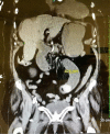
Gallstone ileus is a rare disease that accounts for 1-4% of intestinal obstructions. Almost exclusively a condition in the older female population, it is a difficult diagnosis to make. We report the case of gallstone ileus in a 94-year-old Caucasian female, who presented to the emergency department with acute-onset nausea, coffee-ground emesis, lack of bowel movement, and abdominal distension. On CT scan, the diagnosis of gallstone ileus was made by the presence of a cholecystoduodenal fistula, pneumobilia, and small bowel obstruction. Emergent laparotomy with a one-stage procedure of enterolithotomy and stone removal by milking the bowel distal to the stone were performed. The postoperative course was uneventful until postoperative day 4 when the patient was found tachycardic, lethargic, and unresponsive. We reviewed the literature on the diagnosis and treatment of gallstone ileus.
Keywords: Cholecystoduodenal fistula; Enterolithotomy; Gallstone ileus; Intestinal obstruction; Pneumobilia.
Figures


Similar articles
-
Gallstone Ileus: An Unusual Cause of Intestinal Obstruction.Cureus. 2020 Mar 15;12(3):e7284. doi: 10.7759/cureus.7284. Cureus. 2020. PMID: 32206475 Free PMC article.
-
Gallstone ileus: report of two cases and review of the literature.World J Gastroenterol. 2007 Feb 28;13(8):1295-8. doi: 10.3748/wjg.v13.i8.1295. World J Gastroenterol. 2007. PMID: 17451220 Free PMC article. Review.
-
An unusual case of subacute small bowel obstruction - Gallstone ileus.Int J Surg Case Rep. 2022 Mar;92:106820. doi: 10.1016/j.ijscr.2022.106820. Epub 2022 Feb 8. Int J Surg Case Rep. 2022. PMID: 35189458 Free PMC article.
-
Endoscopic and surgical treatment of jejunal gallstone ileus caused by cholecystoduodenal fistula: A case report.World J Clin Cases. 2023 Jun 16;11(17):4159-4167. doi: 10.12998/wjcc.v11.i17.4159. World J Clin Cases. 2023. PMID: 37388782 Free PMC article.
-
Gallstone ileus: case report and literature review.World J Gastroenterol. 2013 Sep 7;19(33):5586-9. doi: 10.3748/wjg.v19.i33.5586. World J Gastroenterol. 2013. PMID: 24023505 Free PMC article. Review.
Cited by
-
Gallstone Ileus: Clinical Presentation and Radiological Diagnosis.Cureus. 2023 Jul 18;15(7):e42059. doi: 10.7759/cureus.42059. eCollection 2023 Jul. Cureus. 2023. PMID: 37476299 Free PMC article.
-
Gallstone ileus 16 years postcholecystectomy?Radiol Case Rep. 2025 Aug 22;20(11):5668-5675. doi: 10.1016/j.radcr.2025.07.060. eCollection 2025 Nov. Radiol Case Rep. 2025. PMID: 40894996 Free PMC article.
-
Gallstone Ileus Caused by Migration of Gallstone Through Cholecystoduodenal Fistula Resulting in Small Bowel Obstruction.Cureus. 2023 Apr 21;15(4):e37962. doi: 10.7759/cureus.37962. eCollection 2023 Apr. Cureus. 2023. PMID: 37096199 Free PMC article.
-
Gallstone Ileus: An Unusual Cause of Intestinal Obstruction.Cureus. 2020 Mar 15;12(3):e7284. doi: 10.7759/cureus.7284. Cureus. 2020. PMID: 32206475 Free PMC article.
-
Gallstone Ileus: An Improbable Cause of Mechanical Small Bowel Obstruction.Cureus. 2020 Nov 12;12(11):e11460. doi: 10.7759/cureus.11460. Cureus. 2020. PMID: 33329958 Free PMC article.
References
-
- Harris J, Evers M. Small intestine. In: Townsend C, Beauchamp RD, Evers B, Mattox K, editors. Sabiston Textbook of Surgery. ed 20. Philadelphia: Elsevier Saunders; 2016. chapt 49.
-
- Abou-Saif A, Al-Kawas FH. Complications of gallstone disease: Mirizzi syndrome, cholecystocholedochal fistula, and gallstone ileus. Am J Gastroenterol. 2002;97:249–254. - PubMed
-
- Kurtz RJ, Heimann TM, Kurtz AB. Gallstone ileus: a diagnostic problem. Am J Surg. 1983;146:314–317. - PubMed
-
- Clavien PA, Richon J, Burgan S, Rohner A. Gallstone ileus. Br J Surg. 1990;77:737–742. - PubMed
-
- Reisner RM, Cohen JR. Gallstone ileus: a review of 1,001 reported cases. Am Surg. 1994;60:441–446. - PubMed
Publication types
LinkOut - more resources
Full Text Sources
Other Literature Sources

