

A Case of Basaloid Squamous Cell Carcinoma of Polypoid Type in the Esophagus
- PMID: 29033778
- PMCID: PMC5637102
- DOI: 10.1159/000479312
A Case of Basaloid Squamous Cell Carcinoma of Polypoid Type in the Esophagus
Abstract
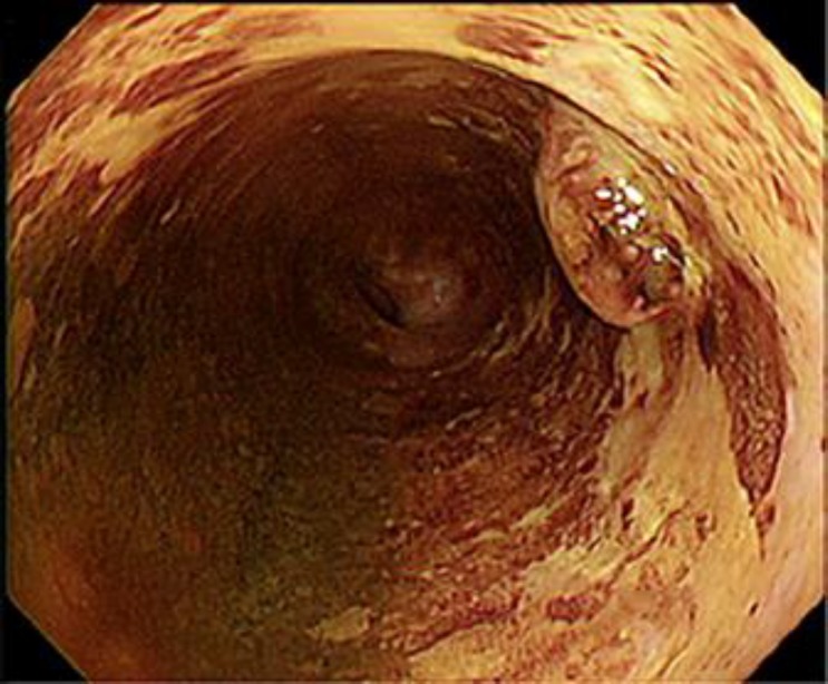
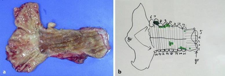
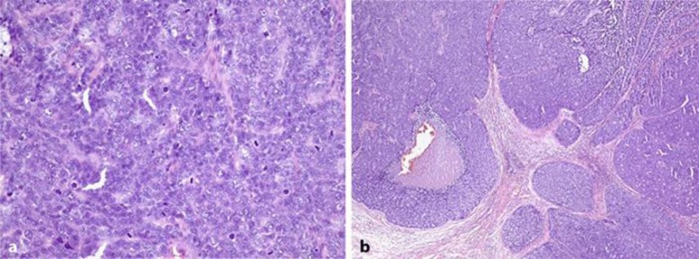
Basaloid squamous cell carcinoma of the esophagus is very rare. Further, polypoid type of esophageal cancer is also rare. We have recently treated a case of basaloid squamous cell carcinoma which presented as a 1.3-cm esophageal polyp. A 48-year-old woman was referred to our hospital because of a polypoid mass at 36 cm distance from the upper incisor on endoscopic examination, and the result of the biopsy was basaloid squamous cell carcinoma. The patient underwent Ivor Lewis operation with lymph node dissection. Two basaloid squamous cell carcinomas, of 1.3 and 0.4 cm, were diagnosed in the final pathologic examination. Regular periodic follow-up showed no evidence of recurrence or metastasis in the 5-month postoperative period.
Keywords: Esophagus; Polyps; Squamous cell carcinoma; Surgery.
Figures
References
-
- Cardesa A, Zidar N, Ereño C. Basaloid squamous cell carcinoma. In: Barnes L, Eveson JW, Reichart P, Sidrasky D, Kleihues P, Sobin LH, editors. Pathology and Genetics of Head and Neck Tumours. World Health Organization Classification of Tumours. Lyon: IARC Press; 2005. pp. 124–125.
-
- Wain SL, Kier R, Wollmer RT, et al. Basaloid-squamous carcinoma of the tongue, hypopharynx, and larynx: report of 10 cases. Hum Pathol. 1986;17:1158–1166. - PubMed
-
- Noguchi H, Naomoto Y, Haisa M, Yamatsuji T, Shigemitsu K, Shirakawa Y, et al. Two cases of superficial basaloid squamous carcinoma of the esophagus. Dis Esophagus. 2003;16:342–345. - PubMed
-
- Olmsted WW, Lichstein JE, Hyams V. Polypoid epithelial malignancies of the esophagus. AJR Am J Roentgenol. 1983;140:921–925. - PubMed
Publication types
LinkOut - more resources
Full Text Sources
Other Literature Sources
