. 2017 Sep 29:11:281.
doi: 10.3389/fncel.2017.00281. eCollection 2017.

Wnt5a Promotes Cortical Neuron Survival by Inhibiting Cell-Cycle Activation

Affiliations

Wnt5a Promotes Cortical Neuron Survival by Inhibiting Cell-Cycle Activation

Li Zhou et al. Front Cell Neurosci. .

Abstract

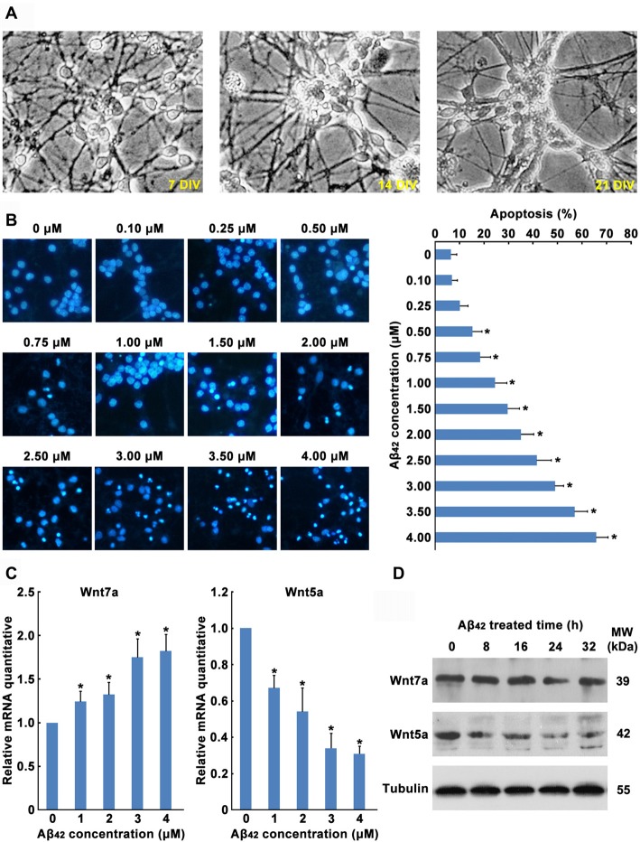
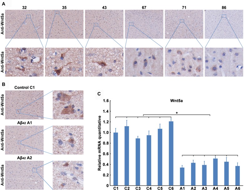
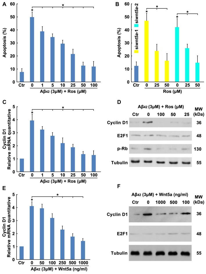
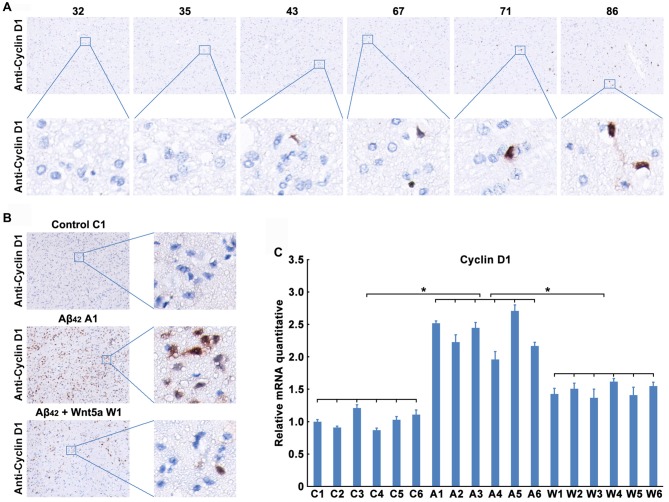
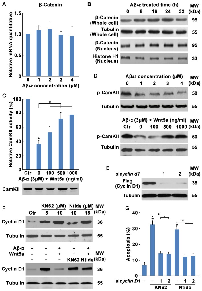
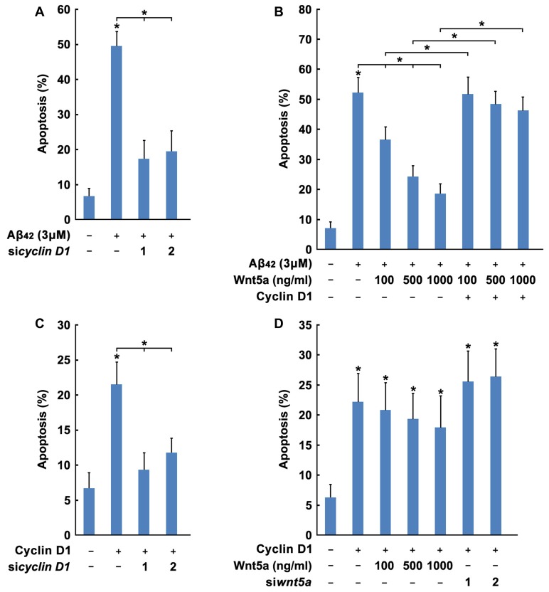
β-Amyloid protein (Aβ) is thought to cause neuronal loss in Alzheimer's disease (AD). Aβ treatment promotes the re-activation of a mitotic cycle and induces rapid apoptotic death of neurons. However, the signaling pathways mediating cell-cycle activation during neuron apoptosis have not been determined. We find that Wnt5a acts as a mediator of cortical neuron survival, and Aβ42 promotes cortical neuron apoptosis by downregulating the expression of Wnt5a. Cell-cycle activation is mediated by the reduced inhibitory effect of Wnt5a in Aβ42 treated cortical neurons. Furthermore, Wnt5a signals through the non-canonical Wnt/Ca2+ pathway to suppress cyclin D1 expression and negatively regulate neuronal cell-cycle activation in a cell-autonomous manner. Together, aberrant downregulation of Wnt5a signaling is a crucial step during Aβ42 induced cortical neuron apoptosis and might contribute to AD-related neurodegeneration.

Keywords: Alzheimer’s disease; Cyclin D1; Wnt5a; apoptosis; cell-cycle activation; cortical neuron; β-Amyloid protein.


Figures

Figure 1
Figure 1
42 promotes the decrease of Wnt5a expression. (A) Cortical neurons cultured in vitro for 7, 14 and 21 days. (B) DIV7 cortical neurons were treated with Aβ42 at indicated concentrations for 24 h. Then neurons were stained for nucleus (Hoechst 33258, Blue). Apoptosis was determined by the percentage of cells that were had pyknotic nuclei. (C) DIV7 cortical neurons were treated with Aβ42 at indicated concentrations for 8 h, total RNA was extracted and analyzed by Q-PCR. (D) DIV7 cortical neurons were treated with 3 μM Aβ42 at indicated times, total protein was extracted and analyzed by Western blotting. DIV, days in vitro; MW, molecular weight; kDa, kilodalton. All data in this figure represent the means ± SEM of three independent experiments. *P < 0.05.
Figure 2
Figure 2
42 inhibited the expression of Wnt5a in vivo. (A) Wnt5a immunoreactivity was primarily detected in the adjacent normal brain tissues of malignant glioma from 32, 35, 43, 67, 71 and 86 years old patients (magnification of upper panel: ×40; magnification of lower panel: ×400). (B,C) A solution of Aβ42 (3 nmol/10 μl) or Aβ42 (3 nmol/10 μl) combined with recombinant Wnt5a protein (6 nmol/10 μl) was injected into the lateral ventricles of mice. Seven days after treatment, Wnt5a immunoreactivity was primarily detected in mouse brain tissues (B, control C1: control group mouse 1; Aβ42 A1: Aβ42 treated group mouse 1; Aβ42 A2: Aβ42 treated group mouse 2; magnification of left panel: ×40; magnification of right panel: ×400), total RNA was extracted and analyzed by Q-PCR (C, C1–C6: control group mouse 1–6; A1–A6: Aβ42 treated group mouse 1–6). All data in this figure represent the means ± SEM of three independent experiments. *P < 0.05.
Figure 3
Figure 3
Wnt5a downregulation contributes to Aβ42 induced cortical neuron apoptosis. (A) HEK239 cells were transfected with Flag-Wnt5a and Wnt5a siRNAs for 12 h, then cells were subjected to immunoblot detection. (B) DIV6 cortical neurons were transfected with GFP plasmids and Wnt5a siRNAs for 24 h. Immunofluorescence detection of Wnt5a of neurons appearing in Red, then neurons were stained for nucleus (Blue; left panel). The efficiency of Wnt5a siRNAs were estimated by a score of 0–3 based on the red fluorescence area in the cytoplasm of neurons (grade 0, <5%; grade 1, 5%–33%; grade 2, 33%–66%; and grade 3, >66%; right panel). (C) DIV6 cortical neurons were transfected with GFP plasmids and Wnt5a siRNAs for 12 h following 50 μM Z-VAD treatment for another 12 h, and then neurons were stained for nucleus (Blue). Apoptosis was determined by the percentage of GFP positive cells that were had pyknotic nuclei. (D) DIV6 cortical neurons were transfected with GFP plasmids, Wnt5a plasmids and Wnt5a siRNAs for 24 h, and then neurons were stained for nucleus (Blue). Apoptosis was determined by the percentage of GFP positive cells that were had pyknotic nuclei. (E) DIV6 cortical neurons were treated with 3 μM Aβ42 with or without recombinant Wnt5a protein at indicated concentrations for 24 h, and then neurons were stained for nucleus (Blue). Apoptosis was determined by the percentage of cells that were had pyknotic nuclei (upper panel); or cells were subjected to immunoblot detection (lower panel). MW, molecular weight; kDa, kilodalton. All data in this figure represent the means ± SEM of three independent experiments. *P < 0.05.
Figure 4
Figure 4
Wnt5a inhibits the expression of Cyclin D1. (A) DIV6 cortical neurons were treated with 3 μM Aβ42 with or without Roscovitine (ROS) at indicated concentrations for 24 h, then apoptosis was determined. (B) DIV6 cortical neurons were treated with Wnt5a siRNAs and ROS at indicated concentrations for 24 h, then apoptosis was determined. (C) DIV6 cortical neurons were treated with 3 μM Aβ42 with or without ROS at indicated concentrations for 12 h, total RNA was extracted and analyzed by Q-PCR. (D) DIV6 cortical neurons were treated with 3 μM Aβ42 with or without ROS at indicated concentrations for 16 h, total protein was extracted and analyzed by Western blotting. (E) DIV6 cortical neurons were treated with 3 μM Aβ42 with or without recombinant Wnt5a protein at indicated concentrations for 12 h, total RNA was extracted and analyzed by Q-PCR. (F) DIV6 cortical neurons were treated with 3 μM Aβ42 with or without recombinant Wnt5a protein at indicated concentrations for 16 h, total protein was extracted and analyzed by Western blotting. MW, molecular weight; kDa, kilodalton. All data in this figure represent the means ± SEM of three independent experiments. *P < 0.05.
Figure 5
Figure 5
Wnt5a inhibits the expression of Cyclin D1 in vivo. (A) Cyclin D1 immunoreactivity was primarily detected in the adjacent normal brain tissues of malignant glioma from 32, 35, 43, 67, 71 and 86 years old patients (magnification of upper panel: ×40; magnification of lower panel: ×400). (B,C) A solution of Aβ42 (3 nmol/10 μl) or Aβ42 (3 nmol/10 μl) combined with recombinant Wnt5a protein (6 nmol/10 μl) was injected into the lateral ventricles of mice. Seven days after treatment, Cyclin D1 immunoreactivity was primarily detected in mouse brain tissues (B, control C1: control group mouse 1; Aβ42 A1: Aβ42 treated group mouse 1; Aβ42 + Wnt5a W1: Aβ42 and Wnt5a treated group mouse 1; magnification of left panel: ×40; magnification of right panel: ×400), total RNA was extracted and analyzed by Q-PCR (C, C1–C6: control group mouse 1–6; A1–A6: Aβ42 treated group mouse 1–6; W1–W6: Aβ42 and Wnt5a treated group mouse 1–6). All data in this figure represent the means ± SEM of three independent experiments. *P < 0.05.
Figure 6
Figure 6
Wnt5a/CaMKII pathway down-regulates the expression of Cyclin D1. (A) DIV7 cortical neurons were treated with Aβ42 at indicated concentrations for 12 h, total RNA was extracted and analyzed by Q-PCR. (B) DIV7 cortical neurons were treated with 3 μM Aβ42 at indicated times, total protein or nuclear protein was extracted and analyzed by Western blotting. (C) DIV6 cortical neurons were treated with 3 μM Aβ42 with or without recombinant Wnt5a protein at indicated concentrations for 12 h. CamKII activity was determined using SignaTECT® Calcium/Calmodulin-Dependent Protein Kinase Assay System (Upper panel). The input total CamKII was analyzed by Western blotting (Lower panel). (D) DIV7 cortical neurons were treated with Aβ42 at indicated concentrations (Upper panel) or treated with 3 μM Aβ42 with/without recombinant Wnt5a protein at indicated concentrations (Lower panel) for 16 h, total protein was extracted and analyzed by Western blotting. (E) DIV7 cortical neurons were treated with KN62 or Calmodulin Kinase II Ntide, Myristoylated (Ntide) at indicated concentrations for 16 h (Left panel), or DIV7 cortical neurons were treated with 3 μM Aβ42 with/without 500 ng/ml recombinant Wnt5a protein or 10 μM KN62 or 15 μM Ntide as indicated for 16 h (Right panel), total protein was extracted and analyzed by Western blotting. (F) HEK239 cells were transfected with Flag-Cyclin D1 and Cyclin D1 siRNAs for 12 h, then cells were subjected to immunoblot detection. (G) DIV7 cortical neurons were treated with 10 μM KN62 or 15 μM Ntide and transfected with or without Cyclin D1 siRNAs for 24 h (Left panel), then apoptosis was determined. MW, molecular weight; kDa, kilodalton. All data in this figure represent the means ± SEM of three independent experiments. *P < 0.05.
Figure 7
Figure 7
Wnt5a promotes cortical neuron survival by inhibiting Cyclin D1 expression. (A) DIV6 cortical neurons were transfected with GFP plasmids and Cyclin D1 siRNAs for 12 h, and treated with 3 μM Aβ42 for another 12 h. Apoptosis was determined by the percentage of GFP positive cells that were had pyknotic nuclei. (B) DIV6 cortical neurons were transfected with GFP plasmids, Flag-Cyclin D1 plasmids and Cyclin D1 siRNAs for 24 h. Apoptosis was determined by the percentage of GFP positive cells that were had pyknotic nuclei. (C) DIV6 cortical neurons were transfected with or without GFP plasmids, Flag-Cyclin D1 plasmids or control plasmids for 12 h, then treated with 3 μM Aβ42 with/without recombinant Wnt5a protein at indicated concentrations for 24 h. Apoptosis was determined by the percentage of GFP positive cells that were had pyknotic nuclei. (D) DIV6 cortical neurons were transfected with or without GFP plasmids, Flag-Cyclin D1 plasmids or Cyclin D1 siRNAs for 12 h, then treated with with/without recombinant Wnt5a protein at indicated concentrations for 24 h. Apoptosis was determined by the percentage of GFP positive cells that were had pyknotic nuclei. MW, molecular weight; kDa, kilodalton. All data in this figure represent the means ± SEM of three independent experiments. *P < 0.05.

Similar articles

Cited by

References

    1. Arendt T., Rödel L., Gartner U., Holzer M. (1996). Expression of the cyclin-dependent kinase inhibitor p16 in Alzheimer’s disease. Neuroreport 7, 3047–3049. 10.1097/00001756-199611250-00050 - DOI - PubMed
    1. Bach S., Knockaert M., Reinhardt J., Lozach O., Schmitt S., Baratte B., et al. . (2005). Roscovitine targets, protein kinases and pyridoxal kinase. J. Biol. Chem. 280, 31208–31219. 10.1074/jbc.m500806200 - DOI - PubMed
    1. Becker E. B., Bonni A. (2004). Cell cycle regulation of neuronal apoptosis in development and disease. Prog. Neurobiol. 72, 1–25. 10.1016/j.pneurobio.2003.12.005 - DOI - PubMed
    1. Biswas S. C., Shi Y., Vonsattel J.-P. G., Leung C. L., Troy C. M., Greene L. A. (2007). Bim is elevated in Alzheimer’s disease neurons and is required for β-amyloid-induced neuronal apoptosis. J. Neurosci. 27, 893–900. 10.1523/jneurosci.3524-06.2007 - DOI - PMC - PubMed
    1. Bitler B. G., Nicodemus J. P., Li H., Cai Q., Wu H., Hua X., et al. . (2011). Wnt5a suppresses epithelial ovarian cancer by promoting cellular senescence. Cancer Res. 71, 6184–6194. 10.1158/0008-5472.can-11-1341 - DOI - PMC - PubMed