. 2017 Sep 29:11:304.
doi: 10.3389/fncel.2017.00304. eCollection 2017.

Spontaneous and Evoked Activity from Murine Ventral Horn Cultures on Microelectrode Arrays

Affiliations

Spontaneous and Evoked Activity from Murine Ventral Horn Cultures on Microelectrode Arrays

Bryan J Black et al. Front Cell Neurosci. .

Abstract

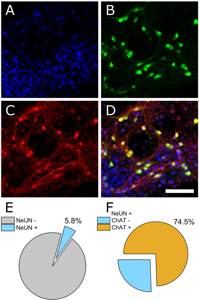
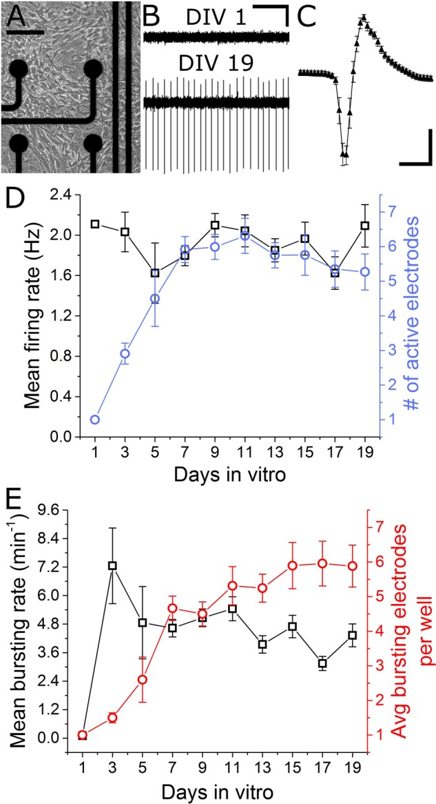
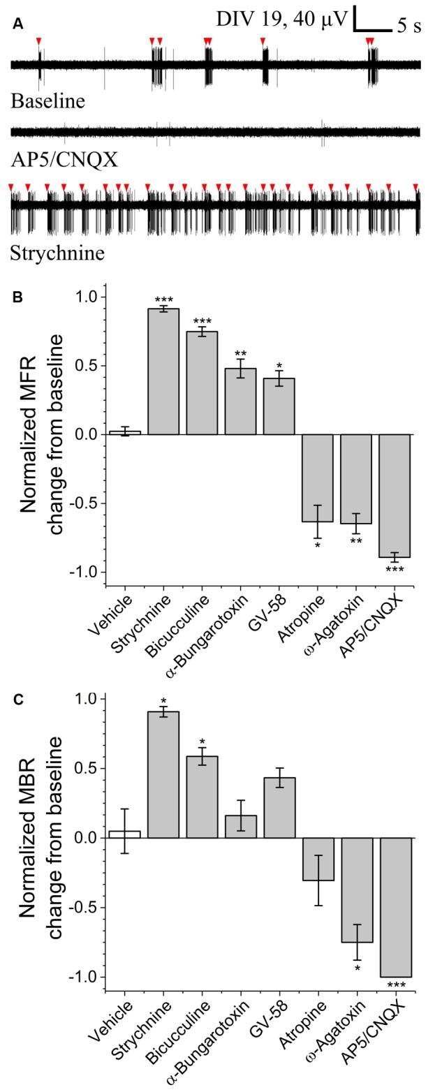
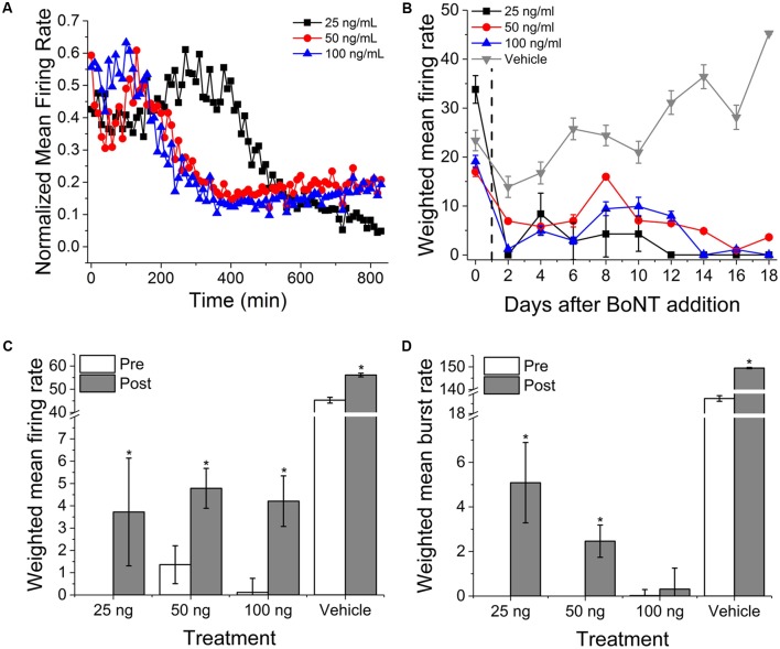
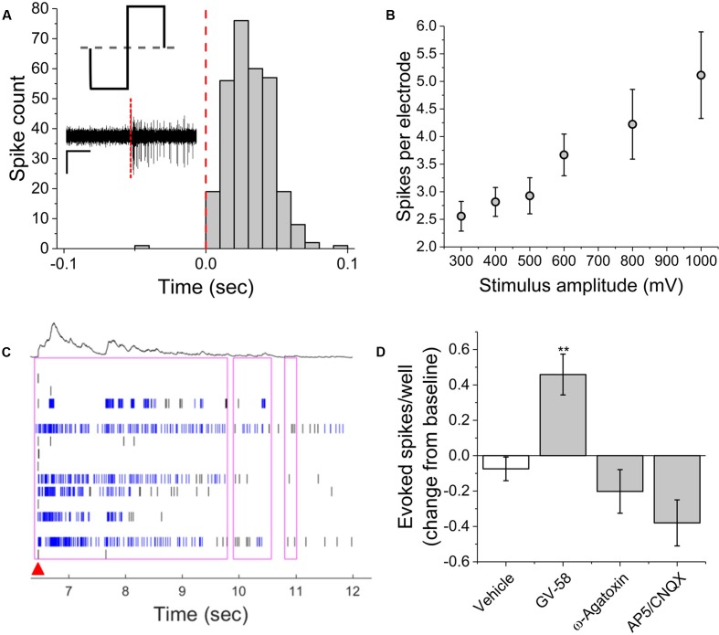
Motor neurons are the site of action for several neurological disorders and paralytic toxins, with cell bodies located in the ventral horn (VH) of the spinal cord along with interneurons and support cells. Microelectrode arrays (MEAs) have emerged as a high content assay platform for mechanistic studies and drug discovery. Here, we explored the spontaneous and evoked electrical activity of VH cultures derived from embryonic mouse spinal cord on multi-well plates of MEAs. Primary VH cultures from embryonic day 15-16 mice were characterized by expression of choline acetyltransferase (ChAT) by immunocytochemistry. Well resolved, all-or-nothing spontaneous spikes with profiles consistent with extracellular action potentials were observed after 3 days in vitro, persisting with consistent firing rates until at least day in vitro 19. The majority of the spontaneous activity consisted of tonic firing interspersed with coordinated bursting across the network. After 5 days in vitro, spike activity was readily evoked by voltage pulses where a minimum amplitude and duration required for excitation was 300 mV and 100 μs/phase, respectively. We characterized the sensitivity of spontaneous and evoked activity to a host of pharmacological agents including AP5, CNQX, strychnine, ω-agatoxin IVA, and botulinum neurotoxin serotype A (BoNT/A). These experiments revealed sensitivity of the cultured VH to both agonist and antagonist compounds in a manner consistent with mature tissue derived from slices. In the case of BoNT/A, we also demonstrated intoxication persistence over an 18-day period, followed by partial intoxication recovery induced by N- and P/Q-type calcium channel agonist GV-58. In total, our findings suggest that VH cultures on multi-well MEA plates may represent a moderate throughput, high content assay for performing mechanistic studies and for screening potential therapeutics pertaining to paralytic toxins and neurological disorders.

Keywords: BoNT/A; electrical stimulation; in vitro models; microelectrode array; motor neuron; neural activity modulation; neural activity recording; ventral horn.


Figures

FIGURE 1
FIGURE 1
Immunocytochemical staining to determine neuronal and non-neuronal culture populations. (A) DAPI staining indicates all cell nuclei. (B) NeUN indicates neuronal nuclei. (C) Choline acetyltransferase (ChAT). (D) Merged image. (E) Quantification of co-expression of DAPI and NeUN versus DAPI+ counts (percentage of neuronal cells). (F) Of the neuronal population, 74.5% were ChAT positive.
FIGURE 2
FIGURE 2
Emergence of spontaneous VH network activity. (A) Adhesion and physical network formation on MEA substrates (DIV 4, scale bar indicates 100 μm). (B) Traces of continuous filtered data on DIV 1 and DIV 19, illustrating the emergence of well-resolved, high-SNR signals (horizontal/vertical scale bar indicates 1 s/10 μV). (C) Average and SD trace of single characteristic unit consistent with time-scale and amplitude of extracellular action potentials (horizontal/vertical scale bar indicates 1 ms/10 μV). (D) Mean firing rate and number of active electrodes per well over 19 days in vitro. (E) Mean bursting rate and average number of bursting electrodes per well over 19 days in vitro. Error bars represent SEM.
FIGURE 3
FIGURE 3
Pharmacological sensitivity of VH networks on MEAs. (A) Characteristic continuous filtered traces of baseline activity (DIV 19) and in the presence of AP5/CNQX or strychnine. Bursts indicated by red arrows. DoS normalized mean firing rates (B) and mean burst rates (C) for various pharmacological treatments and vehicle. p < 0.05, ∗∗p < 0.01, ∗∗∗p < 0.001.
FIGURE 4
FIGURE 4
Onset and persistence of VH network inhibition due to BoNT/A intoxication. (A) Normalized mean firing rates over 14 h (840 min, 10 min bins) following perfusion with 25, 50, and 100 ng/ml BoNT/A. (B) Weighted mean firing rates for 18 days following BoNT/A perfusion. BoNT/A removed after 24 h (indicated by black dashed line). Weighted mean firing rates (C) and burst rates (D) pre- and post-incubation with 20 μM GV-58. p < 0.05.
FIGURE 5
FIGURE 5
Single-channel and network activity evoked by electrical stimulation. (A) Summed peri-event histogram for five consecutive pulses of cathodic-leading charge balanced stimuli (1000 mV at 100 μs per phase). Inset illustrates charge-balanced wave stimulus waveform and shows characteristic filtered continuous trace immediately prior to and following stimulus (red dashed line). (B) Amplitude dependence of evoked spikes recorded on stimulating electrode. (C) Characteristic raster plot illustrating network evoked activity due to single stimulating electrode. Red arrow indicates time-stamp of stimulation. (D) Evoked spikes per well in the presence of pharmacological agents. ∗∗p < 0.01.

References

    1. Alvarez F. J., Fyffe R. E. W. (2007). The continuing case for the Renshaw cell. J. Physiol. 584 31–45. 10.1113/jphysiol.2007.136200 - DOI - PMC - PubMed
    1. Barthélémy-Requin M., Bévengut M., Portalier P., Ternaux J. P. (2006). Release of glutamate by the embryonic spinal motoneurons of rat positively regulated by acetylcholine through the nicotinic and muscarinic receptors. Neurochem. Int. 49 584–592. 10.1016/j.neuint.2006.04.016 - DOI - PubMed
    1. Beaudet M.-J., Yang Q., Cadau S., Blais M., Bellenfant S., Gros-Louis F., et al. (2015). High yield extraction of pure spinal motor neurons, astrocytes and microglia from single embryo and adult mouse spinal cord. Sci. Rep. 5:16763 10.1038/srep16763 - DOI - PMC - PubMed
    1. Beske P. H., Scheeler S. M., Adler M., McNutt P. M. (2015). Accelerated intoxication of GABAergic synapses by botulinum neurotoxin A disinhibits stem cell-derived neuron networks prior to network silencing. Front. Cell. Neurosci. 9:159 10.3389/fncel.2015.00159 - DOI - PMC - PubMed
    1. Burgen A. S., Dickens F., Zatman L. J., Hospital M. (1948). The action of botulinum toxin on the neuro-muscular junction. J. Physiol. 109 10–24. 10.1113/jphysiol.1949.sp004364 - DOI - PMC - PubMed