. 2017 Sep 28:5:89.
doi: 10.3389/fcell.2017.00089. eCollection 2017.

Maternally Contributed Folate Receptor 1 Is Expressed in Ovarian Follicles and Contributes to Preimplantation Development

Affiliations

Maternally Contributed Folate Receptor 1 Is Expressed in Ovarian Follicles and Contributes to Preimplantation Development

Trine Strandgaard et al. Front Cell Dev Biol. .

Abstract

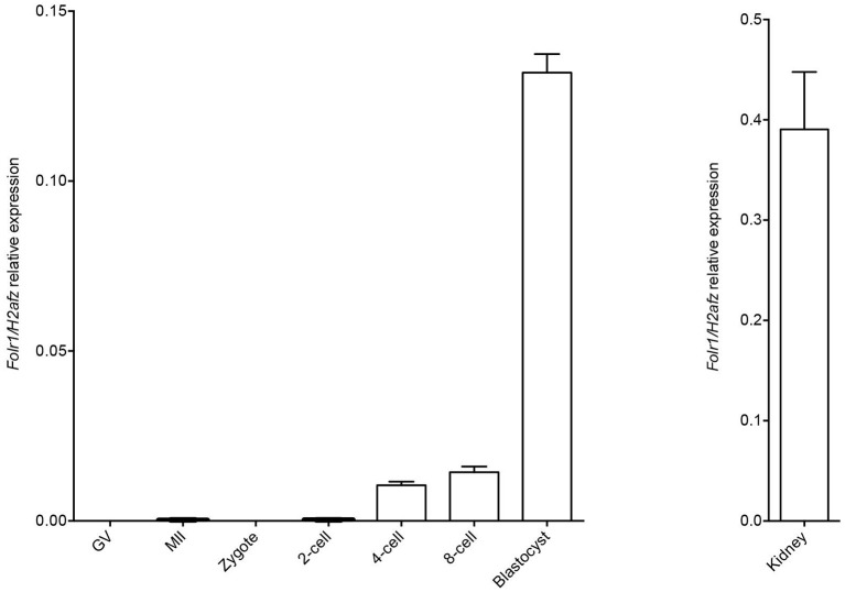
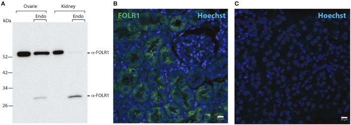
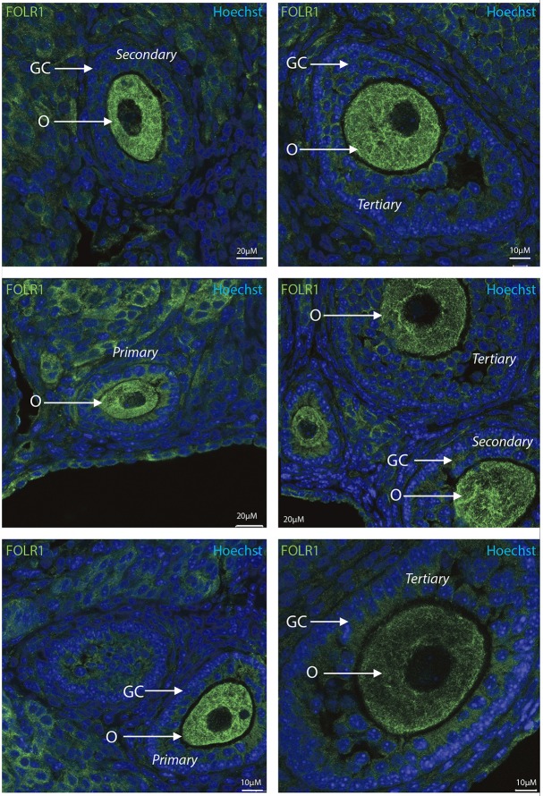
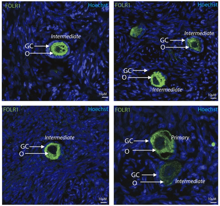
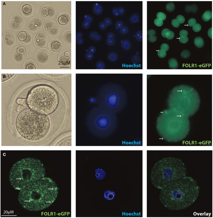
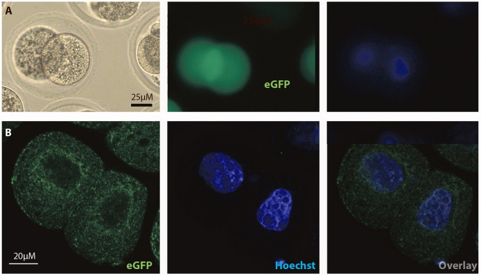
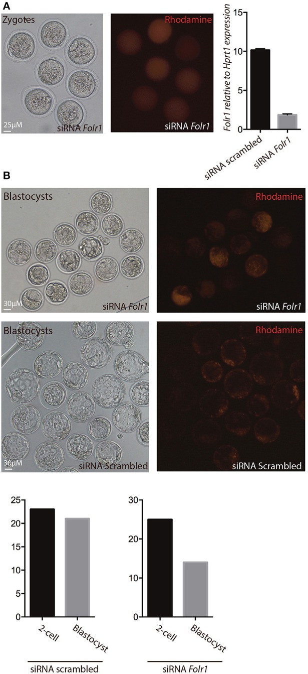
Folates have been shown to play a crucial role for proper development of the embryo as folate deficiency has been associated with reduced developmental capacity such as increased risk of fetal neural tube defects and spontanous abortion. Transcripts encoding the reduced folate carrier RFC1 (SLC19A1 protein) and the high-affinity folate receptor FOLR1 are expressed in oocytes and preimplantation embryos, respectively. In this study, we observed maternally contributed FOLR1 protein during mouse and human ovarian follicle development, and 2-cell mouse embryos. In mice, FOLR1 was highly enriched in oocytes from primary, secondary and tertiary follicles, and in the surrounding granulosa cells. Interestingly, during human follicle development, we noted a high and specific presence of FOLR1 in oocytes from primary and intermediate follicles, but not in the granulosa cells. The distribution of FOLR1 in follicles was noted as membrane-enriched but also seen in the cytoplasm in oocytes and granulosa cells. In 2-cell embryos, FOLR1-eGFP fusion protein was detected as cytoplasmic and membrane-associated dense structures, resembling the distribution pattern observed in ovarian follicle development. Knock-down of Folr1 mRNA function was accomplished by microinjection of short interference (si)RNA targeting Folr1, into mouse pronuclear zygotes. This revealed a reduced capacity of Folr1 siRNA-treated embryos to develop to blastocyst compared to the siRNA-scrambled control group, indicating that maternally contributed protein and zygotic transcripts sustain embryonic development combined. In summary, maternally contributed FOLR1 protein appears to maintain ovarian functions, and contribute to preimplantation development combined with embryonically synthesized FOLR1.

Keywords: blastocyst; folate receptor 1; follicle development; preimplantation development; siRNA.


Figures

Figure 1
Figure 1
Folr1 gene expression in oocytes and preimplantation embryos. Folr1 expression and relative abundan in GV and MII oocytes and preimplantation embryos, as indicated. Kidney tissue was included as a positive control. Folr1 expression levels were normalized by H2afz and relative expression displayed. Data are presented as mean standard deviation SD (bars) of triplicate measurements including standard deviations.
Figure 2
Figure 2
Western blotting and immunohistochemistry controls. (A) Western blot of ovary and kidney lysates [with and without Endoglycosidase H (Endo) treatment] from adult mice with antibodies against FOLR1. (B) Immunofluorescence staining of kidney from adult mice using antibodies against the FOLR1 (green) (C) or without antibody against FOLR1, counterstained with Hoechst (blue) for nuclear stain. Scale bars: 10 μm.
Figure 3
Figure 3
Intraovarian distribution of FOLR in mouse ovary tissue. FOLR1 localized to oocytes in primary, secondary and tertiary follicles. Hoechts staining identifies the nucleus of cells in the slides and the surrounding granulosa cells. o, oocytes; GC, granulosa cells. Scale bars; 10 or 20 μm, as indicated.
Figure 4
Figure 4
Intraovarian distribution of FOLR in human ovary tissue. FOLR1 localized to oocytes in primary and intermediate follicles. Hoechts staining identifies the nucleus of cells in the slides and the surrounding granulosa cells. o, oocytes; GC, granulosa cells. Scale bars; 10 μm, as indicated.
Figure 5
Figure 5
Intracellular distribution of FOLR1-eGFP in mouse 2-cell embryos. Intracelluar immunofluorescent localization and distribution of FOLR1-eGFP in 2-cell mouse embryos using microscopic (A,B) confocal imaging (C). Scale bars 20 or 25 μM, as indicated.
Figure 6
Figure 6
Intracellular distribution of eGFP in mouse 2-cell embryos. Intracelluar immunofluorescent localization and distribution of eGFP in 2-cell mouse embryos using (A) microscopic and (B) confocal imaging. Scale bars 20 or 25 μM, as indicated.
Figure 7
Figure 7
siRNA-mediated knock-down of Folr1 in mouse zygotes. (A) siRNA probes targeting Folr1 (or scrambled control (not shown) was microinjected in combination with rhodamine-conjugated dextrane, in order to identify injected embryos. At the 2-cell stage, qPCR showed that Folr1 transcript is efficiently reduced after Folr1 RNAi. (B) After in vitro culture, siRNA-injected embryos (both RNAi scrambled and Folr1) developed into blastocyst, however the capacity to form blastocysts was reduced in embryos that received siRNA targeting Folr1 (56%) compared to the control RNAi group (92%). Scale bars 25 or 30 μM, as indicated.

Similar articles

Cited by

References

    1. Albertsen M., Teperek M., Elholm G., Fuchtbauer E. M., Lykke-Hartmann K. (2010). Localization and differential expression of the Kruppel-associated box zinc finger proteins 1 and 54 in early mouse development. DNA Cell Biol. 29, 589–601. 10.1089/dna.2010.1040 - DOI - PubMed
    1. Altmae S., Stavreus-Evers A., Ruiz J. R., Laanpere M., Syvanen T., Yngve A., et al. . (2010). Variations in folate pathway genes are associated with unexplained female infertility. Fertil. Steril. 94, 130–137. 10.1016/j.fertnstert.2009.02.025 - DOI - PubMed
    1. Berrocal-Zaragoza M. I., Fernandez-Ballart J. D., Murphy M. M., Cavalle-Busquets P., Sequeira J. M., Quadros E. V. (2009). Association between blocking folate receptor autoantibodies and subfertility. Fertil. Steril. 91, 1518–1521. 10.1016/j.fertnstert.2008.08.104 - DOI - PMC - PubMed
    1. Bianchi E., Doe B., Goulding D., Wright G. J. (2014). Juno is the egg Izumo receptor and is essential for mammalian fertilization. Nature 508, 483–487. 10.1038/nature13203 - DOI - PMC - PubMed
    1. Blencowe H., Cousens S., Modell B., Lawn J. (2010). Folic acid to reduce neonatal mortality from neural tube disorders. Int. J. Epidemiol. 39(Suppl. 1), i110–i121. 10.1093/ije/dyq028 - DOI - PMC - PubMed

LinkOut - more resources