. 2017 Sep 28:4:160.
doi: 10.3389/fvets.2017.00160. eCollection 2017.

Misoprostol Inhibits Lipopolysaccharide-Induced Pro-inflammatory Cytokine Production by Equine Leukocytes

Affiliations

Misoprostol Inhibits Lipopolysaccharide-Induced Pro-inflammatory Cytokine Production by Equine Leukocytes

Emily Medlin Martin et al. Front Vet Sci. .

Abstract

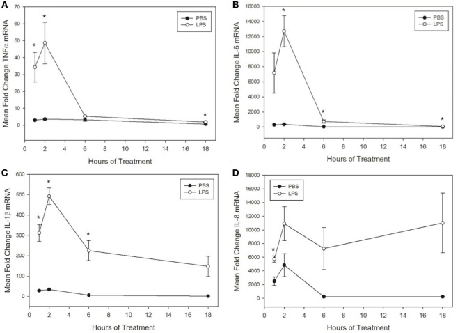
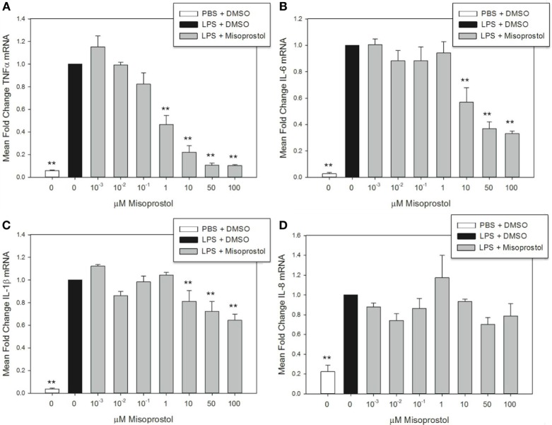
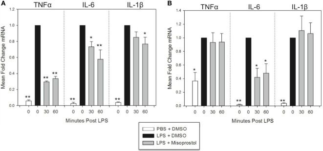
Pro-inflammatory cytokines including tumor necrosis factor α (TNFα), IL-1β, IL-6, and IL-8 are potent immune mediators that exacerbate multiple equine diseases such as sepsis and laminitis. Unfortunately, safe and effective cytokine-targeting therapies are lacking in horses; therefore, novel mechanisms of inhibiting cytokine production are critically needed. One potential mechanism for inhibiting cytokine synthesis is elevation of intracellular cyclic AMP (cAMP). In human leukocytes, intracellular cAMP production is induced by activation of E-prostanoid (EP) receptors 2 and 4. These receptors can be targeted by the EP2/4 agonist and prostaglandin E1 analog, misoprostol. Misoprostol is currently used as a gastroprotectant in horses but has not been evaluated as a cytokine-targeting therapeutic. Thus, we hypothesized that misoprostol treatment would inhibit pro-inflammatory cytokine production by lipopolysaccharide (LPS)-stimulated equine leukocytes in an in vitro inflammation model. To test this hypothesis, equine leukocyte-rich plasma (LRP) was collected from 12 healthy adult horses and used to model LPS-mediated inflammatory signaling. LRP was treated with varying concentrations of misoprostol either before (pretreated) or following (posttreated) LPS stimulation. LRP supernatants were assayed for 23 cytokines using an equine-specific multiplex bead immunoassay. Leukocytes were isolated from LRP, and leukocyte mRNA levels of four important cytokines were evaluated via RT-PCR. Statistical differences between treatments were determined using one-way RM ANOVA (Holm-Sidak post hoc testing) or Friedman's RM ANOVA on Ranks (SNK post hoc testing), where appropriate (p < 0.05, n = 3-6 horses). These studies revealed that misoprostol pre- and posttreatment inhibited LPS-induced TNFα and IL-6 protein production in equine leukocytes but had no effect on IL-8 protein. Interestingly, misoprostol pretreatment enhanced IL-1β protein synthesis following 6 h of LPS stimulation, while misoprostol posttreatment inhibited IL-1β protein production after 24 h of LPS stimulation. At the mRNA level, misoprostol pre- and posttreatment inhibited LPS-induced TNFα, IL-1β, and IL-6 mRNA production but did not affect IL-8 mRNA. These results indicate that misoprostol exerts anti-inflammatory effects on equine leukocytes when applied before or after a pro-inflammatory stimulus. However, the effects we observed were cytokine-specific and sometimes differed at the mRNA and protein levels. Further studies are warranted to establish the inhibitory effects of misoprostol on equine cytokine production in vivo.

Keywords: anti-inflammatory; chemokine; horse; inflammation; leukocyte; non-steroidal anti-inflammatory drug.


Figures

Figure 1
Figure 1
Lipopolysaccharide (LPS) increases tumor necrosis factor α (TNFα), IL-6, IL-1β, and IL-8 mRNA levels in equine leukocytes. Equine leukocyte-rich plasma was stimulated with 100 ng/mL LPS or vehicle (sterile PBS) for 1, 2, 6, or 18 h. mRNA was isolated, and levels of (A) TNFα, (B) IL-6, (C) IL-1β, and (D) IL-8 mRNA were quantified using real-time PCR with each sample assayed in triplicate. Data are expressed as mean ± SEM fold change in mRNA levels over untreated cells at 0 h (not shown). *p < 0.05 indicates a significant difference between LPS-stimulated cells and time-matched unstimulated controls via paired t-test; n = 4.
Figure 2
Figure 2
Misoprostol pretreatment decreases tumor necrosis factor α (TNFα), IL-6, and IL-1β mRNA levels in lipopolysaccharide (LPS)-stimulated equine leukocytes. Equine leukocyte-rich plasma was pretreated with various concentrations of misoprostol or vehicle (0.05% DMSO) for 30 min, followed by stimulation with 100 ng/mL LPS or PBS for 2 h. mRNA was isolated, and levels of (A) TNFα, (B) IL-6, (C) IL-1β, and (D) IL-8 mRNA were quantified via real-time PCR with each sample assayed in triplicate. Data are expressed as mean ± SEM fold change in mRNA levels versus LPS-stimulated cells pretreated with the misoprostol vehicle (denoted as 0 µM misoprostol). **p < 0.001 and *p < 0.05 indicate a significant difference compared to LPS-stimulated cells pretreated with the misoprostol vehicle (black bar) via one-way RM ANOVA; n = 3.
Figure 3
Figure 3
Misoprostol treatment 30 and 60 min following lipopolysaccharide (LPS) stimulation inhibits tumor necrosis factor α (TNFα), IL-6, and IL-1β mRNA production in equine leukocytes. Equine leukocyte-rich plasma was stimulated with 100 ng/mL LPS or vehicle (sterile PBS), followed 30 or 60 min later by treatment with 100 µM misoprostol or vehicle (0.05% DMSO). Cells were incubated for (A) 2 h or (B) 6 h, and mRNA was isolated. Levels of TNFα, IL-6, and IL-1β mRNA were assessed via real-time PCR with each sample assayed in triplicate. Data are expressed as mean ± SEM fold change in mRNA levels versus LPS-stimulated cells that were posttreated with the misoprostol vehicle (denoted as 0 µM misoprostol). **p < 0.001 and *p < 0.05 indicate a significant deference compared to LPS-stimulated cells that were posttreated with the misoprostol vehicle (black bars) via one-way RM ANOVA; n = 6 [(A), 2 h incubation] or n = 4 [(B), 6 h incubation].

Similar articles

Cited by

References

    1. Kolaczkowska E, Kubes P. Neutrophil recruitment and function in health and inflammation. Nat Rev Immunol (2013) 13:159–75.10.1038/nri3399 - DOI - PubMed
    1. Radi ZA, Kehrli MEJ, Ackermann MR. Cell adhesion molecules, leukocyte trafficking, and strategies to reduce leukocyte infiltration. J Vet Intern Med (2001) 15:516–29.10.1111/j.1939-1676.2001.tb01586.x - DOI - PubMed
    1. Chaudhry H, Zhou J, Zhong Y, Ali MM, McGuire F, Nagarkatti PS, et al. Role of cytokines as a double-edged sword in sepsis. In Vivo (2013) 27:669–84. - PMC - PubMed
    1. Wojdasiewicz P, Poniatowski XUA, Szukiewicz D. The role of inflammatory and anti-inflammatory cytokines in the pathogenesis of osteoarthritis. Mediators Inflamm (2014) 2014:1–19.10.1155/2014/561459 - DOI - PMC - PubMed
    1. Műzes G, Molnár B, Tulassay Z, Sipos F. Changes of the cytokine profile in inflammatory bowel diseases. World J Gastroenterol (2012) 18:5848–61.10.3748/wjg.v18.i41.5848 - DOI - PMC - PubMed

LinkOut - more resources