

miR-1 suppresses the proliferation and promotes the apoptosis of esophageal carcinoma cells by targeting Src
- PMID: 29034995
- PMCID: PMC5727306
- DOI: 10.1002/cam4.1214
miR-1 suppresses the proliferation and promotes the apoptosis of esophageal carcinoma cells by targeting Src
Abstract
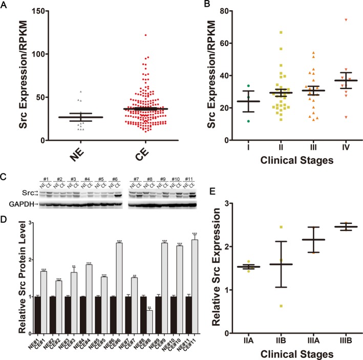
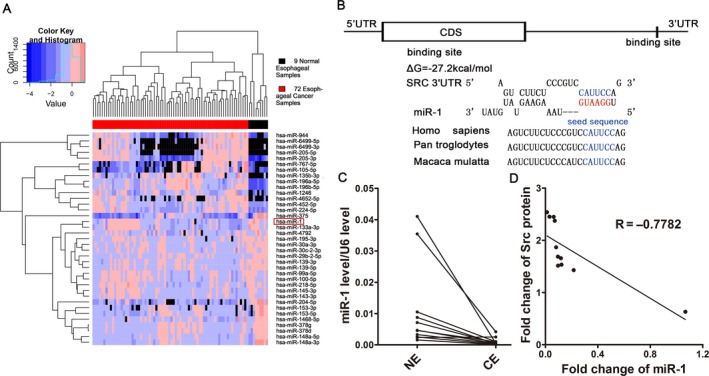
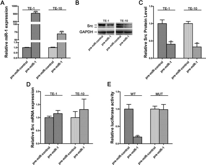
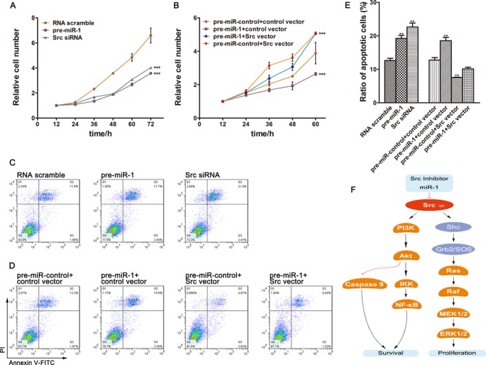
Nonreceptor tyrosine kinase c-Src, also known as Src, is a potent oncogene involved in a series of biological processes including cell growth, differentiation, and apoptosis; however, its expression pattern and function in esophageal cancer is poorly addressed. In this study, abnormal overexpression of Src protein was observed in esophageal cancer tissues, which fuelled the speculation that microRNA-mediated posttranscriptional regulatory mechanism might be involved. Bioinformatic analyses were applied to identify miRNAs that could potentially target Src. miR-1 was predicted and further validated as a direct repressor of Src. Moreover, we manipulated knockdown and overexpression experiment on TE-1 and TE-10 cells to demonstrate miR-1 suppressed proliferation and promoted apoptosis in esophageal cancer cells by inhibiting Src. Taken together, this study underlines a negative regulatory mechanism in which miR-1 serves as a suppressor of Src in esophageal cancer cells and may provide insights into novel therapeutic approaches for esophageal cancer.
Keywords: Src; apoptosis; esophageal cancer; miR-1; proliferation.
© 2017 The Authors. Cancer Medicine published by John Wiley & Sons Ltd.
Figures
Similar articles
-
miR-203 suppresses the proliferation and migration and promotes the apoptosis of lung cancer cells by targeting SRC.PLoS One. 2014 Aug 20;9(8):e105570. doi: 10.1371/journal.pone.0105570. eCollection 2014. PLoS One. 2014. PMID: 25140799 Free PMC article.
-
Overexpression of Csk-binding protein decreases growth, invasion, and migration of esophageal carcinoma cells by controlling Src activation.World J Gastroenterol. 2015 Feb 14;21(6):1814-20. doi: 10.3748/wjg.v21.i6.1814. World J Gastroenterol. 2015. PMID: 25684946 Free PMC article.
-
MiR-451 inhibits proliferation of esophageal carcinoma cell line EC9706 by targeting CDKN2D and MAP3K1.World J Gastroenterol. 2015 May 21;21(19):5867-76. doi: 10.3748/wjg.v21.i19.5867. World J Gastroenterol. 2015. PMID: 26019450 Free PMC article.
-
Mir-192 suppresses apoptosis and promotes proliferation in esophageal aquamous cell caicinoma by targeting Bim.Int J Clin Exp Pathol. 2015 Jul 1;8(7):8048-56. eCollection 2015. Int J Clin Exp Pathol. 2015. PMID: 26339371 Free PMC article.
-
Down-Regulation of miR-2053 Inhibits the Development and Progression of Esophageal Carcinoma by Targeting Fyn-Related Kinase (FRK).Dig Dis Sci. 2020 Oct;65(10):2853-2862. doi: 10.1007/s10620-019-06015-5. Epub 2020 Jan 1. Dig Dis Sci. 2020. PMID: 31894485
Cited by
-
Identification and validation of hub microRNAs dysregulated in esophageal squamous cell carcinoma.Aging (Albany NY). 2020 May 15;12(10):9807-9824. doi: 10.18632/aging.103245. Epub 2020 May 15. Aging (Albany NY). 2020. PMID: 32412911 Free PMC article.
-
Construction of long non-coding RNA- and microRNA-mediated competing endogenous RNA networks in alcohol-related esophageal cancer.PLoS One. 2022 Jun 15;17(6):e0269742. doi: 10.1371/journal.pone.0269742. eCollection 2022. PLoS One. 2022. PMID: 35704638 Free PMC article.
-
Up-regulation of androgen receptor by heat shock protein 27 and miR-1 induces pathogenesis of androgenic alopecia.Zhong Nan Da Xue Xue Bao Yi Xue Ban. 2022 Jan 28;47(1):72-78. doi: 10.11817/j.issn.1672-7347.2022.210131. Zhong Nan Da Xue Xue Bao Yi Xue Ban. 2022. PMID: 35545365 Free PMC article. Chinese, English.
-
Long noncoding antisense RNA FAM83A-AS1 promotes lung cancer cell progression by increasing FAM83A.J Cell Biochem. 2019 Jun;120(6):10505-10512. doi: 10.1002/jcb.28336. Epub 2019 Jan 18. J Cell Biochem. 2019. PMID: 30659636 Free PMC article.
-
Synthetic Evaluation of MicroRNA-1-3p Expression in Head and Neck Squamous Cell Carcinoma Based on Microarray Chips and MicroRNA Sequencing.Biomed Res Int. 2021 Aug 24;2021:6529255. doi: 10.1155/2021/6529255. eCollection 2021. Biomed Res Int. 2021. PMID: 34485523 Free PMC article.
References
-
- Torre, L. A. , Bray F., Siegel R. L., Ferlay J., Lortet‐Tieulent J., and Jemal A.. 2015. Global cancer statistics, 2012. CA Cancer J. Clin. 65:87–108. - PubMed
-
- Pennathur, A. , Gibson M. K., Jobe B. A., and Luketich J. D.. 2013. Oesophageal carcinoma. Lancet 381:400–412. - PubMed
-
- Kim, V. N. 2005. MicroRNA biogenesis: coordinated cropping and dicing. Nat. Rev. Mol. Cell Bio. 6:376–385. - PubMed
MeSH terms
Substances
LinkOut - more resources
Full Text Sources
Other Literature Sources
Medical
Molecular Biology Databases
Miscellaneous
