. 2017 Nov;98(11):2837-2849.
doi: 10.1099/jgv.0.000903. Epub 2017 Oct 16.

A pUL25 dimer interfaces the pseudorabies virus capsid and tegument

Affiliations

A pUL25 dimer interfaces the pseudorabies virus capsid and tegument

Yun-Tao Liu et al. J Gen Virol. 2017 Nov.

Abstract

Inside the virions of α-herpesviruses, tegument protein pUL25 anchors the tegument to capsid vertices through direct interactions with tegument proteins pUL17 and pUL36. In addition to promoting virion assembly, both pUL25 and pUL36 are critical for intracellular microtubule-dependent capsid transport. Despite these essential roles during infection, the stoichiometry and precise organization of pUL25 and pUL36 on the capsid surface remain controversial due to the insufficient resolution of existing reconstructions from cryo-electron microscopy (cryoEM). Here, we report a three-dimensional (3D) icosahedral reconstruction of pseudorabies virus (PRV), a varicellovirus of the α-herpesvirinae subfamily, obtained by electron-counting cryoEM at 4.9 Å resolution. Our reconstruction resolves a dimer of pUL25 forming a capsid-associated tegument complex with pUL36 and pUL17 through a coiled coil helix bundle, thus correcting previous misinterpretations. A comparison between reconstructions of PRV and the γ-herpesvirus Kaposi's sarcoma-associated herpesvirus (KSHV) reinforces their similar architectures and establishes important subfamily differences in the capsid-tegument interface.

Keywords: CryoEM; pUL17; pUL25 dimer; pUL36 (VP1/2); pseudorabies virus; tegument proteins.


Figures
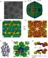
Fig. 1.
Fig. 1.
CryoEM reconstruction of PRV at 4.9 Å resolution. (a) CryoEM image of PRV virions. (b) 3D reconstruction of the PRV virion at 4.9 Å, coloured radially. The left and right halves of the capsid are rendered at a contour level (CL) of four and two times standard deviations above the mean density (σ), respectively. The write box and the black dashed box demark a hexon and a vertex region containing five CATCs, respectively. (c, d) Top and bottom views of the hexon as demarked by the write box in (b). H, hexon; Tb and Te, triplexes. (e) A slab of the PRV MCP upper domain (MCPud) density map (transparent grey) fitted with the HSV-1 MCPud atomic model (blue ribbon; PDB ID: 1NO7) (f) Front view of the CATC-crowning penton as demarked by the box in (b). One of the five CATC complexes is highlighted in orange. 5, fivefold axis; Ta and Tc, triplexes; H, Hexon. (g) Two different views of the CATC containing two intertwining globular heads (orange), coiled-coil (purple) and V-shaped density (yellow).
Fig. 2.
Fig. 2.
Each CATC contains a dimer of pUL25. (a, b) CATC in two different views. For clarity, only densities for the pUL25 dimer are displayed in (b) to better reveal the N-terminal segments of both monomers of the pUL25 dimer. (c) One density slab of the boxed region of (a) revealing the excellent match of α-helices in the X-ray models of the HSV-1 pUL25 globular domain (ribbon) and the PRV cryoEM density map. (d) An example of bulky aromatic side-chains in the cryoEM map: aromatic side-chains H482 and Y489 of the helix (H1, 475–497 aa) of the upper monomer of the pUL25 dimer. (e) Secondary structure prediction of PRV pUL25 N-terminal domain. Two helices (H2, 41–109 aa; H3, 142–152 aa) and short β-strands (yellow) are present in pUL25 N-terminal 1–160 aa. (f, g) The in situ (full-length) cryoEM structure and schematic model of the upper pUL25 (f) and lower pUL25 (g) segmented from the PRV map, with each consisting of an N-terminal loop (1–40 aa), a long helix (H2) and a hypothetical path of the unresolved loop (110–124 aa), and the C-terminal globular domain. The green asterisks mark the location of 124aa in pUL25.
Fig. 3.
Fig. 3.
Structure of pUL17 and the assignment of the pUL36 stem α-helices. (a) CATC structure of PRV with the pUL17 density shown in yellow. (b) pUL17 bridges two triplexes, Ta and Tc. The pUL17 are coloured yellow and the triplexes are coloured in light blue. The pUL17 density contains the V-shaped density and a stem helix. The V-shaped density can be divided into Ta and Tc binding domains. (c) All four α-helices (H1-4) that are longer than 20 Å, as identified from the secondary structural prediction of pUL17. (d) Two different views of pUL17 density. Of the four helices (H1-4, coloured pink) found in the map, three (H1–H4) are located in the V-shaped density. (e) Helix wheel diagram of H4 helix. Yellow, red and blue circles represent amino acids with large hydrophobic, negatively charged and positively charged side-chains, respectively. The white circles are amino acids with small hydrophobic (alanine) and polar uncharged (serine) side-chains. (f) CATC structure with the same orientation as in (a). Two helices of pUL36 are highlighted in purple.
Fig. 4.
Fig. 4.
Comparison of CATC interactions with penton in PRV and KSHV. (a) When the fivefold axes of the PRV and KSHV maps are aligned, the N-terminal helix is both longer and 20° further bent for the PRV pUL25 (orange) subunit in PRV compared to its KSHV homologue, the pORF19 (semi-transparent grey). The density of the pUL25 globular domain rotated 80° further than that of the pORF19. (b) The upper pUL25 interacts with two MCP subunits on penton. The HSV-1 pUL25 and two atomic models of the MCP upper domain (PDB ID: 1NO7) [46] are coloured red and blue, respectively. The density of the pUL25 and MCP is displayed in transparent red and blue, respectively, in the left figure. (c, d) The slabs of density at the same positions of PRV (a) and KSHV (b). The atomic models (ribbons) of HSV-1 pUL25 and MCPud were fitted into the density maps. KSHV contains the smallest capsid protein SCP (green) at the corresponding location of the upper pUL25 subunit. (e, f) Side views of the upper pUL25 and pORF19 interacting with penton capsid proteins in PRV (e) and KSHV (f), respectively.
Fig. 5.
Fig. 5.
Comparison of CATC interactions with hexon in PRV and KSHV. (a, b) Top view of a CATC and the CATC-interacting hexon in PRV (a) and KSHV (b). The hexon is shown in cyan, with light and heavy shades distinguishing the subunit boundaries. (c, d) Slice views of the boxed regions of (a, b) showing only two MCP subunits in CATC-interacting hexon in PRV (c) and KSHV (d). MCPud atomic models of HSV-1 are fitted in the map as ribbons. (e) The extended hairpin (β strand–loop– β strand motif) structure of the PRV MCP segment interacting with pUL17 as inferred from the fit HSV-1 MCPud model (left) and from the secondary structure prediction of the corresponding sequence of PRV MCP (right). (f) PRV pUL17 structure superimposed with its KSHV homologue (semi-transparent cyan), pORF32, revealing a 40° bend.
Fig. 6.
Fig. 6.
Schematic illustration of interactions of CATC in PRV (a) and KSHV (b). Two copies of pUL25 (pORF19) located on the capsid vertices in both PRV and KSHV. One of the pORF19 globular domains in KSHV that is not observed in the density map is shown in a fuzzy shape, and the other pORF19 binds one penton MCP. In PRV, the lack of SCP on the PRV penton and the longer N-terminal region of pUL25 seem to have enabled each pUL25 to bind two penton MCPs. The PRV pUL17 binds to two hexon MCP and it bends 40° compared to KSHV pORF32, which only binds to one hexon MCP.

Similar articles

Cited by

References

    1. Zerboni L, Sen N, Oliver SL, Arvin AM. Molecular mechanisms of varicella zoster virus pathogenesis. Nat Rev Microbiol. 2014;12:197–210. doi: 10.1038/nrmicro3215. - DOI - PMC - PubMed
    1. Pomeranz LE, Reynolds AE, Hengartner CJ. Molecular biology of pseudorabies virus: impact on neurovirology and veterinary medicine. Microbiol Mol Biol Rev. 2005;69:462–500. doi: 10.1128/MMBR.69.3.462-500.2005. - DOI - PMC - PubMed
    1. Kramer T, Enquist LW. Directional spread of alphaherpesviruses in the nervous system. Viruses. 2013;5:678–707. doi: 10.3390/v5020678. - DOI - PMC - PubMed
    1. Smith G. Herpesvirus transport to the nervous system and back again. Annu Rev Microbiol. 2012;66:153–176. doi: 10.1146/annurev-micro-092611-150051. - DOI - PMC - PubMed
    1. Antinone SE, Smith GA. Retrograde axon transport of herpes simplex virus and pseudorabies virus: a live-cell comparative analysis. J Virol. 2010;84:1504–1512. doi: 10.1128/JVI.02029-09. - DOI - PMC - PubMed

MeSH terms

Substances