. 2017 Nov 1;127(11):4207-4215.
doi: 10.1172/JCI96036. Epub 2017 Oct 16.

mTORC1 stimulates phosphatidylcholine synthesis to promote triglyceride secretion

Affiliations

mTORC1 stimulates phosphatidylcholine synthesis to promote triglyceride secretion

William J Quinn 3rd et al. J Clin Invest. .

Abstract

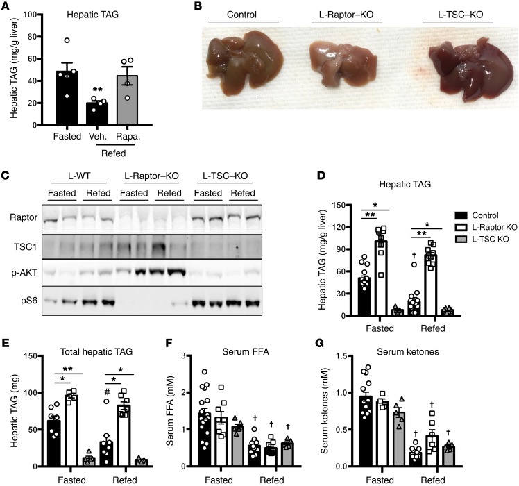
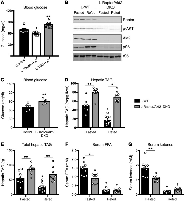
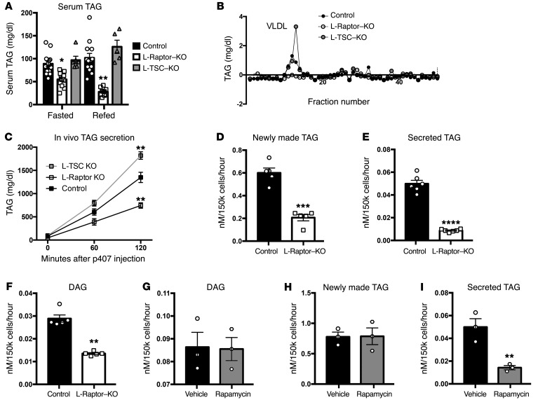
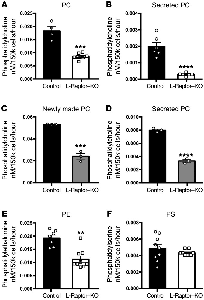
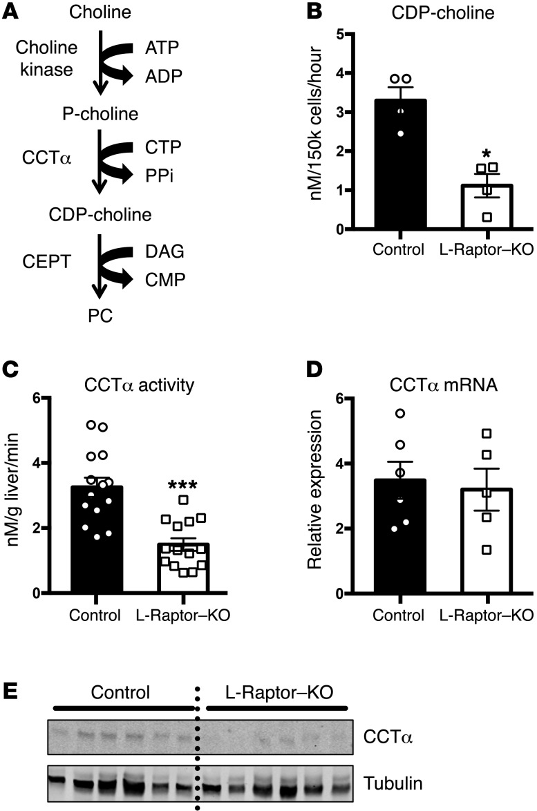
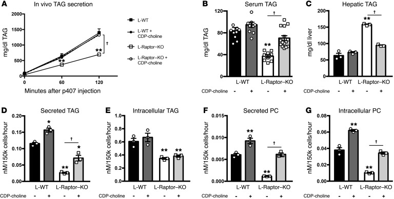
Liver triacylglycerol (TAG) synthesis and secretion are closely linked to nutrient availability. After a meal, hepatic TAG formation from fatty acids is decreased, largely due to a reduction in circulating free fatty acids (FFA). Despite the postprandial decrease in FFA-driven esterification and oxidation, VLDL-TAG secretion is maintained to support peripheral lipid delivery and metabolism. The regulatory mechanisms underlying the postprandial control of VLDL-TAG secretion remain unclear. Here, we demonstrated that the mTOR complex 1 (mTORC1) is essential for this sustained VLDL-TAG secretion and lipid homeostasis. In murine models, the absence of hepatic mTORC1 reduced circulating TAG, despite hepatosteatosis, while activation of mTORC1 depleted liver TAG stores. Additionally, mTORC1 promoted TAG secretion by regulating phosphocholine cytidylyltransferase α (CCTα), the rate-limiting enzyme involved in the synthesis of phosphatidylcholine (PC). Increasing PC synthesis in mice lacking mTORC1 rescued hepatosteatosis and restored TAG secretion. These data identify mTORC1 as a major regulator of phospholipid biosynthesis and subsequent VLDL-TAG secretion, leading to increased postprandial TAG secretion.


Conflict of interest statement

Conflict of interest: M. Wan is currently an employee of Agios Pharmaceuticals. M.J. Birnbaum is an employee of Pfizer Inc. P.M. Titchenell receives research funding from Pfizer Inc.

Figures

Figure 1
Figure 1. mTORC1 activity is both required and sufficient to induce hepatic steatosis.
Fasted 8- to 12-week-old C57BL/6 mice were injected with 20 mg/kg of rapamycin (A) liver TAG; n = 4. Veh., vehicle; Rapa, rapamycin. (BG) Six- to ten-week-old Raptorfl/fl and Tsc1fl/fl animals were injected with either AAV-GFP (control, black) or AAV-CRE (L-Raptor–KO, white; L-TSC–KO, gray) for 2 weeks prior to sacrifice. Cohorts were either fasted for 18 hours or fasted and refed for 4 hours. (B) Livers from mice of the indicated genotypes. (C) Immunoblot for indicated proteins. (D) Hepatic TAG was measured. (E) Total hepatic TAG normalized to liver. (F) Serum FFAs. (G) Serum ketones. n = 5–12 per group. *P < 0.05; **P < 0.01 vs. control. #P < 0.05 vs. fasted (when comparing refed vs. fasting). P < 0.01 vs. fasted (when comparing refed vs. fasting). Two-way ANOVA.
Figure 2
Figure 2. Loss of mTORC1 increases hepatic TAG independently of Akt.
Six- to ten-week-old Raptorfl/fl and Tsc1fl/fl animals were injected with either AAV-GFP (control, black) or AAV-CRE (L-Raptor–KO, white; L-TSC–KO, gray) for 2 weeks prior to sacrifice. (A) Fasting blood glucose. n = 5. (BG) Six- to ten-week-old Akt2/Raptorfl/fl animals were injected with either AAV-GFP (control, black) or AAV-CRE (L-Akt2/Raptor–DKO, gray) for 2 weeks prior to sacrifice. (C) Fasting blood glucose. (D) Hepatic TAG was measured. (E) Total Hepatic TAG normalized to liver. (F) Serum FFAs. (G) Serum ketones. n = 4–8. *P < 0.05; **P < 0.01 vs. control. #P < 0.05 vs. fasted (when comparing refed vs. fasting). P < 0.01 vs. fasted (when comparing refed vs. fasting). Two-way ANOVA.
Figure 3
Figure 3. mTORC1 cell autonomously regulates TAG secretion.
Six- to ten-week-old Raptorfl/fl and Tsc1fl/fl animals were injected with either AAV-GFP (control, black) or AAV-CRE (L-Raptor–KO, white; L-TSC–KO, gray) for 2 weeks prior to sacrifice. (A) Serum TAG levels. n = 5–9. (B) Serum was subjected to FPLC analysis, and triglyceride content was measured in each of the eluted fractions. (C) Triglyceride secretion rates were determined in fasted animals by blocking triglyceride uptake via i.p. injection of poloxamer 407 and measuring the accumulation of triglyceride in the serum over time. n = 4–6 per group. (DF) Six- to ten-week-old Raptorfl/fl animals were injected with AAV-GFP (black) or AAV-Cre (white) and rested 2 weeks to allow for gene excision. Hepatocytes were isolated and metabolically labeled for 4 hours in culture with 3H-glycerol. Medium and cellular fractions were separated. Samples were then fractionated by TLC and compared with lipid standards. (D) Intracellular TAG. (E) Secreted TAG. n = 6. (F) Intracellular DAG. n = 6. C57BL/6 hepatocytes were isolated and metabolically labeled with 3H-glycerol in the presence (white) or absence (black) of rapamycin (10 ng/ml) for 4 hours. Medium and cellular fractions were separated. Samples were then fractionated by TLC and compared with lipid standards. (G) Intracellular DAG. n = 3. (H) Intracellular TAG. n = 3. (I) Secreted TAG. n = 3. For hepatocyte studies in L-Raptor–KO or control, hepatocytes from 4 to 9 mice per group were isolated and technical replicates pooled. Data represent 4 to 9 individual mice per group. For hepatocyte studies using rapamycin, hepatocytes from 3 mice per group were isolated and technical replicates pooled. Data represent 3 individual mice per condition. *P < 0.05; **P < 0.01; ***P < 0.001; ****P < 0.0001 vs. control condition using 2-way ANOVA (A and C) or Student’s t test (DI).
Figure 4
Figure 4. mTORC1 activity is required for PC biosynthesis and secretion.
Six- to ten-week-old Raptorfl/fl animals were injected with either AAV-GFP (control, black) or AAV-CRE (L-Raptor–KO, white) for 2 weeks prior to sacrifice. Hepatocytes were isolated and metabolically labeled for 4 hours in culture with 3H-glycerol. Medium and cellular fractions were separated. Samples were then fractionated by TLC and compared with lipid standards. (A) Intracellular PC. n = 4–6. (B) Secreted PC in the medium. n = 4–6. C57BL/6 hepatocytes were isolated and metabolically labeled with 3H-glycerol in the presence (white) or absence (black) of rapamycin (10 ng/ml) for 4 hours. Medium and cellular fractions were separated. Samples were then fractionated by TLC and compared with lipid standards. (C) Intracellular PC. n = 3. (D) Secreted PC in the medium. n = 3. (E) Intracellular PE in L-Raptor–KO. n = 6–9. (F) Intracellular phosphatidylserine (PS) in L-Raptor–KO. n = 6–9. For hepatocyte studies in L-Raptor–KO or control, hepatocytes from 4 to 9 mice per group were isolated and technical replicates pooled. Data represent 4 to 9 individual mice per condition. For hepatocyte studies using rapamycin, hepatocytes from 3 mice per group were isolated and technical replicates pooled. Data represent 3 individual mice per condition. **P < 0.01; ***P < 0.001; ****P < 0.0001 vs. control condition using Student’s t test.
Figure 5
Figure 5. CCTα is regulated posttranscriptionally by mTORC1 activity.
Six- to ten-week-old Raptorfl/fl animals were injected with either AAV-GFP (control, black) or AAV-CRE (L-Raptor–KO, white) for 2 weeks prior to sacrifice. (A) Schematic of the Kennedy pathway. (B) Generation of CDP-choline was measured following incubation with 14C-choline chloride for 4 hours. n = 4. (C) CCTα activity was measured in hepatocyte lysates in the presence of excess CTP and 14C-phosphocholine. n = 12–13. (D) CCTα mRNA was interrogated by quantitative RT-PCR. n = 5–6. (E) CCTα protein was measured by Western blot. Data are representative of 6 animals. *P < 0.05; ***P < 0.001 vs. control using Student’s t test.
Figure 6
Figure 6. Restoration of PC synthesis in the absence of mTORC1 is sufficient to regulate TAG secretion in vivo.
Six- to ten-week-old Raptorfl/fl animals were injected with either AAV-GFP (control, black) or AAV-CRE (L-Raptor–KO, white) and injected daily with saline (solid) or 150 mg/kg CDP-choline for 2 weeks. (A) Triglyceride secretion rates were determined in fasted animals by blocking triglyceride uptake via i.p. injection of poloxamer 407 and measuring the accumulation of triglyceride in the serum over time. n = 9–10. (B) Fasting serum triglyceride levels were measured. n = 9–10. (C) Fasting hepatic triglyceride levels were measured. n = 3. Isolated hepatocytes from treatment groups were cultured with 3H-glycerol ± CDP-choline (150 mg/kg) (DG). (D) Secreted TAG was measured in the medium. n = 3. (E) Intracellular TAG was measured. n = 3. (F) Newly made secreted PC was measured in the medium. n = 3. (G) Intracellular newly made PC was measured. n = 3. For hepatocyte studies, hepatocytes from 3 mice were isolated and technical replicates pooled. Data represent 3 individual mice per condition. *P < 0.05; **P < 0.01 vs. control using 1-way ANOVA. P < 0.01 using 1-way ANOVA.

References

    1. Adamovich Y, Aviram R, Asher G. The emerging roles of lipids in circadian control. Biochim Biophys Acta. 2015;1851(8):1017–1025. - PubMed
    1. Browning JD, Baxter J, Satapati S, Burgess SC. The effect of short-term fasting on liver and skeletal muscle lipid, glucose, and energy metabolism in healthy women and men. J Lipid Res. 2012;53(3):577–586. doi: 10.1194/jlr.P020867. - DOI - PMC - PubMed
    1. Lambert JE, Parks EJ. Postprandial metabolism of meal triglyceride in humans. Biochim Biophys Acta. 2012;1821(5):721–726. - PMC - PubMed
    1. Tiwari S, Siddiqi SA. Intracellular trafficking and secretion of VLDL. Arterioscler Thromb Vasc Biol. 2012;32(5):1079–1086. doi: 10.1161/ATVBAHA.111.241471. - DOI - PMC - PubMed
    1. Sparks JD, Sparks CE, Adeli K. Selective hepatic insulin resistance, VLDL overproduction, and hypertriglyceridemia. Arterioscler Thromb Vasc Biol. 2012;32(9):2104–2112. doi: 10.1161/ATVBAHA.111.241463. - DOI - PubMed

Substances