. 2017 Nov;23(11):1352-1361.
doi: 10.1038/nm.4415. Epub 2017 Oct 9.

Targeting glioma stem cells through combined BMI1 and EZH2 inhibition

Affiliations

Targeting glioma stem cells through combined BMI1 and EZH2 inhibition

Xun Jin et al. Nat Med. 2017 Nov.

Abstract

Glioblastomas are lethal cancers defined by angiogenesis and pseudopalisading necrosis. Here, we demonstrate that these histological features are associated with distinct transcriptional programs, with vascular regions showing a proneural profile, and hypoxic regions showing a mesenchymal pattern. As these regions harbor glioma stem cells (GSCs), we investigated the epigenetic regulation of these two niches. Proneural, perivascular GSCs activated EZH2, whereas mesenchymal GSCs in hypoxic regions expressed BMI1 protein, which promoted cellular survival under stress due to downregulation of the E3 ligase RNF144A. Using both genetic and pharmacologic inhibition, we found that proneural GSCs are preferentially sensitive to EZH2 disruption, whereas mesenchymal GSCs are more sensitive to BMI1 inhibition. Given that glioblastomas contain both proneural and mesenchymal GSCs, combined EZH2 and BMI1 targeting proved more effective than either agent alone both in culture and in vivo, suggesting that strategies that simultaneously target multiple epigenetic regulators within glioblastomas may be effective in overcoming therapy resistance caused by intratumoral heterogeneity.


Conflict of interest statement

COMPETING FINANCIAL INTERESTS

JNR received an honorarium from PTC Therapeutics as an advisory board member. No other author declares competing financial interests.

Figures
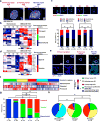
Figure 1
Figure 1. Anatomical distribution of transcriptional profiles in glioblastoma
(a) T1-weighted MRI images of multiregional glioblastoma sampling process (representative of four patients) for qPCR and immunofluorescence analysis. (b) A heatmap of molecular subtype maker expression in three multiregional glioblastoma samples and four non-malignant human brain samples. Z-scores were calculated from qPCR ΔCt values. (c) Immunofluorescence images (representative of 10 fields for each region) showing SOX2 (red), CD44 (green), OLIG2 (red), and YKL40 (green) positive cells in multiregional glioblastoma samples. Scale bar, 25 μm. SOX2, CD44, OLIG2, and YKL40 positive cells were compared by Chi-square test. **, p < 0.001. (d) Heatmap showing the expression of markers of hypoxia (CA9, GLUT3, GLUT1, HIF1α, MCT1, MCT4, and LDH5) and vascular (CD31, VEGFR2, CD34, ACTIN, and VEGFB) in three multiregional glioblastoma samples and four non-malignant human brain samples. Z-scores were calculated from qPCR ΔCt values. (e) Immunofluorescence images (representative from three fields for each region) showing vWF (red) and CA9 (green) positive locations in multiregional glioblastoma samples. Scale bar upper panels, 25 μm. Scale bar lower panels, 10 μm. (f) Heatmap showing Z-scores of each glioblastoma subtype signature normalized within each patient sample set determined via ssGSEA for each RNA-sample in the Anatomic Structure Study dataset (RNA-sample n = 122; patient n = 10) from the Ivy Glioblastoma Atlas Project (Ivy GAP) database. The corresponding histological feature for each RNA-sample is labeled above: Pseudopallisading cells around necrosis (PSEU); microvascular proliferative region (MV); cellular tumor (CT); leading edge (LE); infiltrating tumor (IT). (g, h) Chi-square test of glioblastoma histological feature distributions among transcriptional profiles and molecular subtype distribution among histological structures, respectively. **, p < 0.001.
Figure 2
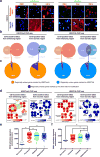
Figure 2. Epigenetic GSC signatures in multi-regional primary specimens
(a) Immunofluorescence images showing CD31 (red), CA9 (red), CD15 (red), H3K27Me3 (green), and H2AK119Ub (green) positive location and cells in multiregional glioblastoma samples. Scale bar, 25 μm. (b) Overlap in regional specific H3K27me3 or H2AK119Ub binding genes in CW2451- and CW2473-derived CD15 positive glioma cells. Venn diagrams showing overlaps between region specific peaks derived from H3K27me3 or H2AK119Ub ChIP-seq experiments on two primary glioblastoma specimens. (c) Pie graph showing fraction of regional unique target genes by H3K27Me3 or H2AK119Ub from the overlapping region specific genes shown in (b). (d) Gene ontology bubble plots showing gene signatures enriched in the overlapping region specific peaks shown in (b). (e, f) Enrichment levels of EZH2 (e) or BMI1 (f) activation signatures in transcriptional subgroups from the TCGA glioblastoma dataset (n = 10, Non-tumor; n = 161, Proneural; n = 209, Classical; n = 167, Mesenchymal). Data shows the median value (black bars). **, p < 0.01; by one-way ANOVA with Tukey’s method for multiple comparisons.
Figure 3
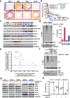
Figure 3. Differential polycomb repressive complex function in glioblastoma subgroups
(a) Representative images of glioblastoma tissue microarray samples (n = 96) showing the expression of EZH2, BMI1, CD44, and OLIG2. Scale bar, 500 μm. (b) Kaplan-Meier curve of patient survival stratified by EZH2 and BMI1 protein expression level from a glioblastoma tissue microarray. Log-rank p-values were used to determine statistics significance. (c) Cell lysates were prepared from neural progenitor cells (NPC1, NPC2, and NPC3), proneural GSCs 670 (PN11, PN23, PN1919, and PN3691), and mesenchymal GSCs (MES20, MES3565, MES28, MES738). Protein was resolved by SDS-PAGE. Immunoblot for performed for BMI1, H2K119Ub, EZH2, H3K27me3, and markers for mesenchymal (CD44 and YKL40) and proneural (OLIG2 and SOX2) glioblastoma. (d) Immunoprecipitation followed by immunoblot was performed for BMI1 polyubiquitination in PN1919 and MES28 cells in presence or absence of lactacystin treatment for 5 hours (Lacta, 10 μM). Right panel: quantification of BMI1 polyubiquitination by Image J. Data normalized by input loading controls. (e) Rank-ordered list of correlation coefficients (r values) between ubiquitin ligases and BMI1 activation or inhibition signatures in TCGA glioblastoma samples. (f) BMI1 polyubiquitination by RNF144A in PN1919 cells after transduction with shRNA control (shCTRL) or shRNF144A (shRNF144A-1099 and shRNF144A-3112) was examined by immunoprecipitation of BMI1 followed by immunoblotting for BMI1 or ubiquitin in presence or absence of lactacystin treatment for 5 hours (Lacta, 10 μM). Levels of BMI1, RNF144A, and TUBULIN were measured by immunoblot in the input whole cell lysates. (g) Neural progenitor cells (NPC1), proneural GSCs (PN1919 and PN3691), and mesenchymal GSCs (MES20 and MES28) were grown under baseline, low-glucose, hypoxia, or combined conditions. Levels of RNF144A, BMI1, EZH2, CD44, YKL40, OLIG2, and SOX2 proteins were measured by immunoblot. (h) Cell viability of neural progenitor cells (NPC1), proneural GSCs (PN11, PN23, PN1919, and PN3691), and mesenchymal GSCs (MES20, MES28, MES3565, and MES738) were determined under baseline, low-glucose, hypoxia, or combined conditions. Data are presented as mean ± SEM. *, p < 0.05; by Wilcoxon and Mann-Whitney t-test.
Figure 4
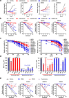
Figure 4. Differential efficacy of BMI1 and EZH2 inhibitors against glioblastoma subgroups
(a) Cell growth rates of GSCs transduced with shRNA control (shCNTRL), EZH2-knockdown (shEZH2-950 and shEZH2-2450) or BMI1-knockdown (shBMI1-880 and shBMI1-939) (n = 5 per group and time point). Data shows mean ± SEM. **, p < 0.01; by one-way ANOVA with Dunnett’s multiple-comparison test. (b) In vitro limited dilution assay of GSCs transduced with shRNA control (shCNTRL), EZH2-knockdown (shEZH2-950 and shEZH2-2450) or BMI1-knockdown (shBMI1-880 and shBMI1-939) (n = 15 per group). **, p < 0.01; by Chi-square test. (c) Cell viability curves of neural progenitors (NPC1), proneural GSCs (PN11, PN-JK2, PN-MMK1, PN1919, and PN1914.2), or mesenchymal GSCs (MES20, MES28, MES3565, MES-MN1, MES3128, and MES738) after treatment with increasing concentrations of (left) BMI1 inhibitor (BMI1-i, PTC596) or (right) EZH2 inhibitor (EZH2-i, EPZ6438). (d) IC50 of BMI1-i (left graph) or EZH2-i (right graph) for indicated cells. (e) In vitro limited dilution assay of PN1919, PN3691, MES738, and MES20 cells after treatment with vehicle control, 10 nM BMI1-i, 5 μM EZH2-i, or the combined treatment (n = 15 per group). **, p < 0.01; by Chi-square test.
Figure 5
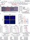
Figure 5. In vivo therapeutic efficacy of combined pharmacologic inhibition of BMI1 and EZH2 on subtype-mixed glioblastoma model
(a) Experimental design for in vivo effects of BMI1 (PTC596) and EZH2 (EPZ6438) inhibitors on xenograft of mixed proneural and mesenchymal GSCs. (b) Bioluminescence images of mice bearing mixed proneural and mesenchymal xenografts derived from luciferase-expressing PN1919 and MES20 cells, showing the effect of combined treatment of 10 mg/kg BMI1-i per week and 350 mg/kg EZH2-i thrice weekly on tumor growth. Right panels: Quantification of bioluminescence signals during 45 days of treatment in mice implanted with luciferase-expressing PN1919 and MES20 cells. The signals were normalized to day 10 signaling intensity for each mouse (n = 10 per group and time point). *, p < 0.05; **, p < 0.01; by one-way ANOVA with Tukey’s method for multiple comparisons. (c) Whole brain images showing distribution of PN1919 (red) and MES20 (green) population in the intermediate tumor samples (day 48). Right panel: Quantification of relative area of occupied by PN1919 (red), MES20 (green), and total tumor population in the intermediate tumor samples. Vehicle control, n = 10; EZH2-i alone, n = 15; BMI1-i alone, n = 14; combined treatment, n = 10. *, p < 0.01; **, p < 0.01; by one-way ANOVA with Tukey’s method for multiple comparisons. (d, e) Kaplan-Meier survival curve of orthotopic tumor-bearing mice with PN1919 (d) or MES20 (e) cells under the combined treatment of 12.5 mg/kg BMI1-i and 350 mg/kg 710 EZH2-i (n = 5 per group). Log-rank p-values were used to determine statistics significance. (f) Kaplan-Meier survival curve of subtype-mixed orthotopic tumor-bearing mice with PN1919 and MES20 cells upon the combined treatment of 10 mg/kg BMI1-i per week and 350 mg/kg EZH2-i thrice weekly (n = 8 per group). Log-rank p-values were used to determine statistics significance.

References

    1. Wen PY, Kesari S. Malignant Gliomas in Adults. N Engl J Med. 2008;359:492–507. - PubMed
    1. Singh SK, et al. Identification of human brain tumour initiating cells. Nature. 2004;432:396–401. - PubMed
    1. Galli R, et al. Isolation and Characterization of Tumorigenic, Stem-like Neural Precursors from Human Glioblastoma. Cancer Res. 2004;64:7011–7021. - PubMed
    1. Jin X, et al. Blockade of EGFR signaling promotes glioma stem-like cell invasiveness by abolishing ID3-mediated inhibition of p27KIP1 and MMP3 expression. Cancer Lett. 2013;328:235–242. - PubMed
    1. Bao S, et al. Glioma stem cells promote radioresistance by preferential activation of the DNA damage response. Nature. 2006;444:756–760. - PubMed

MeSH terms

Substances