. 2017 Dec 15;26(24):4896-4905.
doi: 10.1093/hmg/ddx370.

The role of the ER stress-response protein PERK in rhodopsin retinitis pigmentosa

Affiliations

The role of the ER stress-response protein PERK in rhodopsin retinitis pigmentosa

Dimitra Athanasiou et al. Hum Mol Genet. .

Abstract

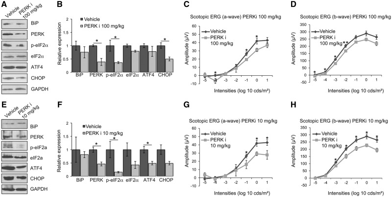
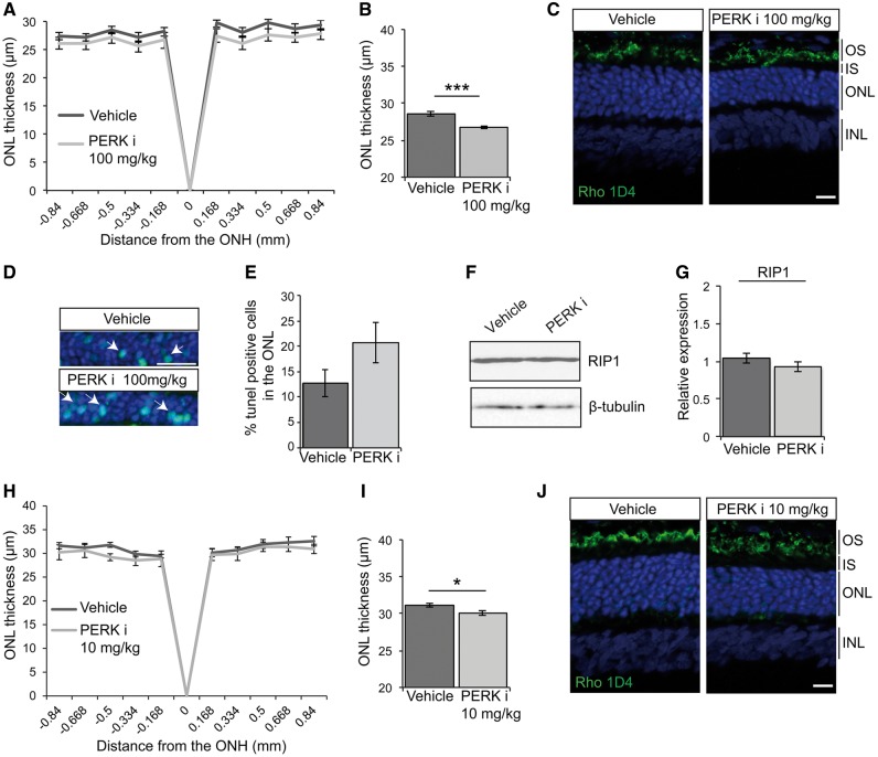
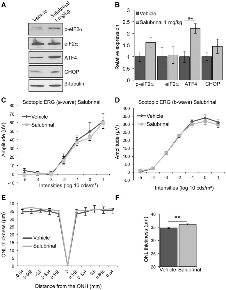
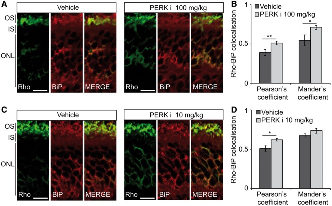
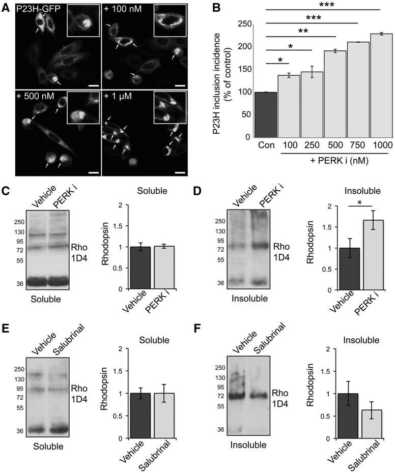
Mutations in rhodopsin, the light-sensitive protein of rod cells, are the most common cause of dominant retinitis pigmentosa (RP), a type of inherited blindness caused by the dysfunction and death of photoreceptor cells. The P23H mutation, the most frequent single cause of RP in the USA, causes rhodopsin misfolding and induction of the unfolded protein response (UPR), an adaptive ER stress response and signalling network that aims to enhance the folding and degradation of misfolded proteins to restore proteostasis. Prolonged UPR activation, and in particular the PERK branch, can reduce protein synthesis and initiate cell death through induction of pro-apoptotic pathways. Here, we investigated the effect of pharmacological PERK inhibition on retinal disease process in the P23H-1 transgenic rat model of retinal degeneration. PERK inhibition with GSK2606414A led to an inhibition of eIF2α phosphorylation, which correlated with reduced ERG function and decreased photoreceptor survival at both high and low doses of PERK inhibitor. Additionally, PERK inhibition increased the incidence of inclusion formation in cultured cells overexpressing P23H rod opsin, and increased rhodopsin aggregation in the P23H-1 rat retina, suggesting enhanced P23H misfolding and aggregation. In contrast, treatment of P23H-1 rats with an inhibitor of eIF2α phosphatase, salubrinal, led to improved photoreceptor survival. Collectively, these data suggest the activation of PERK is part of a protective response to mutant rhodopsin that ultimately limits photoreceptor cell death.


Figures

Figure 1.
Figure 1.
PERK inhibition reduces visual responses in P23H-1 rats. P23H-1 rats were treated from P21-P35 with either GSK2606414A (PERKi) or vehicle. (A, E) Representative western blot of retina lysates of P36 P23H-1 rats treated with 100 mg/kg (A) or 10 mg/kg (E) PERKi or vehicle for BiP, PERK, p-eIF2α, eIF2α, ATF4 and CHOP. GAPDH was used as a loading control. (B, F) Quantification of expression levels of BiP, PERK, p-eIF2α, total eIF2α, ATF4 and CHOP in P23H-1 rats after treatment with 100 mg/kg (B) or 10 mg/kg (F) PERKi. Densitometric analysis was used to calculate the levels of these proteins relative to vehicle; values are mean ± SEM n ≥ 4. (C, D) Scotopic ERG a-wave (C) and b-wave (D) amplitude results of P23H-1 rats (P36) treated from P21-P35 with either 100 mg/kg PERKi (n = 8) or vehicle (n = 6). (G, H) Scotopic ERG a-wave (G) and b-wave (H) amplitude results of P23H-1 rats (P36) treated from P21-P35 with either 10 mg/kg PERKi (n = 8) or vehicle (n = 6). Values are mean ± SEM, *P < 0.5, **P < 0.01, Student's t test.
Figure 2.
Figure 2.
PERK inhibition reduces photoreceptor survival in P23H-1 rats. P23H-1 rats were treated from P21-P35 with either 100 mg/kg (A–G) or 10 mg/kg (H–J) PERKi or vehicle. (A, H) Spider plot showing P23H-1 ONL thickness at P36 after PERKi (n = 6) or vehicle (n = 6) assessed by OCT. (B, I) Mean ONL thickness across the whole retina (E). Values are mean ± SEM. *P < 0.05, ***P < 0.001, Student’s t-test. (C, J) Representative images of the retina from P23H-1 (P36) rats treated with vehicle or PERKi. Cryosections were stained with anti-rhodopsin antibody 1D4 (green), and DAPI (blue) as indicated. (D, E) TUNEL staining in the ONL of P23H-1 rats treated with 100 mg/kg PERKi or vehicle treated. Arrows highlight positive TUNEL cells. Scale bar 10 µm. (D) Percentage of TUNEL positive cells in the ONL quantified by scoring n = 10 images each from 2 vehicle-treated rats and 2 PERKi-treated rats (100 mg/kg). Immunoblotting (F) and quantification (G) relative to vehicle of RIP1 immunoreactivity in PERKi (100mg/kg) and vehicle treated retinal lysates, β-tubulin was used as a loading control. Scale bar 20 µm.
Figure 3.
Figure 3.
Salubrinal enhances photoreceptor survival in P23H-1 rats. P23H-1 rats were treated from P21-P35 with either 1 mg/kg salubrinal or vehicle. (A) Representative western blot of retina lysates of P23H-1 rats treated with salubrinal or vehicle for p-eIF2α, eIF2α, ATF4, and CHOP. β-tubulin was used as a loading control. (B) Quantification of expression levels of p-eIF2α, total eIF2α, PERK ATF4 and CHOP relative to vehicle. (C, D) Scotopic ERG a-wave (C) and b-wave (D) amplitude results of P23H-1 rats (P36) treated from P21-P35 with either salubrinal (n = 8) or vehicle (n = 9). (E) Spider plot showing P23H-1 ONL thickness at P36 after salubrinal (n = 5) or vehicle (n = 4) assessed by OCT. (F) Mean ONL thickness across the whole retina Values are mean ± SEM, **P < 0.01, Student’s t-test.
Figure 4.
Figure 4.
PERK inhibition increases rhodopsin ER localisation in P23H-1 rats. P23H-1 rats were treated from P21-P35 with either (A, B) 100 mg/kg or (C, D) 10 mg/kg PERKi or vehicle. (A, C) Immunohistochemistry of rhodopsin (green) and BiP (red) immunoreactivity in the ONL and inner segment (IS). Scale bar 10 µm. (B, D) Rhodopsin-BiP co-localisation quantified by calculating the Pearson's and Mander's co-localisation coefficients using the JaCOP plug-in and ImageJ software, n = 18 images each from 3 vehicle-treated mice and 3 PERKi-treated mice. Values are means ± SEM, *P < 0.05, ***P < 0.001, unpaired two-sided Student's t test.
Figure 5.
Figure 5.
PERK inhibition increases P23H rhodopsin aggregation. (A, B) SK-N-SH cells were transfected with P23H-GFP rod opsin. Three hours post-transfection and after 2 h recovery in serum cells were either left untreated or treated with 100, 250, 500, 750 mM and 1μM of PERKi for 18 h prior to fixation. (A) Representative confocal images of P23H-GFP untreated rod opsin or treated with 100mM, 500 mM and 1 μM PERKi as indicated. Scale bar 10 μm. Magnified cell images are shown in insets. (Β) The incidence of inclusion formation of P23H-GFP in the absence and presence of PERKi at the indicated concentrations was assessed by scoring the percentage of cells with rod opsin P23H-GFP inclusions in 8 fields of ∼100 transfected cells. (C–F) Retinae of P23H-1 rats treated from P21-P35 with either 100 mg/kg PERKi (C,D), or salubrinal (E, F) or vehicle were analysed by a sedimentation assay. Fractions were immunoblotted with the 1D4 antibody against rhodopsin. Densitometric analysis was used to calculate the levels of soluble rhodopsin (C,E) relative to the vehicle treated and insoluble rhodopsin (D,F) relative to the vehicle after normalisation to soluble rhodopsin. Values are means ± SEM, n ≥ 4 (biological replicates). Error bars represent standard error, *P < 0.5, **P < 0.01, ***P < 0.001 Students t-test.

References

    1. Dryja T.P., McGee T.L., Reichel E., Hahn L.B., Cowley G.S., Yandell D.W., Sandberg M.A., Berson E.L. (1990) A point mutation of the rhodopsin gene in one form of retinitis pigmentosa. Nature, 343, 364–366. - PubMed
    1. Mendes H.F., van der Spuy J., Chapple J.P., Cheetham M.E. (2005) Mechanisms of cell death in rhodopsin retinitis pigmentosa: implications for therapy. Trends Mol. Med., 11, 177–185. - PubMed
    1. Illing M.E., Rajan R.S., Bence N.F., Kopito R.R. (2002) A rhodopsin mutant linked to autosomal dominant retinitis pigmentosa is prone to aggregate and interacts with the ubiquitin proteasome system. J. Biol. Chem., 277, 34150–34160. - PubMed
    1. Saliba R.S., Munro P.M., Luthert P.J., Cheetham M.E. (2002) The cellular fate of mutant rhodopsin: quality control, degradation and aggresome formation. J. Cell Sci., 115, 2907–2918. - PubMed
    1. Lin J.H., Li H., Yasumura D., Cohen H.R., Zhang C., Panning B., Shokat K.M., Lavail M.M., Walter P. (2007) IRE1 signaling affects cell fate during the unfolded protein response. Science, 318, 944–949. - PMC - PubMed

Publication types

MeSH terms

Substances