Review
. 2018 Apr 9;20(5):608-620.
doi: 10.1093/neuonc/nox183.

Glioma CpG island methylator phenotype (G-CIMP): biological and clinical implications

Affiliations
Review

Glioma CpG island methylator phenotype (G-CIMP): biological and clinical implications

Tathiane M Malta et al. Neuro Oncol. .

Abstract

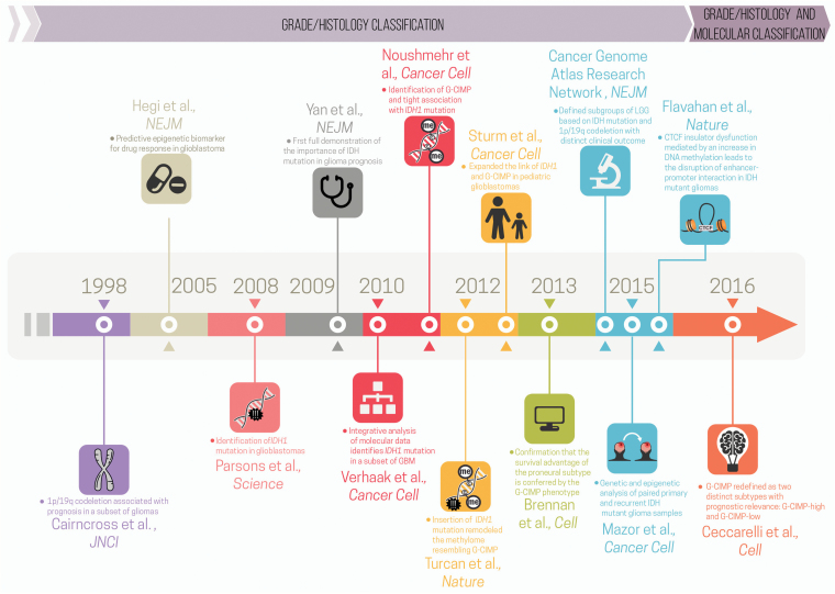
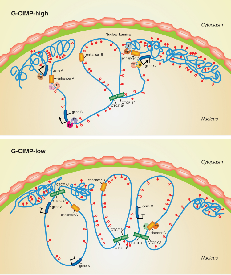
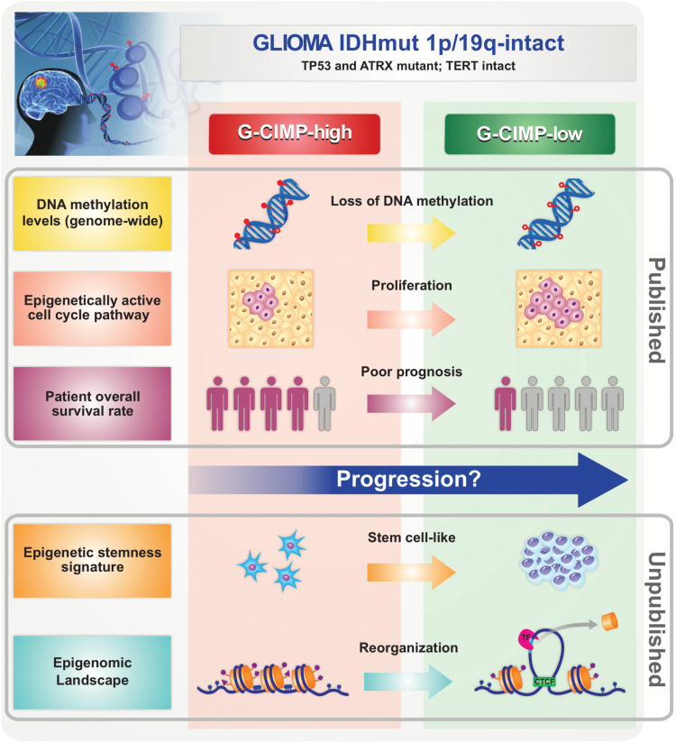
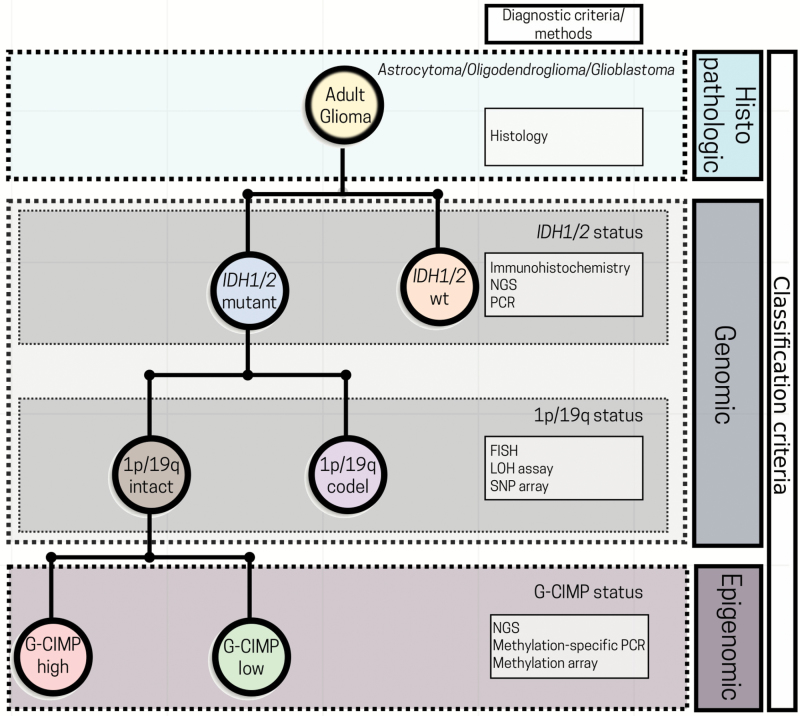
Gliomas are a heterogeneous group of brain tumors with distinct biological and clinical properties. Despite advances in surgical techniques and clinical regimens, treatment of high-grade glioma remains challenging and carries dismal rates of therapeutic success and overall survival. Challenges include the molecular complexity of gliomas, as well as inconsistencies in histopathological grading, resulting in an inaccurate prediction of disease progression and failure in the use of standard therapy. The updated 2016 World Health Organization (WHO) classification of tumors of the central nervous system reflects a refinement of tumor diagnostics by integrating the genotypic and phenotypic features, thereby narrowing the defined subgroups. The new classification recommends molecular diagnosis of isocitrate dehydrogenase (IDH) mutational status in gliomas. IDH-mutant gliomas manifest the cytosine-phosphate-guanine (CpG) island methylator phenotype (G-CIMP). Notably, the recent identification of clinically relevant subsets of G-CIMP tumors (G-CIMP-high and G-CIMP-low) provides a further refinement in glioma classification that is independent of grade and histology. This scheme may be useful for predicting patient outcome and may be translated into effective therapeutic strategies tailored to each patient. In this review, we highlight the evolution of our understanding of the G-CIMP subsets and how recent advances in characterizing the genome and epigenome of gliomas may influence future basic and translational research.


Figures

Fig. 1
Fig. 1
CIMP subtypes in human cancer. This illustration depicts aberrant DNA methylation changes at specific genomic loci in normal and tumor cells, especially in CIMP tumors. Each DNA strand represents one individual methylome. Methylated CpG sites in normal state are represented in blue, non-CIMP tumor DNA methylation gain in yellow, and aberrant DNA hypermethylation in CIMP tumors in red (modified from Weisenberger).
Fig. 2
Fig. 2
Timeline of major milestones in integrating genomics and epigenomics data that directly or indirectly uncovered glioma molecular and clinical phenotypes associated with G-CIMP subsets. Each milestone is indicated by marker papers that reported key molecular findings with clinical implications, along with a bullet summarizing their contribution. The timeline is broken up by the 2016 WHO publication (before and after) (modified from the original copy-free design by Freepik).
Fig. 3
Fig. 3
Chromatin changes in the progression of G-CIMP-high to G-CIMP-low tumors. This illustration shows a model of chromatin reorganization during the progression from G-CIMP-high to G-CIMP-low. G-CIMP-low (lower panel) shows a loss of DNA methylation at specific loci causing disruption of CTCF binding sites, reorganization of chromatin, and dysregulation of gene expression.
Fig. 4
Fig. 4
Overview of major discoveries that define G-CIMP-high and G-CIMP-low glioma subsets. G-CIMP-high and G-CIMP-low tumors share the following genomic alterations: IDH-mutant–1p/19q intact, telomerase reverse transcriptase promoter wildtype, and alpha thalassemia/mental retardation syndrome X-linked and tumor protein 53 mutant. However, the G-CIMP-low subset defined a subgroup of IDH-mutant 1p/19q intact gliomas associated with DNA demethylation. Changes in chromatin architecture led to an imbalance between the insulators and enhancers and the consequent activation of cell-cycle related genes, the increase in stemness features, and poor clinical outcome compared with G-CIMP-high gliomas (cartoon representation, not to scale).
Fig. 5
Fig. 5
Perspectives in incorporating G-CIMP subsets into the current 2016 WHO glioma classification algorithm. A simplified diagram for glioma classification based on histological, genetic, and epigenetic features. The incorporation of G-CIMP subsets further refined glioma classification. NGS: next-generation sequencing; PCR: polymerase chain reaction; FISH: fluorescent in situ hybridization; LOH: loss of heterozygosity; SNP: single-nucleotide polymorphism; wt: wildtype (modified from the original copy-free design by Freepik).

References

    1. Louis DN, Perry A, Reifenberger G et al. . The 2016 World Health Organization Classification of Tumors of the Central Nervous System: a summary. Acta Neuropathol. 2016;131(6):803–820. - PubMed
    1. Masui K, Mischel PS, Reifenberger G. Molecular classification of gliomas. Handb Clin Neurol. 2016;134:97–120. - PubMed
    1. Siegal T. Clinical relevance of prognostic and predictive molecular markers in gliomas. Adv Tech Stand Neurosurg 2016;(43):91–108. - PubMed
    1. Taylor JW, Chi AS, Cahill DP. Tailored therapy in diffuse gliomas: using molecular classifiers to optimize clinical management. Oncology (Williston Park). 2013;27(6):504–514. - PubMed
    1. Gittleman H, Lim D, Kattan MW et al. . An independently validated nomogram for individualized estimation of survival among patients with newly diagnosed glioblastoma: NRG Oncology RTOG 0525 and 0825. Neuro Oncol 2016;19(5):669–677. - PMC - PubMed

Publication types