. 2017 Dec 15;26(24):4861-4872.
doi: 10.1093/hmg/ddx364.

Progranulin-mediated deficiency of cathepsin D results in FTD and NCL-like phenotypes in neurons derived from FTD patients

Affiliations

Progranulin-mediated deficiency of cathepsin D results in FTD and NCL-like phenotypes in neurons derived from FTD patients

Clarissa Valdez et al. Hum Mol Genet. .

Abstract

Frontotemporal dementia (FTD) encompasses a group of neurodegenerative disorders characterized by cognitive and behavioral impairments. Heterozygous mutations in progranulin (PGRN) cause familial FTD and result in decreased PGRN expression, while homozygous mutations result in complete loss of PGRN expression and lead to the neurodegenerative lysosomal storage disorder neuronal ceroid lipofuscinosis (NCL). However, how dose-dependent PGRN mutations contribute to these two different diseases is not well understood. Using iPSC-derived human cortical neurons from FTD patients harboring PGRN mutations, we demonstrate that PGRN mutant neurons exhibit decreased nuclear TDP-43 and increased insoluble TDP-43, as well as enlarged electron-dense vesicles, lipofuscin accumulation, fingerprint-like profiles and granular osmiophilic deposits, suggesting that both FTD and NCL-like pathology are present in PGRN patient neurons as compared to isogenic controls. PGRN mutant neurons also show impaired lysosomal proteolysis and decreased activity of the lysosomal enzyme cathepsin D. Furthermore, we find that PGRN interacts with cathepsin D, and that PGRN increases the activity of cathepsin D but not cathepsins B or L. Finally, we show that granulin E, a cleavage product of PGRN, is sufficient to increase cathepsin D activity. This functional relationship between PGRN and cathepsin D provides a possible explanation for overlapping NCL-like pathology observed in patients with mutations in PGRN or CTSD, the gene encoding cathepsin D. Together, our work identifies PGRN as an activator of lysosomal cathepsin D activity, and suggests that decreased cathepsin D activity due to loss of PGRN contributes to both FTD and NCL pathology in a dose-dependent manner.


Figures

Figure 1.
Figure 1.
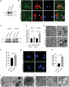
FTD-linked PGRN mutant neurons exhibit both FTD and NCL-like pathology. (A) PGRN expression in PGRN WT and mutant iPSC-derived neurons (day 35 post-differentiation) using NSE, a neuronal marker, as a loading control. (B) Immunofluorescence of TDP-43 and neuronal marker β-III tubulin in PGRN WT and mutant neuronal cultures (day 35 post-differentiation). PGRN mutant neurons have decreased nuclear TDP-43 (white arrows) as compared to PGRN WT neurons (yellow arrows). (C) Insoluble TDP-43 in PGRN WT and mutant neuron lysates (days 35 and 100 post-differentiation) shown by Western blot analysis using vinculin as a loading control. (D) Quantification of insoluble TDP-43 in PGRN WT and mutant neuron lysates (days 35 and 100 post-differentiation) (n = 3). (E) Electron dense vesicles in PGRN WT and mutant neurons (day 130 post-differentiation) visualized through electron microscopy analysis. Scale bar, 500 nM. (F) Quantification of electron-dense vesicle size in PGRN WT and mutant neurons (day 130 post-differentiation) (n = 6–8 cells/line). (G) Autofluorescent puncta in PGRN WT and mutant neurons (day 130 post-differentiation). (H) Quantification of lipofuscin puncta/cell in PGRN WT and mutant neurons (day 130 post-differentiation) (n = 3, 30–50 cells/experiment). Electron microscopy analysis of PGRN mutant neurons (day 130 post-differentiation) showed (I) fingerprint-like profiles and (J) granular osmiophilic deposits (n = 6–8 cells/line). Scale bar, 500 nM. Student’s t-test, unpaired, two-tailed statistical analysis was performed. Error bars represent S.E.M. of total experiments. n.s., not significant, *P < 0.05, ****P < 0.0001.
Figure 2.
Figure 2.
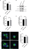
iPSC-derived PGRN mutant neurons have impaired lysosomal function. (A) Lysosomal proteolysis measured in PGRN WT and mutant cortical neurons (day 35 post-differentiation) through pulse-chase analysis (n = 6). (B) LAMP1 and LAMP2 expression in PGRN WT and mutant neuron lysates (day 35 post-differentiation) shown by Western blot analysis using β-III tubulin, a neuronal marker, as a loading control. Quantification of (C) LAMP1 and (D) LAMP2 expression in PGRN WT and mutant neuron lysates (day 35 post-differentiation) (n = 3). (E) Immunofluorescence of LAMP1 in PGRN WT and mutant neurons (day 35 post-differentiation). (F) Quantification of LAMP1 immunofluorescence intensity in PGRN WT and mutant neurons (day 35 post-differentiation) (n = 3, 95–114 cells/experiment). Student’s t-test, unpaired, two-tailed statistical analysis was performed. Error bars represent S.E.M. of total experiments, n.s., not significant, *P < 0.05.
Figure 3.
Figure 3.
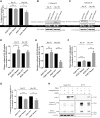
iPSC-derived PGRN mutant neurons have reduced cathepsin D activity. (A) Cathepsin D activity in PGRN WT and mutant cortical neuron lysates (days 35 and 100 post-differentiation) (n = 3). Immature and mature (B) cathepsin D and (C) cathepsin B expression in PGRN WT and mutant neuron lysates (days 35 and 100 post-differentiation) shown by Western blot analysis using β-III tubulin, a neuronal marker, as a loading control. Quantification of (D) mature and (E) immature cathepsin D expression in PGRN WT and mutant neuron lysates (days 35 and 100 post-differentiation) (n = 3). (F) Ratio of mature: immature cathepsin D in PGRN WT and mutant neuron lysates (days 35 and 100 post-differentiation) (n = 3). (G) Cathepsin D activity per mature cathepsin D expression in PGRN WT and mutant neuron lysates (35 and 100 days post-differentiation) (n = 3). (H) Co-immunoprecipitation assay in HEK293 cells co-expressing cathepsin D-V5 and PGRN show cathepsin D and PGRN interaction (n = 3). Student’s t-test, unpaired, two-tailed statistical analysis was performed. Error bars represent S.E.M. of total experiments, n.s., not significant, *P < 0.05, **P < 0.01, ***P < 0.001.
Figure 4.
Figure 4.
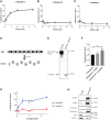
PGRN and granulin E are activators of cathepsin D activity. An in vitro dose-dependent (A) cathepsin D, (B) cathepsin B and (C) cathepsin L activity assay using recombinant cathepsins and recombinant PGRN (n = 3). (D) Structure of the human PGRN protein. PGRN is a precursor protein that can be enzymatically cleaved into 7.5 individual granulin motifs by the protease elastase. (E) Western blot analysis demonstrates effective cleavage of recombinant PGRN (0.12 nM) after incubation with elastase to obtain granulin sample. (F) Cathepsin D activity assay using recombinant cathepsin D incubated with either recombinant PGRN or granulins (n = 3). (G) A dose-dependent cathepsin D activity assay using recombinant cathepsin D incubated with either recombinant granulin E or granulin C (n = 3). (H) Western blot analysis of total cell lysate and lysosome-enriched samples in HEK293 cells (n = 3). Student’s t-test, unpaired, two-tailed statistical analysis was performed. Error bars represent S.E.M of total experiments, n.s., not significant, *P <0.05.

Similar articles

Cited by

References

    1. Onyike C.U., Diehl-Schmid J. (2013) The epidemiology of frontotemporal dementia. Int. Rev. Psychiatry, 25, 130–137. - PMC - PubMed
    1. Vieira R.T., Caixeta L., Machado S., Silva A.C., Nardi A.E., Arias-Carrion O., Carta M.G. (2013) Epidemiology of early-onset dementia: a review of the literature. Clin. Pract. Epidemiol. Ment. Health, 9, 88–95. - PMC - PubMed
    1. Chow T.W., Miller B.L., Hayashi V.N., Geschwind D.H. (1999) Inheritance of frontotemporal dementia. Arch. Neurol., 56, 817–822. - PMC - PubMed
    1. Rascovsky K., Hodges J.R., Knopman D., Mendez M.F., Kramer J.H., Neuhaus J., van Swieten J.C., Seelaar H., Dopper E.G., Onyike C.U. (2011) Sensitivity of revised diagnostic criteria for the behavioural variant of frontotemporal dementia. Brain, 134, 2456–2477. - PMC - PubMed
    1. Wood E.M., Falcone D., Suh E., Irwin D.J., Chen-Plotkin A.S., Lee E.B., Xie S.X., Van Deerlin V.M., Grossman M. (2013) Development and validation of pedigree classification criteria for frontotemporal lobar degeneration. JAMA Neurol., 70, 1411–1417. - PMC - PubMed

MeSH terms