. 2017 Nov 16;45(20):11989-12004.
doi: 10.1093/nar/gkx852.

The basic tilted helix bundle domain of the prolyl isomerase FKBP25 is a novel double-stranded RNA binding module

Affiliations

The basic tilted helix bundle domain of the prolyl isomerase FKBP25 is a novel double-stranded RNA binding module

David Dilworth et al. Nucleic Acids Res. .

Abstract

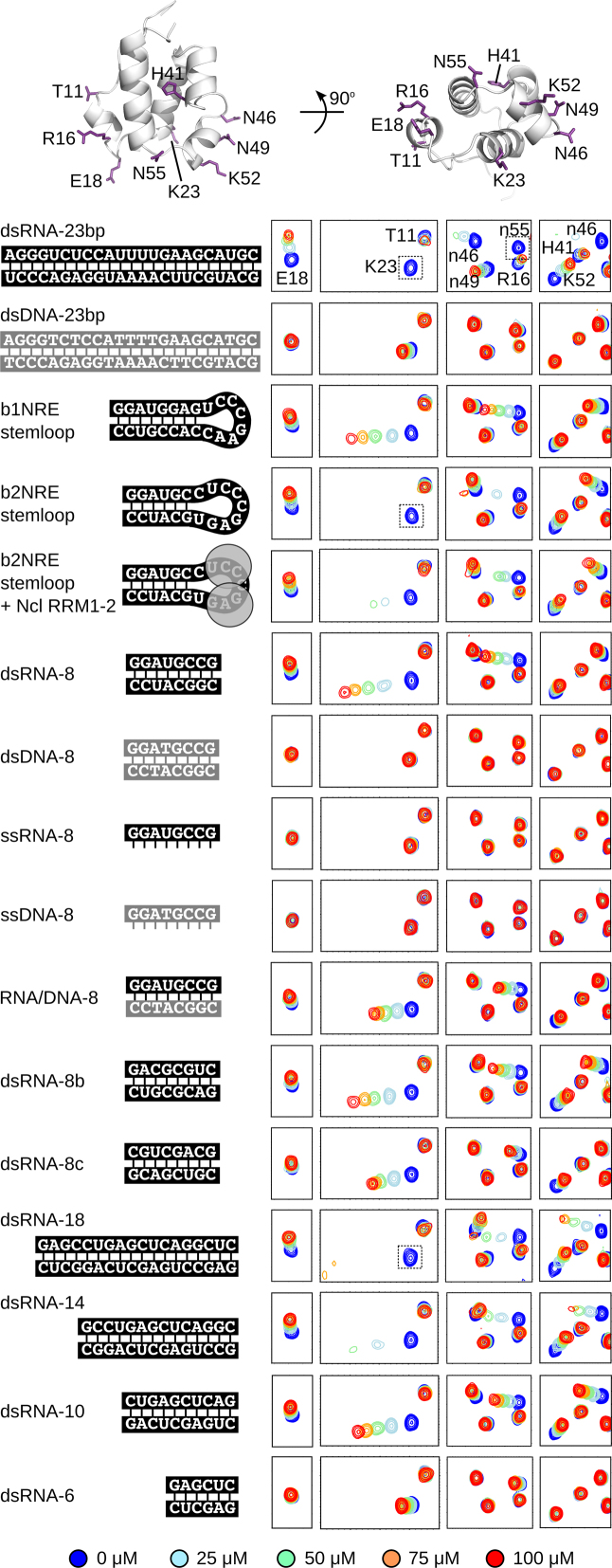
Prolyl isomerases are defined by a catalytic domain that facilitates the cis-trans interconversion of proline residues. In most cases, additional domains in these enzymes add important biological function, including recruitment to a set of protein substrates. Here, we report that the N-terminal basic tilted helix bundle (BTHB) domain of the human prolyl isomerase FKBP25 confers specific binding to double-stranded RNA (dsRNA). This binding is selective over DNA as well as single-stranded oligonucleotides. We find that FKBP25 RNA-association is required for its nucleolar localization and for the vast majority of its protein interactions, including those with 60S pre-ribosome and early ribosome biogenesis factors. An independent mobility of the BTHB and FKBP catalytic domains supports a model by which the N-terminus of FKBP25 is anchored to regions of dsRNA, whereas the FKBP domain is free to interact with neighboring proteins. Apart from the identification of the BTHB as a new dsRNA-binding module, this domain adds to the growing list of auxiliary functions used by prolyl isomerases to define their primary cellular targets.


Figures

Figure 1.
Figure 1.
FKBP25 associates with ribosome biogenesis factors and other proteins in an RNA-dependent manner. (A) Schematic of BioID-based identification of cellular proteins proximal to FKBP25. (B) Streptavidin-HRP western blot of whole cell extracts and streptavidin capture from U2OS cells stably expressing an FKBP25 biotin ligase fusion or biotin ligase control, incubated for 24 hours in media containing 50 μM biotin. (C) Mass spectrometry identification of biotinylated proteins enriched in FKBP25-BirA streptavidin purifications relative to BirA control. The number of significant peptides identified relative to fold change is shown. (D) Enriched gene ontologies by molecular function. (E) FKBP25 3xFLAG-tagged co-immunoprecipitated material with and without pre-treatment with RNaseA analyzed by SDS-PAGE and visualized by silver stain. Empty vector cell lines are shown as a control. (F) Mass spectrometry analysis of proteins enriched in the FKBP25-FLAG sample relative to control, for samples either untreated or pre-treated with RNaseA. (G) Summarized gene ontology analysis by biological process. (H) Overlap in identified proteins between the BioID and FLAG co-immunoprecipitation experiments.
Figure 2.
Figure 2.
FKBP25 requires RNA for nucleolar localization. (A) Western blot analysis of U2OS cells following CSK treatment, with an additional Triton wash or a combination of Triton and RNaseA treatment. (B) Confocal microscopy of FKBP25 and the nucleolar-specific upstream binding factor (UBF). Cells were either untreated (CSK), pre-extracted with Triton (CSK + T), or pre-extracted with Triton and treated with RNaseA (CSK + T + R) prior to fixation. Scale bar is 10 μm. (C) Epi-fluorescence microscopy of FKBP25 in cells treated with DMSO or the RNA polymerase I inhibitor actinomycin D (10 nM for 4 h) and pre-extracted with CSK-T. Scale bar is 5 μm.
Figure 3.
Figure 3.
The BTHB domain displays binding preference for dsRNA over dsDNA. (A) 15N-HSQC spectra regions corresponding to 100 μM 15N-labeled full-length FKBP25 (in black) superimposed by the spectra with 100 μM added 23-bp dsDNA (left, in red) or 100 μM added 23-bp dsRNA (right, in red). Crosspeaks corresponding to three lysine residues from the BTHB domain (K22, K23, K48) and one from the FKBP domain (K154) have been annotated. Most crosspeaks corresponding to the BTHB domain broaden below detection upon addition of dsRNA-23bp. Full spectra in Supplementary Figure S3. (B and C) Similar analysis with the isolated BTHB domain (residues 1–74) and the isolated FKBP domain (residues 108–224). Full spectra in Supplementary Figure S3. (D and E) Combined 1HN and 15N chemical shift perturbation for backbone amides in 100 μM samples of full-length FKBP25, BTHB domain or FKBP domain constructs upon addition of (D) 100 μM dsRNA-23bp or (E) dsDNA-23bp. Residue crosspeaks that are not observed in the ligand-bound complexes due to line-broadening are indicated with a grey bar. The secondary structure of FKBP25 is illustrated at the top.
Figure 4.
Figure 4.
Binding preference for full-length FKBP25. (AE) Electrophoretic mobility shift assays with 100 nM Cy3-labelled oligonucleotides and varying concentrations of FKBP25 from 0.5 to 500 μM. The first and last lanes of each gel do not contain protein. The migration of the free oligonucleotides as well as defined complexes are indicated. The asterisk denotes a minor contaminating species from the oligonucleotide synthesis.
Figure 5.
Figure 5.
Binding preference for the isolated BTHB domain. Regions of NMR spectra following titration of various ligands into 100 μM 15N-labelled FKBP25(1–74). The unbound spectrum is shown in blue, and the spectra following addition of 25, 50, 75 and 100 μM ligand are coloured light blue, light green, orange and red, respectively. The location of residues from the BTHB with annotated crosspeaks are indicated (top panel; atomic coordinates from PDB ID 2KFV) (23). Annotations with a small letter n correspond to the asparagine sidechain amide crosspeaks. Dotted boxes indicate peaks that broaden below detection upon addition of ligand. Full spectra are in Supplementary Figure S4.
Figure 6.
Figure 6.
Mutation of key lysine residues reduces in vitro and cellular RNA-binding. (A) Residue amides in FKBP25(1–74) that are strongly and moderately affected by titration with dsRNA-10 are coloured in orange and light orange, respectively. Orientations of the domain in the image are the same as in Figure 5. Chemical shift perturbation details are in Supplementary Figure S8A. (B) NMR spectra following titration of dsRNA-10 into 100 μM 15N-labeled FKBP25(1–74) with the K22M/K23M or K48/K52A mutations. Colours as in Figure 5. (C and D) Electrophoretic mobility shift assays with varying concentrations of full-length FKBP25 (K22M/K23M) as in Figure 4A and B. (E) Western blot analysis of FLAG-tagged FKBP25 constructs (wild-type and the K22M/K23M mutant) relative to endogenous FKBP25 (empty vector control) in HEK 293 cells. Antibodies against α-tubulin, FLAG-tag and FKBP25 correspond to the loading control, detection of FKBP25 constructs, and detection of both endogenous and FKBP25 constructs, respectively. (F) FLAG-affinity capture of cells expressing an empty vector control, wild-type FKBP25, or the K22M/K23M mutant with analysis by western blot using antibodies against the FKBP25-interacting proteins Parp1, nucleolin and RPS6. FKBP25 construct expression verified by antibodies against the FLAG tag. (G) RNA cross-linking IP (CLIP) experiment with wild-type FKBP25 or the K22M/K23M mutant in HEK293 cells, with DNAse pre-treatment coupled with variable amounts of RNase A.
Figure 7.
Figure 7.
The BTHB and FKBP domains of FKBP25 are structurally independent. (A) 15N relaxation measurements of 15N-labelled FKBP25 at 298K using a 700 MHz NMR spectrometer. Heteronuclear {1H}15N NOE measurements for backbone amide nitrogen identify residues that are conformationally rigid (values greater than 0.5) or flexible (less than 0.5) at the ps-ns timescale. The ratio of T1 over T2 values results with differing values of 9 ± 1 and 15 ± 3 for the BTHB and FKBP domains, respectively. The secondary structure of FKBP25 is illustrated at the top. (B) Incorporation of the nitroxide compound TEMPO to the FKBP25 mutant Q14C, T69C and T151C. Distance-dependent attenuation of crosspeak intensities caused by the paramagnetic nitroxide are shown, and are restricted to intra-domain effects. Full analysis in Supplementary Figure S6. (C) The two domains of FKBP25 do not form a single structural unit in solution, but are only connected by the flexible linker.
Figure 8.
Figure 8.
Model of FKBP25 interacting with dsRNA. (A) Surface representation of 23-bp dsDNA and dsRNA with main-chain phosphates coloured black and the major and minor grooves labeled. (B) Comparison of 6, 8 and 10 bp dsDNA ligands. Phosphates on opposing sides of the major groove require a minimum length of 8 bp. In longer ligands the BTHB domain is able to bind in multiple registers or slide along the major groove. (C) Docking model of BTHB bound to an A-form conformation of dsRNA-10. Further details in Supplementary Figure S7A. (D) Schematic model of FKBP25 bound to dsRNA.

Similar articles

Cited by

References

    1. Schmid F.X. Prolyl isomerase: enzymatic catalysis of slow protein-folding reactions. Annu. Rev. Biophys. Biomol. Struct. 1993; 22:123–142. - PubMed
    1. Schmid F.X., Mayr L.M., Mücke M., Schönbrunner E.R.. Prolyl isomerases: role in protein folding. Adv. Protein Chem. 1993; 44:25–66. - PubMed
    1. Heitman J., Movva N.R., Hall M.N.. Proline isomerases at the crossroads of protein folding, signal transduction, and immunosuppression. New Biol. 1992; 4:448–460. - PubMed
    1. Sinkins W.G., Goel M., Estacion M., Schilling W.P.. Association of Immunophilins with Mammalian TRPC Channels. J. Biol. Chem. 2004; 279:34521–34529. - PubMed
    1. Lummis S.C.R., Beene D.L., Lee L.W., Lester H.A., Broadhurst R.W., Dougherty D.A.. Cis–trans isomerization at a proline opens the pore of a neurotransmitter-gated ion channel. Nature. 2005; 438:248–252. - PubMed

MeSH terms