. 2017 Oct 16;15(1):42.
doi: 10.1186/s12964-017-0198-6.

A novel protein derived from lamprey supraneural body tissue with efficient cytocidal actions against tumor cells

Affiliations

A novel protein derived from lamprey supraneural body tissue with efficient cytocidal actions against tumor cells

Yue Pang et al. Cell Commun Signal. .

Erratum in

Abstract

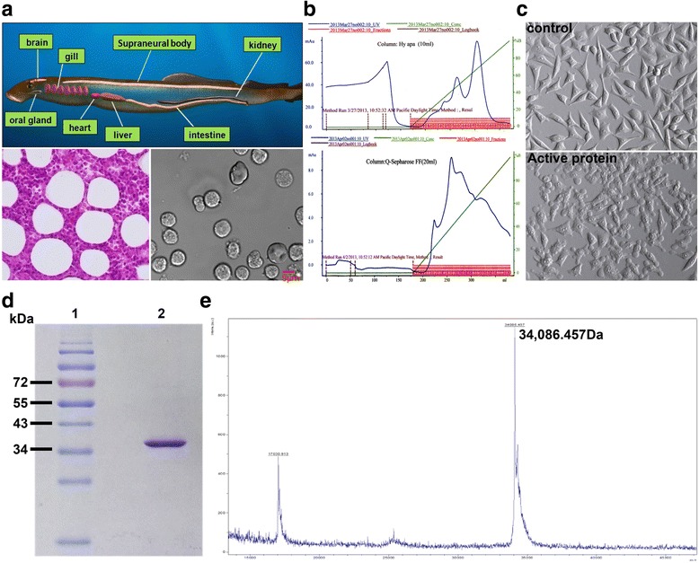
Background: In previous research, we found that cell secretion from the adult lamprey supraneural body tissues possesses cytocidal activity against tumor cells, but the protein with cytocidal activity was unidentified.

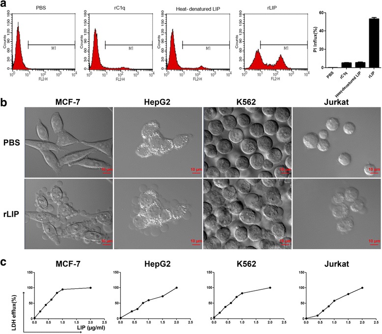
Methods: A novel lamprey immune protein (LIP) as defense molecule was first purified and identified in jawless vertebrates (cyclostomes) using hydroxyapatite column and Q Sepharose Fast Flow column. After LIP stimulation, morphological changes of tumor cells were analysed and measured whether in vivo or in vitro.

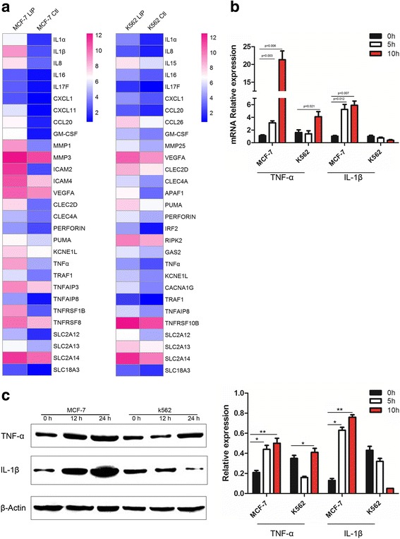
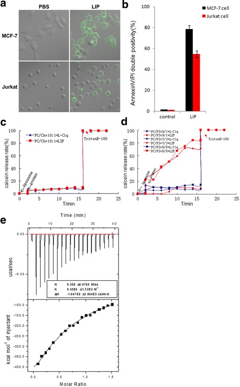
Results: LIP induces remarkable morphological changes in tumor cells, including cell blebbing, cytoskeletal alterations, mitochondrial fragmentation and endoplasmic reticulum vacuolation, and most of the cytoplasmic and organelle proteins are released following treatment with LIP. LIP evokes an elevation of intracellular calcium and inflammatory molecule levels. Our analysis of the cytotoxic mechanism suggests that LIP can upregulate the expression of caspase 1, RIPK1, RIP3 to trigger pyroptosis and necroptosis. To examine the effect of LIP in vivo, tumor xenograft experiments were performed, and the results indicated that LIP inhibits tumor growth without damage to mice. In addition, the cytotoxic action of LIP depended on the phosphatidylserine (PS) content of the cell membrane.

Conclusions: These observations suggest that LIP plays a crucial role in tumor cell survival and growth. The findings will also help to elucidate the mechanisms of host defense in lamprey.

Keywords: Cytotoxic activity; Inflammatory; LIP; Lamprey; Phosphatidylserine.


Conflict of interest statement

Ethics approval and consent to participate

The animal experiments were performed in accordance with the regulations of the Animal Welfare and Research Ethics Committee of the Institute of Dalian Medical University’s Animal Care protocol (Permit Number: SCXK2008-0002).

Consent for publication

Not applicable.

Competing interests

The authors declare that they have no competing interests.

Publisher’s Note

Springer Nature remains neutral with regard to jurisdictional claims in published maps and institutional affiliations.

Figures

Fig. 1
Fig. 1
Identification of active protein from supraneural body in the lamprey. a Tissue of the supraneural body by hematoxylin and eosin (left pane). Types of cells from the supraneural body (right pane). b Macro-Prep Ceramic Hydroxyapatite column of flow-through and eluted fraction. – green line, KPB gradient; − blue line, absorbance at 280 nm; □ activity protein(upper pane). Q Sepharose Fast Flow column of flow-through and eluted fraction. – green line, KCl gradient; − blue line, absorbance at 280 nm;□ activity protein (low pane). c MCF-7 cells were seeded at 5 × 104 cells in a 96-well plate. Each of the purified fractions were added to MCF-7 cells, and photographs were taken after the 30 min using optical microscope. (Magnification: 100×). d Determination of the purity and molecular mass of LIP via SDS-PAGE under reduced conditions. Lane 1, protein molecular markers; lane 2, native active LIP. e Identification of the molecular weight of LIP through MALDI-TOF mass spectroscopy
Fig. 2
Fig. 2
Sequences analysis and the predicted structure of the LIP. a LIP have the N-terminal β-prism lectin module and the C-terminal aerolysin module, respectively. b Sequence alignment analysis
Fig. 3
Fig. 3
Cytocidal activity of recombinant LIP. a MCF-7 cells were incubated with unrelated protein purified from E.coli (rC1q), heat-denatured LIP (0.5 μg/mL) and recombinant LIP (0.5 μg/mL) for 12 h at 37 °C. Cell death was analyzed by PI staining and by flow cytometry. PBS-treatment cells were used as a negative control (left pane). Histogram showing statistics of the above results (right pane). All experiments were repeated at least three times with similar results. b The cells were incubated with 0.5 μg/mL LIP at 37 °C and visualized by Live-Cell Imaging. c Cytocidal activity of LIP against cultured tumor cells. A total of 2 × 104 cells were preincubated at 37 °C for 20 h and then treated with LIP (final concentrations, 0.2 μg to 2 μg) for 12 h at 37 °C. Cytocidal activity of LIP was determined using the LDH cytotoxicity assay kit
Fig. 4
Fig. 4
Morphological changes in cells upon exposure to LIP. a Live-cell fluorescence images of MCF-7 cells treated with FITC-tagged LIP. MCF-7 cells were incubated in medium containing 0.5 μg CellMask™ orange plasma membrane stain for 4 min and then incubated with FITC-tagged LIP (1.0 μg/ml). The cells were observed using an Olympus FluoView FV1000 confocal microscope and photographed at the indicated time points. (Magnification: 63×). b Microtubule and mitochondrial fragmentation and ER vacuolation. MCF-7 cells were incubated with or without LIP (0.5 μg/mL) at 37 °C for 12 h. The cells were stained with a monoclonal antibody against tubulin and incubated in medium containing 30 nM MitoTracker Red or ER tracker. Merged images of cells double-stained with DAPI are shown. c Leakage of various proteins from the cytosol and organelles. MCF-7 cells were incubated with (+) or without (−) 0.5 μg/mL LIP at 37 °C for 12 h. The culture medium and cells were independently collected, and four marker proteins in each fraction were separated by SDS-PAGE and detected by western blotting using appropriate antibodies. M and C indicate the medium and cell fractions, respectively. d Elevation of intracellular calcium concentrations in MCF-7 cells following LIP treatment. Each bar represents the mean value from three determinations with the standard deviation (SD). Data (mean ± SD) with asterisks significantly differ (**P < 0.01) between treatments
Fig. 5
Fig. 5
LIP can significantly increase the expression of inflammatory molecules in MCF-7 cells. a Heat map representation of candidate genes involved in the pathways induced by LIP. Blue and red colors represent low-to-high expression levels, and the color scales correspond to the expression values of the microarray. b Q-PCR analysis of inflammatory molecule (TNF-α, IL-1β) expression in MCF-7 and K562 cells incubated with LIP for different times. Total RNA was quantified by qRT-PCR and normalized to gapdh expression. c Western blot analysis of inflammatory factor expression in MCF-7 and K562 cells. Western blot analysis for the expression of TNF-α & IL-1β in MCF-7 and K562 cells incubated with LIP for different times. β-actin served as a loading control(left pane). Histogram showing statistics of the above results (right pane). Means ± SDs are shown (n = 3 per group). **P < 0.01
Fig. 6
Fig. 6
LIP induced pyroptosis or necroptosis pathway. Western blot analysis of RIPK1, RIP3 and Caspase-1 proteins expression using specific antibodies. β-actin was used as a loading control. Histogram showing statistics of the above results. Data are presented as the mean ± SEM of three independent experiments performed in duplicate. Three types of cell lines showed different increases in the levels of cellular proteins in response to LIP stimulation. *P < 0.05, **P < 0.01
Fig. 7
Fig. 7
The effect of LIP treatment on cancer tissues and non-cancer tissues in vivo. a Mice with tumor growths were injected intratumorally (i.t.) 20 μg/kg rLIP or PBS. b-d Tumor growth of tumor-bearing nude mice were treated with either PBS or rLIP (n = 11 per group). Tumor size (b), tumor weight (c), body weight (d) of tumor-bearing nude mice. e Effect of LIP treatment on tumor tissues from xenografts. The cancer tissue pieces were treated with 2.5% glutaraldehyde for 24 h at 4 °C, and then a thin section of cancer tissues was observed via transmission electron microscopy (TEM). Red arrows, normal mitochondria; black arrows, mitochondria distension. f HE and IHC staining demonstrated that LIP induced the tumor cells death in vivo, as indicated by the expression of Ki67 and LIP, TUNEL-positive cells, and F4/80-positive cells. g Effect of LIP on non-cancer tissues from xenografts. Five different non-cancer tissues sections were stained by hematoxylin/eosin (H&E)
Fig. 8
Fig. 8
LIP induces exposure of phosphatidylserine and binds phosphatidylserine. a LIP induces exposure of phosphatidylserine. Cells were treated with LIP for 30 min. The cells were then imaged using Zeiss LSM 780 inverted microscope after staining for annexin-V-FITC. b LIP induces cell death in MCF-7 cells and Jurkat cells. Cells were treated with LIP for 12 h. The cells were then subjected to flow cytometric analysis after staining for annexin-V-FITC and propidium iodide, as mentioned in the methods. Shown are the data from three independent experiments. c Effect of LIP on a liposome membrane composed of a mixture of PC and CHL containing calcein. d Effect of LIP on a liposome membrane composed of a mixture of PC and PS containing calcein. e Calorimetric measurements of the LIP interaction with PS

Similar articles

Cited by

References

    1. Gadhamsetty S, Marée AF, Beltman JB, de Boer RJ. A general functional response of cytotoxic T lymphocyte-mediated killing of target cells. Biophys J. 2014;106(8):1780–1791. doi: 10.1016/j.bpj.2014.01.048. - DOI - PMC - PubMed
    1. Petranovic D, Pilcic G, Valkovic T, Sotosek Tokmadzic V, Laskarin G. Perforin- and granulysin-mediated cytotoxicity and interleukin 15 play roles in neurocognitive impairment in patients with acute lymphoblastic leukaemia. Med Hypotheses. 2014;83(1):122–126. doi: 10.1016/j.mehy.2014.03.024. - DOI - PubMed
    1. Wu F, Chen L, Liu X, Wang H, Su P, Han Y, Feng B, Qiao X, Zhao J, Ma N, Liu H, Zheng Z, Li Q. Lamprey variable lymphocyte receptors mediate complement-dependent cytotoxicity. J Immunol. 2013;190(3):922–930. doi: 10.4049/jimmunol.1200876. - DOI - PubMed
    1. Apiratikul N, Penglong T, Suksen K, Svasti S, Chairoungdua A, Yingyongnarongkula B. In vitro delivery of curcumin with cholesterol-based cationic liposomes. Bioorg Khim. 2013;39(4):497–503. - PubMed
    1. Pang Y, Wang S, Ba W, Li Q. Cell secretion from the adult lamprey supraneural body tissues possesses cytocidal activity against tumor cells. SpringerPlus. 2015;4:569. doi: 10.1186/s40064-015-1270-6. - DOI - PMC - PubMed

Publication types

MeSH terms