Review
. 2018 Jan;34(1):41-53.
doi: 10.1016/j.tig.2017.09.010. Epub 2017 Oct 13.

Caenorhabditis elegans Dosage Compensation: Insights into Condensin-Mediated Gene Regulation

Affiliations
Review

Caenorhabditis elegans Dosage Compensation: Insights into Condensin-Mediated Gene Regulation

Sarah Elizabeth Albritton et al. Trends Genet. 2018 Jan.

Abstract

Recent work demonstrating the role of chromosome organization in transcriptional regulation has sparked substantial interest in the molecular mechanisms that control chromosome structure. Condensin, an evolutionarily conserved multisubunit protein complex, is essential for chromosome condensation during cell division and functions in regulating gene expression during interphase. In Caenorhabditis elegans, a specialized condensin forms the core of the dosage compensation complex (DCC), which specifically binds to and represses transcription from the hermaphrodite X chromosomes. DCC serves as a clear paradigm for addressing how condensins target large chromosomal domains and how they function to regulate chromosome structure and transcription. Here, we discuss recent research on C. elegans DCC in the context of canonical condensin mechanisms as have been studied in various organisms.

Keywords: X chromosome; condensin; cooperation; dosage compensation; gene regulation; genome organization.


Figures

Figure 1
Figure 1. Structure and binding of the different types of condensin complexes in C. elegans
A) Subunits of the three C. elegans condensin complexes and the DCC proteins are shown. Within a condensin, each SMC protein folds over to form a ~50nm anti-parallel coiled-coiled arm with a hinge and an ATPase domain at opposite ends. Two SMC proteins dimerize at their respective hinge domains, interacting with three CAP subunits at coiled-coil arms and near their ATPase domains. The CAPH (Kleisin) subunit links the two SMC proteins and interacts with two HEAT-domain containing CAP subunits (CAPD and CAPG). Condensins I and II share the same SMC heterodimer and are distinguished by their differing sets of CAP subunits. Condensin DC differs from condensin I by a single SMC4 variant, DPY-27. B) ChIP-seq binding profiles (from [22]) of condensin II (SMC-4 and KLE-2), condensin DC (DPY-27), condensin I/DC (DPY-26) subunits in C. elegans embryos are shown across a representative ~60 kb region on an autosome (left) and on the X (right). The condensin DC specific subunit, DPY-27, shows baseline enrichment on the X, punctuated with peaks previously shown to be enriched at a subset of gene promoters, enhancers, and other intergenic sites including tRNA and ncRNA genes [22].
Figure 2
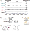
Figure 2. Features of DCC recruitment sites on the C. elegans X chromosome
A) ChIP-seq binding profiles from embryos are shown for condensin DC (blue) and non-condensin (pink) subunits of the C. elegans DCC. This ~250kb region on the X contains a strong (pink) and a weaker recruitment site (grey). Data are from [39]. B) Features of strong recruitment sites (multiple motifs (shown in blue-green) and overlap with HOT-sites) are X-specific. Weaker sites tend to overlap with tRNA genes. C) The 12-bp DCC recruitment motif sequence and a 10-bp sequence motif enriched at autosomal condensin II binding sites. The two motif sequences share a “GCGC” core. D) Long-distance cooperation between recruitment sites located 0.1–1Mb apart may recruit DCC specifically and robustly to large chromosomal domains on the X chromosomes. Autosomes contain fewer potential recruitment sites and thus lack the benefit of cooperation between multiple recruitment sites.
Figure 3
Figure 3. A model for condensin DC ring loading onto the X chromosomes
The DCC subunits required for condensin DC binding are SDC-2, SDC-3, and DPY-30. These recruiter proteins may recognize specific features of the recruitment sites including motif sequence within a nucleosomal context, DNA shape, and transcription factor binding at HOT-sites. High DNA-encoded nucleosome occupancy at the recruitment sites may be involved in nucleosome-mediated cooperativity of the motifs (small green bars), and may also prevent binding of maternally-loaded DCC subunits prior to sdc-2 expression in early embryogenesis. The recruitment sites may be the actual sites of condensin DC ring loading upon removal of nucleosomes. Alternatively, condensin DC may load along the chromosome and then accumulate at the recruitment sites (not depicted). SUMO-SIM interactions within DCC subunits may stabilize complex binding at recruitment sites.
Figure 4
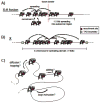
Figure 4. DCC spreading along the X chromosomes
A) DCC spreads into the autosomal sequence of X-to-autosome fusion (X;A) chromosomes. Spreading occurs over 1–3 Mb (dependent on X;A strain), and reduces with distance from the end of the X. B) On the X chromosomes, deletion of a single recruitment site reduces DCC binding across Mb-sized domains surrounded by TAD boundaries. The level of reduction in DCC binding correlates with the distance from the deleted recruitment site, suggesting existence of Mb-sized spreading domains. C) The possible molecular mechanisms by which stable and non-stable DCC complexes may spread along the X chromosomes. The mechanism of spreading may involve ring sliding, loop-extrusion, and/or diffusion/hopping.
Figure 5
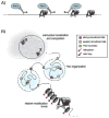
Figure 5. The effect of DCC on X chromosome transcription and structure
A) The DCC reduces RNA Pol II binding to promoters, resulting in an average 2-fold reduction of gene expression across the hermaphrodite X chromosomes. B) During early embryogenesis, X chromosome targeting of the DCC is initiated by a set of strong DCC recruitment sites that are distributed across the ~17Mb X chromosomes. Long-range cooperation between multiple strong and weaker recruitment sites followed by local spreading establishes robust DCC binding across large chromosomal domains. DCC spreading is restricted by TAD boundaries (dotted circles), which are, in part, demarcated by DCC itself. During later embryogenesis, DCC binding results in increased levels of H4K20me1 and decreased levels of H4K16ac on both X chromosomes. As a result, the dosage compensated hermaphrodite X chromosomes become ~40% more compacted compared to autosomes. Lastly, DCC binding influences nuclear localization of the X. Altogether, these observations suggest that a multitude of mechanisms are involved in establishing and maintaining a repressive environment across the hermaphrodite X chromosomes.

References

    1. Hirano T. At the heart of the chromosome: SMC proteins in action. Nat Rev Mol Cell Biol. 2006;7(5):311–22. - PubMed
    1. Gligoris T, Lowe J. Structural Insights into Ring Formation of Cohesin and Related Smc Complexes. Trends Cell Biol. 2016;26(9):680–93. - PMC - PubMed
    1. Merkenschlager M, Nora EP. CTCF and Cohesin in Genome Folding and Transcriptional Gene Regulation. Annu Rev Genomics Hum Genet. 2016;17:17–43. - PubMed
    1. Lau AC, Csankovszki G. Condensin-mediated chromosome organization and gene regulation. Front Genet. 2014;5:473. - PMC - PubMed
    1. Ercan S. Mechanisms of x chromosome dosage compensation. J Genomics. 2015;3:1–19. - PMC - PubMed

LinkOut - more resources