. 2018 Jan:114:1-9.
doi: 10.1016/j.yjmcc.2017.10.005. Epub 2017 Oct 14.

Early effects of Epac depend on the fine-tuning of the sarcoplasmic reticulum Ca2+ handling in cardiomyocytes

Affiliations

Early effects of Epac depend on the fine-tuning of the sarcoplasmic reticulum Ca2+ handling in cardiomyocytes

N Lezcano et al. J Mol Cell Cardiol. 2018 Jan.

Abstract

In cardiac muscle, signaling through cAMP governs many fundamental cellular functions, including contractility, relaxation and automatism. cAMP cascade leads to the activation of the classic protein kinase A but also to the stimulation of the recently discovered exchange protein directly activated by cAMP (Epac). The role of Epac in the regulation of intracellular Ca2+ homeostasis and contractility in cardiac myocytes is still matter of debate. In this study we showed that the selective Epac activator, 8-(4-chloro-phenylthio)-2'-O-methyladenosine-3', 5'-cyclic monophosphate (8-CPT), produced a positive inotropic effect when adult rat cardiac myocytes were stabilized at low [Ca2+]o (0.5mM), no changes at 1mM [Ca2+]o and a negative inotropic effect when [Ca2+]o was increased to 1.8mM. These effects were associated to parallel variations in sarcoplasmic reticulum (SR) Ca2+ content. At all [Ca2+]o studied, 8-CPT induced an increase in Ca2+ spark frequency and enhanced CaMKII autophosphorylation and the CaMKII-dependent phosphorylation of SR proteins: phospholamban (PLN, at Thr17 site) and ryanodine receptor (RyR2, at Ser2814 site). We used transgenic mice lacking PLN CaMKII phosphorylation site (PLN-DM) and knock-in mice with an inactivated CaMKII site S2814 on RyR2 (RyR2-S2814A) to investigate the involvement of these processes in the effects of Epac stimulation. In PLN-DM mice, 8-CPT failed to induce the positive inotropic effect at low [Ca2+]o and RyR2-S2814A mice showed no propensity to arrhythmic events when compared to wild type mice myocytes. We conclude that stimulation of Epac proteins could have either beneficial or deleterious effects depending on the steady-state Ca2+ levels at which the myocyte is functioning, favoring the prevailing mechanism of SR Ca2+ handling (uptake vs. leak) in the different situations.

Keywords: CaMKII-dependent phosphorylations; Epac; Sarcoplasmic reticulum calcium handling.


Conflict of interest statement

Conflict of interest: None declared.

Figures

Figure 1
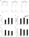
Figure 1. Epac differentially modifies cell shortening, [Ca2+]i transient and relaxation at different [Ca2+]o
A, representative simultaneous recordings of cell shortening (above) and Ca2+ transient (below) in rat myocytes loaded with Fura-2AM and field-stimulated at 1 Hz, before (black traces) and after (red traces) 10 μM 8-CPT application at 0.5, 1 and 1.8 mM [Ca2+]o; B, C, D and E, overall results of sarcomere cell shortening; Ca2+ transient amplitude; time to 50 % relaxation (t1/2) of twitches and time to 50 % decay of Ca2+ transient (t1/2 Ca2+ transient) of rat myocytes before (white bars) and after (black bars) 10 μM 8-CPT. * p<0.05 paired t test vs. before 8-CPT stimulation at each [Ca2+]o. Numbers in bars indicate number of cells of at least 3 rats.
Figure 2
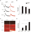
Figure 2. Epac stimulation increases SR Ca2+ content and Ca2+ spark occurrence at different [Ca2+]o
A, typical records of caffeine-triggered Ca2+ transients and B, averaged amplitude of these transients of rat myocytes loaded with Fura-2AM in the absence and presence of 10 μM 8-CPT. C, representative line-scan images of spontaneous Ca2+ sparks of myocytes loaded with Fluo-4 before (Control, left) and after treatment with 10 μM 8-CPT (right) at 0.5; 1 and 1.8 mM [Ca2+]o. D, bar graph showing the measured Ca2+ spark frequency in control cells (white bars) and cells in the presence of 8-CPT (black bars) at different [Ca2+]o. Numbers in bars indicate number of cells from at least 3 rats. * p<0.05 vs. control at each [Ca2+]o. # p<0.05 vs. 1 mM [Ca2+]o in the absence of drug.
Figure 3
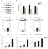
Figure 3. Epac stimulation increases CaMKII-dependent phosphorylation of its substrates via PKC at different [Ca2+]o
Typical immunoblots (A) and overall results (B) of the autophosphorylation of CaMKII (Thr286 site) and site-specific CaMKII phosphorylation of PLN (Thr17) and RyR2 (Ser2814) in rat myocytes incubated in the absence (white bars) or presence of 10 μM 8-CPT (black bars) or in the simultaneous presence of 8-CPT and the PKC inhibitor, 0.5 μM calphostine C (CC, grey bars). * p<0.05 vs. control, in the absence of drug; † p<0.05 vs. 8-CPT. C, D and E, typical immunoblots and overall results of the phosphorylation of myosin binding protein C (MyBP-C, Ser282) and the site-specific PKA phosphorylation of PLN (Ser16) and RyR2 (Ser2808), respectively. Cardiac myocytes incubated with 1 μM Isoproterenol (Iso) were used as positive controls. Experiments in A-E were performed at 1 mM [Ca2+]o. F, G and H, CaMKII phosphorylation of the kinase, PLN and RyR2 at different [Ca2+]o. Numbers in bars indicate number of cells from at least 5 rats. * p<0.05 vs. control at each [Ca2+]o. # p<0.05 vs. 1 mM [Ca2+]o in the absence of drug. Ctrl, Control.
Figure 4
Figure 4. Epac-induced positive inotropic and lusitropic effects are blunted in PLN-DM transgenic mice
A, sarcomere cell shortening; B, Ca2+ transient amplitude; C, time to 50 % relaxation (half relaxation time; t1/2) of twitches and D, time to 50 % decay of calcium transient of WT and PLN-DM mouse myocytes loaded with Fura-2AM and field-stimulated at 1 Hz, before (white bars) and during (black bars) 10 μM 8-CPT application at different [Ca2+]o. * p<0.05 paired t test vs. before 8-CPT stimulation at each [Ca2+]o. Numbers in bars indicate number of cells of at least 5 mice.
Figure 5
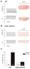
Figure 5. Epac-induced arrhythmogenesis is prevented in RyR2S2814A transgenic mice
Representative recordings of Ca2+ transient and sarcomere cell shortening of myocytes from WT mice (A) and RyR2-S2814A mice (B) before and after 5 min of application of 10 μM 8-CPT at 2.5 mM [Ca2+]o. C, percentage of myocytes in each group that experienced arrhythmic episodes. Numbers in bars indicate number of cells of at least 5 mice per group. * p<0.05 vs. control by Fisher’s exact test for each mice.

Similar articles

Cited by

References

    1. Fujita T, Umemura M, Yokoyama U, Okumura S, Ishikawa Y. The role of Epac in the heart. Cell Mol Life Sci. 2017;74(4):591–606. doi: 10.1007/s00018-016-2336-5. - DOI - PMC - PubMed
    1. Lezoualc’h F, Fazal L, Laudette M, Conte C. Cyclic AMP Sensor Epac proteins and their role in cardiovascular function and disease. Circ Res. 2016;118(5):881–97. doi: 10.1161/CIRCRESAHA.115.306529. - DOI - PubMed
    1. Métrich M, Lucas A, Gastineau M, Samuel JL, Heymes C, Morel E, Lezoualc’h F. Epac mediates beta adrenergic receptor-induced cardiomyocyte hypertrophy. Circ Res. 2008;102:959–965. doi: 10.1161/CIRCRESAHA.107.164947. - DOI - PubMed
    1. Ulucan C, Wang X, Baljinnyam E, Bai Y, Okumura S, Sato M, Minamisawa S, Hirotani S, Ishikawa Y. Developmental changes in gene expression of Epac and its upregulation in myocardial hypertrophy. Am J Physiol Heart Circ Physiol. 2007;293:H1662–H1672. doi: 10.1152/ajpheart.00159.2007. - DOI - PubMed
    1. Oestreich EA, Wang H, Malik S, Kaproth-Joslin KA, Blaxall BC, Kelley GG, Dirksen RT, Smrcka AV. Epac-mediated activation of phospholipase C(epsilon) plays a critical role in beta-adrenergic receptor-dependent enhancement of Ca2+ mobilization in cardiac myocytes. J Biol Chem. 2007;282:5488–5495. doi: 10.1074/jbc.M608495200. - DOI - PubMed

Publication types

MeSH terms

Substances