. 2017 Dec;200(3):325-333.
doi: 10.1016/j.jsb.2017.10.003. Epub 2017 Oct 14.

The structure of the actin-smooth muscle myosin motor domain complex in the rigor state

Affiliations

The structure of the actin-smooth muscle myosin motor domain complex in the rigor state

Chaity Banerjee et al. J Struct Biol. 2017 Dec.

Abstract

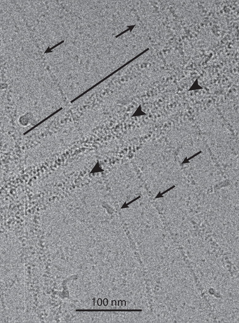
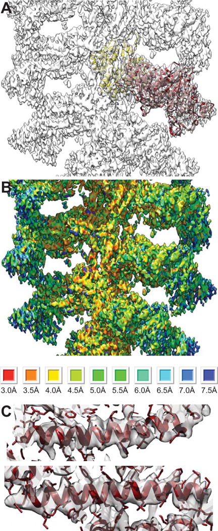
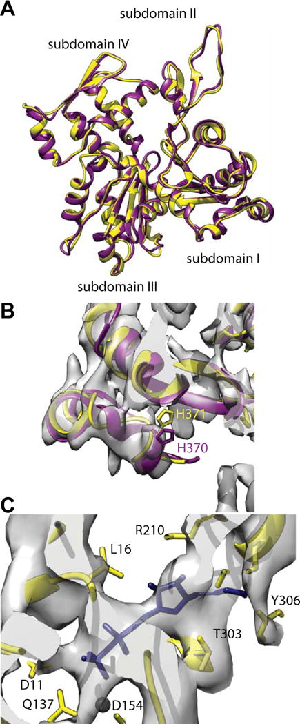
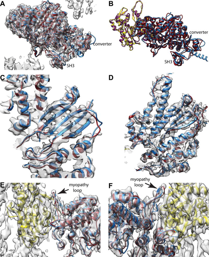
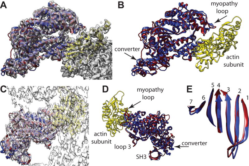
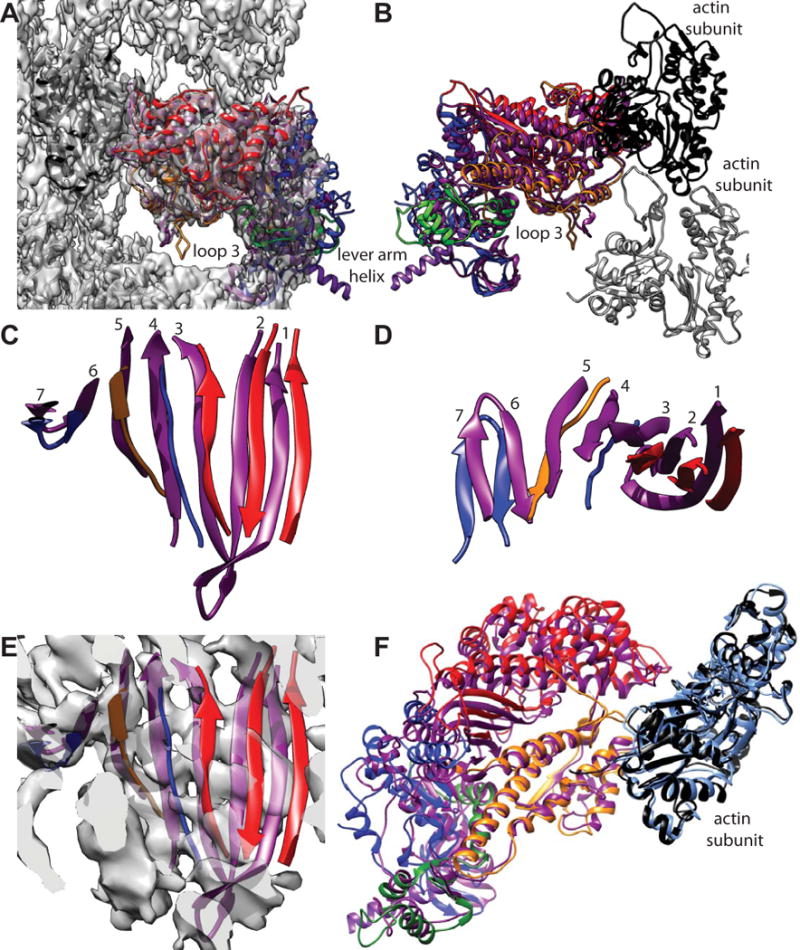
Myosin-based motility utilizes catalysis of ATP to drive the relative sliding of F-actin and myosin. The earliest detailed model based on cryo-electron microscopy (cryoEM) and X-ray crystallography postulated that higher actin affinity and lever arm movement were coupled to closure of a feature of the myosin head dubbed the actin-binding cleft. Several studies since then using crystallography of myosin-V and cryoEM structures of F-actin bound myosin-I, -II and -V have provided details of this model. The smooth muscle myosin II interaction with F-actin may differ from those for striated and non-muscle myosin II due in part to different lengths of important surface loops. Here we report a ∼6 Å resolution reconstruction of F-actin decorated with the nucleotide-free recombinant smooth muscle myosin-II motor domain (MD) from images recorded using a direct electron detector. Resolution is highest for F-actin and the actin-myosin interface (3.5-4 Å) and lowest (∼6-7 Å) for those parts of the MD at the highest radius. Atomic models built into the F-actin density are quite comparable to those previously reported for rabbit muscle actin and show density from the bound ADP. The atomic model of the MD, is quite similar to a recently published structure of vertebrate non-muscle myosin II bound to F-actin and a crystal structure of nucleotide free myosin-V. Larger differences are observed when compared to the cryoEM structure of F-actin decorated with rabbit skeletal muscle myosin subfragment 1. The differences suggest less closure of the 50 kDa domain in the actin bound skeletal muscle myosin structure.

Keywords: ATPase; Electron microscopy; Molecular motor; Single particle.


Figures

Figure 1
Figure 1
Electron micrograph of F-actin decorated with the smooth muscle myosin motor domain. Segments were taken only from the filament region marked by the line. Arrowheads point to bundled filaments. Arrows point to actin filaments completely undecorated with myosin heads.
Figure 2
Figure 2
Overview and resolution of the reconstruction of F-actin decorated with smMD. (A) Overview showing the atomic models of the actin subunit (yellow) and the smMD (dark red). (B) RESMAP image of the reconstruction. Resolution is clearly highest close to the filament axis and lowest at the high radius where the converter and SH3 domain are positioned. RESMAP color ranges are shown at the bottom. (C) Images of a pair of long α-helices from the acto-smMD reconstruction (purple). Density corresponding to large side chains is clearly visible. Top panel helix comprises residues 477–506; bottom panel helix comprises residues 420–450.
Figure 3
Figure 3
Comparison of vertebrate non-muscle γ-actin and skeletal muscle α-actin subunits. (A) Overlay of the fitted actin subunits. Skeletal muscle α-actin is colored yellow and the non-muscle γ-actin subunit colored dark magenta. (B) Region near the C-terminus. The rabbit α-actin subunit used for initiating the fitting did not include four residues at the C-terminus. These were added in later but have not been energy minimized. The C-termini of the non-muscle γ-actin subunit fits the density very well indicating that after refinement, the muscle α-actin C-terminus will likely be very similar. (C) Region near the ADP binding site. Substantial density is present where the nucleotide binds. The black sphere is a magnesium ion for which clear density is not visible. Its presence provides a useful landmark.
Figure 4
Figure 4
Comparison of vertebrate non-muscle and smooth muscle myosin MDs when bound to F-actin. Vertebrate smooth muscle MD is colored dark red; the vertebrate non-muscle MD is colored dodger blue, muscle α-actin is yellow, non-muscle γ-actin is colored dark magenta. (A) View showing the relative difference between converter and SH3 domains plus the reconstruction envelope. (B) Slightly different view from (A) showing the actin subunits and the converter in better profile without the map. (C) View showing five strands of the transducer β-sheet with both the smooth muscle MD (dark red) and non-muscle MD (dodger blue). (D) Similar view as (C) but showing four major α-helices, which align well to the smooth muscle MD density and atomic model. (E) Actin-myosin interface from the “front”. (F) Actin-myosin interface from the back. In all these views, the vertebrate smooth muscle acto-MD and the vertebrate non-muscle acto-MD atomic models are nearly superimposable at this resolution.
Figure 5
Figure 5
Comparison of the actin bound, smMD and the nucleotide-free myosin-V MD crystal structure (PDB 1OE9), which has been aligned to the reconstruction using Chimera’s fitinmap utility. Coloring scheme has the actin subunit yellow, the smMD dark magenta, and the myosin-V MD blue. (A) View down the actin binding cleft showing the excellent fit of the myosin-V crystal structure even though not bound to actin. The myosin-V converter domain has a very similar position and orientation as the smMD converter. (B) Same view direction as panel A but with the map removed to shown the excellent alignment of the myosin-V helices with the corresponding smMD helices and loops. (C) View perpendicular to the helix axis showing the fit of the myosin-V coordinates within the acto-smMD reconstruction. (D) View from the opposite side without the map showing alignment of the myopathy loop, loop 3 and the SH3 domains. (E) View showing the excellent alignment of the 7-stranded transducer β-sheets.
Figure 6
Figure 6
Comparison of acto-smMD with acto-skMD. (A) The two reconstructions shown with the acto-smMD reconstruction. The acto-skMD was aligned to the acto-smMD using the actin subunit coordinates from the acto-skMD atomic model (PDB 5H53), which is colored black, to drive the alignment. There is little difference between the actin subunit atomic models from the two reconstructions. The smMD atomic model is colored purple. The acto-skMD atomic model is colored according to the MD subdomains, which are N-terminal 25 kDa domain (blue), upper 50 kDa domain (red), lower 50 kDa domain (orange), converter domain (green) and the lever arm (magenta). Note that the smMD does not have the lever arm helix. (A) Both atomic models shown within the reconstruction envelope. Many features of the skMD atomic model fall outside of the density envelope of the smMD. The most obvious difference is the position of loop 3 (skeletal residues K567–F579), which falls clearly outside the reconstruction envelope. Loop 3 is part of the lower 50 kDa domain. (B) The atomic models of the skMD and the smMD shown with a pair of actin subunits, one black, the other gray. This view from the opposite direction from that of panel A. (C) Comparison of the transducer β-sheet with the smooth muscle structure shown in purple and the skeletal muscle sheet colored according to subdomain origin. Since the sheet itself is curved, the displacements for strands 1 and 2 are the most obvious. This view direction is from outside the MD looking in towards the actin-binding cleft. The relative displacement has the skeletal β-sheet to the side and on the outside of the smooth β-sheet (roughly looking from the top of panel F towards the bottom). (D) View looking down from the top of panel C. (E) Same view direction as panel C but with the reconstruction envelope showing. Note that the skMD β-sheet mostly falls outside of the corresponding density envelope. (F) View looking down the actin binding cleft showing the actin subunit atomic models from the two reconstructions as well as their MD atomic models. The actin atomic model from the acto-smMD reconstruction is shown in sky blue. Note that the helices of the lower 50 kDa domains overlap well, whereas features of the upper 50 kDa domains overlap poorly. The 25 kDa domains also overlap poorly.

References

    1. Behrmann E, Muller M, Penczek PA, Mannherz HG, Manstein DJ, Raunser S. Structure of the rigor actin-tropomyosin-myosin complex. Cell. 2012;150:327–338. - PMC - PubMed
    1. Clemens DL, Ge P, Lee BY, Horwitz MA, Zhou ZH. Atomic structure of T6SS reveals interlaced array essential to function. Cell. 2015;160:940–951. - PMC - PubMed
    1. Coureux PD, Wells AL, Ménétrey J, Yengo CM, Morris CA, Sweeney HL, Houdusse A. A structural state of the myosin V motor without bound nucleotide. Nature. 2003;425:419–423. - PubMed
    1. Coureux PD, Sweeney HL, Houdusse A. Three myosin V structures delineate essential features of chemo-mechanical transduction. EMBO J. 2004;23:4527–4537. - PMC - PubMed
    1. Criddle A, Geeves M, Jeffries T. The use of actin labelled with N-(1-pyrenyl) iodoacetamide to study the interaction of actin with myosin subfragments and troponin/tropomyosin. Biochem J. 1985;232:343–349. - PMC - PubMed

Publication types

LinkOut - more resources