. 2017 Nov 28;37(24):e00295-17.
doi: 10.1128/MCB.00295-17. Print 2017 Dec 15.

Loss of Extracellular Signal-Regulated Kinase 1/2 in the Retinal Pigment Epithelium Leads to RPE65 Decrease and Retinal Degeneration

Affiliations

Loss of Extracellular Signal-Regulated Kinase 1/2 in the Retinal Pigment Epithelium Leads to RPE65 Decrease and Retinal Degeneration

Aswin Pyakurel et al. Mol Cell Biol. .

Abstract

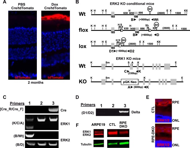
Recent work suggested that the activity of extracellular signal-regulated kinase 1/2 (ERK1/2) is increased in the retinal pigment epithelium (RPE) of age-related macular degeneration (ARMD) patients and therefore could be an attractive therapeutic target. Notably, ERK1/2 pathway inhibitors are used in cancer therapy, with severe and noncharacterized ocular side effects. To decipher the role of ERK1/2 in RPE cells, we conditionally disrupted the Erk1 and Erk2 genes in mouse RPE. The loss of ERK1/2 activity resulted in a significant decrease in the level of RPE65 expression, a decrease in ocular retinoid levels concomitant with low visual function, and a rapid disorganization of RPE cells, ultimately leading to retinal degeneration. Our results identify the ERK1/2 pathway as a direct regulator of the visual cycle and a critical component of the viability of RPE and photoreceptor cells. Moreover, our results caution about the need for a very fine adjustment of kinase inhibition in cancer or ARMD treatment in order to avoid ocular side effects.

Keywords: AP-1; ERK1/2; RPE65; electron microscopy; photoreceptors; retinal degeneration; retinoid.


Figures

FIG 1
FIG 1
Generation and characterization of CTL and RPE-DKO mice. (A) tdTomato fluorescence of cryostat sections of fixed whole-mount eyes from Cre/tdTomato mice injected with either PBS or doxycycline (Dox). (B) Constructs used to generate Erk1-KO and Erk2 conditional mice (Erk1−/− Erk2f/f) and primers used to genotype the mice. Wt, wild type. (C) Representative genotyping of Erk1+/−; Erk2+/f mice (lane 1); Erk1−/−; Erk2f/f mice, called CTL mice (lane 2); and VMD2-rtTA/TRE-Cre; Erk1−/−; Erk2Δ/Δ mice, called RPE-DKO mice, when injected with Dox (lane 3). (D) The specific loss of Erk2 in the RPE is confirmed by the delta fragment present in genomic DNA isolated from RPE cells from Erk1+/−; Erk2+/f mice (lane 1), Erk1−/−; Erk2f/f mice, called CTL mice (lane 2); and VMD2-rtTA/TRE-Cre; Erk1−/−; Erk2Δ/Δ mice, called RPE-DKO mice, when injected with Dox (lane 3). (E) Cryostat sections of fixed whole-mount eyes from CTL and RPE-DKO mice at 1 month, immunostained as indicated. (F) Western blot analysis of ERK1/2 expression in RPE protein lysates from CTL and RPE-DKO mice at 1 month. The ARPE19 protein lysate is used as a positive control for ERK1 expression.
FIG 2
FIG 2
Loss of ERK1/2 in the RPE leads to vision impairment. (A) Representative fundus images of CTL and RPE-DKO mice at 2 or 4 months. (B) Representative OCT images of CTL and RPE-DKO mice at 4 months. (C) Graphs of the scotopic (n = 10) and photopic (n = 4) ERG responses (b wave) of CTL and RPE-DKO mice at 2 months. (*, P < 0.05; **, P < 0.001). The right panel shows the scotopic (50 mcd s/m2) and photopic (10 mcd s/m2) ERG responses of CTL and RPE-DKO mice. (D) Chromatographs and quantification of retinoids (retinol [ROL] and retinal [RAL]) measured in the retina and in the RPE-choroid from mice at 1 month, which were dark adapted for 16 h, or measured in whole eyes (containing the retina, RPE, and choroid) from mice (same conditions) exposed to 1,000 mA of light for 2 h.
FIG 3
FIG 3
Loss of ERK1/2 in the RPE leads to photoreceptor degeneration. (A) Cryostat sections of fixed whole-mount eyes from CTL and RPE-DKO mice at 2 or 4 months, immunostained against different rod and cone markers, as indicated. (B) Flat-mount retinal preparation from CTL and RPE-DKO mice at 4 months, immunostained as indicated. (C) Outer nuclear layer (ONL) and inner nuclear layer (INL) lengths measured manually by using ImageJ. Data represent means ± standard errors of the means of results from four independent experiments. *, P < 0.05. (D) GNAT1 and cone arrestin stainings from CTL and RPE-DKO mice at 1 month. The presence of both photoreceptor markers was observed in the RPE of RPE-DKO mice (white arrows). (E) TUNEL staining of cryostat sections of fixed whole-mount eyes from CTL and RPE-DKO mice at 2 months.
FIG 4
FIG 4
Decreases of outer nuclear layer (ONL), outer segment (OS), and inner segment (IS) lengths from the start of degeneration to complete absence in RPE-DKO mice at 1 year. (A) Electron microscopy images of retina-RPE layers from CTL and RPE-DKO mice at 2 or 4 months. The bottom panel is a magnification of the OS layer of photoreceptors (PR). (B) Measurement of ONL, OS, and IS from electron microscopy images. Data represent means ± standard errors of the means of results from 3 experiments. **, P < 0.008. (C) Cryostat sections of fixed whole-mount eyes from CTL and RPE-DKO mice at 1 year, immunostained as indicated.
FIG 5
FIG 5
Loss of ERK1/2 in the RPE causes specific reductions in the levels of cone markers. (A) Representative images and quantification graphs of retinal protein lysates from CTL and RPE-DKO mice at 1, 2, or 4 months. Data represent means ± standard errors of the means of results from 5 independent experiments (*, P < 0.02; **, P < 0.001). (B) Quantification graphs for qPCR performed on retinal extracts from CTL and RPE-DKO mice at 1, 2, or 4 months. RL8 was used as an internal control to normalize RNA expression. Results are expressed as a percentage of the value for CTL mice and as means ± standard errors of the means of results from 4 independent experiments (*, P < 0.05; **, P < 0.003).
FIG 6
FIG 6
Loss of ERK1/2 in the RPE leads to massive ultrastructural changes in RPE-DKO mice at 1 month. (A) Representative electron microscopy images showing different retinal and RPE cell layers (B, Bruch's membrane; RPE, retinal pigment epithelium; OS, outer segment; IS, inner segment; ONL, outer nuclear layer). (B) Representative electron microscopy image of Bruch's membrane (Mbre) and the underlying RPE cells and measurement of the length of Bruch's membrane (*, P < 0.0001). (C) Representative electron microscopy images of the RPE cell layer and the underlying OS of the PR, measurement of the RPE length, and quantification of the area of mitochondria (stars, mitochondria) (*, P < 0.015). (D) Electron microscopy images showing the accumulation of membrane-enriched phagolysosomes in the RPE of RPE-DKO mice in comparison to CTL mice (white arrows). (E and F) RPE flat mounts from CTL and RPE-DKO mice at 2 months, immunostained against phalloidin and counterstained with DAPI.
FIG 7
FIG 7
ERK1/2 directly regulates RPE65 expression. (A) Cryostat section of fixed whole-mount eyes from CTL and RPE-DKO mice at 1 month, immunostained as indicated. (B) Cryostat section of fixed whole-mount eyes from CTL and RPE-DKO mice at 2 weeks, immunostained as indicated. (C) qPCR analysis of mRNAs for the genes indicated, extracted from the RPE-choroids of CTL and RPE-DKO mice at 1 month. RL8 was used as an internal control to normalize RNA expression. Results are expressed as a percentage of the value for CTL mice and as means ± standard errors of the means of results from 3 retinas. *, P < 0.03. (D) Representative immunoblot and quantification of RPE protein lysates from CTL and RPE-DKO mice at 1 month, immunoblotted as indicated. **, P < 0.0002 (n = 5). (E) Luciferase assay for both RPE65 and LRAT promoters. HEK293 cells were transfected and treated as indicated. Luciferase fluorescence was normalized to β-Gal fluorescence. Data represent means ± standard errors of the means of results from 4 independent experiments. *, P < 0.05 versus pGL2-Basic-RPE65; #, P < 0.05 versus pGL2-Basic. (F) EMSA of the AP-1 complex in nuclei from ARPE19 cells treated or not with U0126. AP-1 DNA binding analysis of nuclear extracts of ARPE19 cells treated with either U0126 or PD0325901. Data represent means ± standard errors of the means of results from 3 independent experiments, expressed as a percentage of the value for the control. *, P < 0.02; **, P < 0.002. (G) AP-1 DNA binding analysis of whole-cell extracts from mice at 1 month. Data represent means ± standard errors of the means of results from 3 independent experiments, expressed as a percentage of the control. *, P < 0.015; **, P < 0.002 (n = 6). Also shown is a representative immunoblot of RPE protein lysates from CTL and RPE-DKO mice at 1 month, showing the expression of C-FOS and FRA-1; tubulin was used as a control.

Similar articles

Cited by

References

    1. Hageman GS, Luthert PJ, Chong NHV, Johnson LV, Anderson DH, Mullins RF. 2001. An integrated hypothesis that considers drusen as biomarkers of immune-mediated processes at the RPE-Bruch's membrane interface in aging and age-related macular degeneration. Prog Retin Eye Res 20:705–732. doi:10.1016/S1350-9462(01)00010-6. - DOI - PubMed
    1. Rodrigues EB. 2007. Inflammation in dry age-related macular degeneration. Ophthalmologica 221:143–152. doi:10.1159/000099293. - DOI - PubMed
    1. Zhou JL, Jang YP, Kim SR, Sparrow JR. 2006. Complement activation by photooxidation products of A2E, a lipofuscin constituent of the retinal pigment epithelium. Proc Natl Acad Sci U S A 103:16182–16187. doi:10.1073/pnas.0604255103. - DOI - PMC - PubMed
    1. Iriyama A, Fujiki R, Inoue Y, Takahashi H, Tamaki Y, Takezawa S, Takeyama K, Jang WD, Kato S, Yanagi Y. 2008. A2E, a pigment of the lipofuscin of retinal pigment epithelial cells, is an endogenous ligand for retinoic acid receptor. J Biol Chem 283:11947–11953. doi:10.1074/jbc.M708989200. - DOI - PubMed
    1. Eldred GE, Lasky MR. 1993. Retinal age pigments generated by self-assembling lysosomotropic detergents. Nature 361:724–726. doi:10.1038/361724a0. - DOI - PubMed

MeSH terms

LinkOut - more resources