. 2017 Nov 8;37(45):10943-10954.
doi: 10.1523/JNEUROSCI.0190-17.2017. Epub 2017 Oct 16.

CB1-Dependent Long-Term Depression in Ventral Tegmental Area GABA Neurons: A Novel Target for Marijuana

Affiliations

CB1-Dependent Long-Term Depression in Ventral Tegmental Area GABA Neurons: A Novel Target for Marijuana

Lindsey Friend et al. J Neurosci. .

Abstract

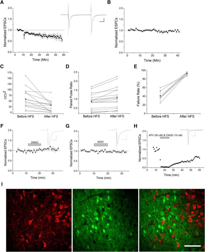
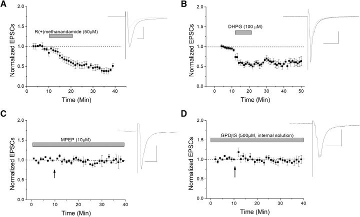
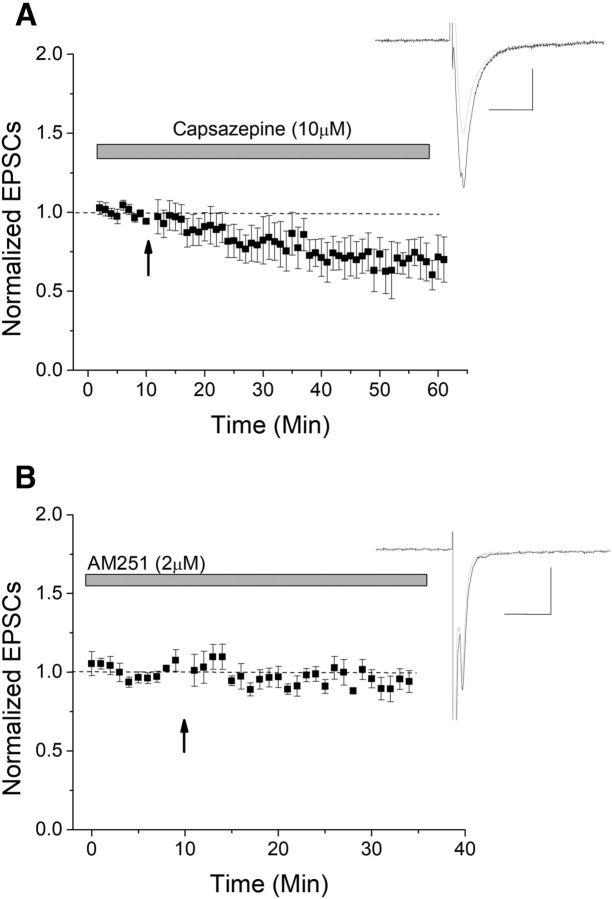
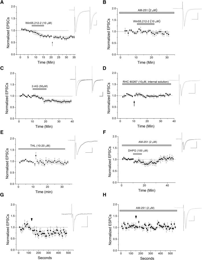
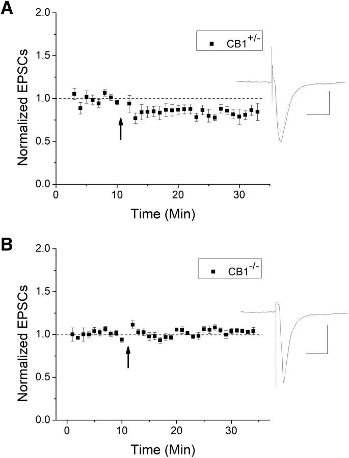
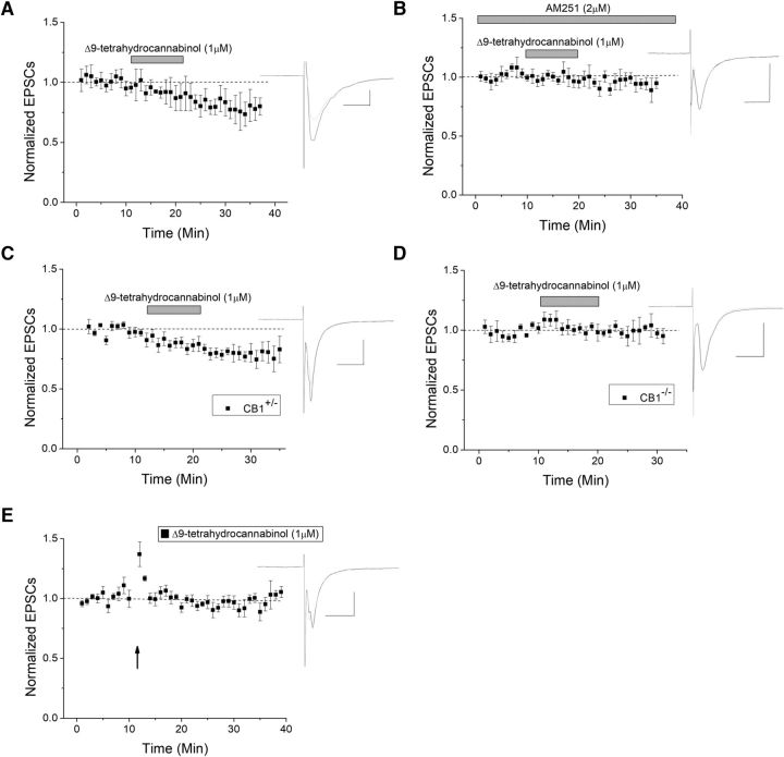
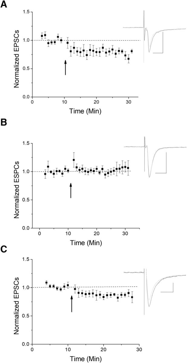
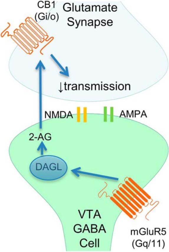
The VTA is necessary for reward behavior with dopamine cells critically involved in reward signaling. Dopamine cells in turn are innervated and regulated by neighboring inhibitory GABA cells. Using whole-cell electrophysiology in juvenile-adolescent GAD67-GFP male mice, we examined excitatory plasticity in fluorescent VTA GABA cells. A novel CB1-dependent LTD was induced in GABA cells that was dependent on metabotropic glutamate receptor 5, and cannabinoid receptor 1 (CB1). LTD was absent in CB1 knock-out mice but preserved in heterozygous littermates. Bath applied Δ9-tetrahydrocannabinol depressed GABA cell activity, therefore downstream dopamine cells will be disinhibited; and thus, this could potentially result in increased reward. Chronic injections of Δ9-tetrahydrocannabinol occluded LTD compared with vehicle injections; however, a single exposure was insufficient to do so. As synaptic modifications by drugs of abuse are often tied to addiction, these data suggest a possible mechanism for the addictive effects of Δ9-tetrahydrocannabinol in juvenile-adolescents, by potentially altering reward behavioral outcomes.SIGNIFICANCE STATEMENT The present study identifies a novel form of glutamatergic synaptic plasticity in VTA GABA neurons, a currently understudied cell type that is critical for the brain's reward circuit, and how Δ9-tetrahydrocannabinol occludes this plasticity. This study specifically addresses a potential unifying mechanism whereby marijuana could exert rewarding and addictive/withdrawal effects. Marijuana use and legalization are a pressing issue for many states in the United States. Although marijuana is the most commonly abused illicit drug, the implications of legalized, widespread, or continued usage are speculative. This study in juvenile-adolescent aged mice identifies a novel form of synaptic plasticity in VTA GABA cells, and the synaptic remodeling that can occur after Δ9-tetrahydrocannabinol use.

Keywords: THC; TRPV1; anandamide; endocannabinoid; mGluR5; withdrawal.


Figures

Figure 1.
Figure 1.
Presynaptic LTD in VTA GABA cells. A, Glutamate currents recorded from GAD67-GFP-positive VTA GABA cells. The 100 Hz HFS induces LTD as EPSC responses were reduced by 21 ± 5.5% at 10–15 min after HFS compared with baseline (n = 14, p ≪ 0.05, baseline compared with postconditioning at 10–15, 15–20, and 20–25 min) and eventually reached >30% depression. All plots are normalized EPSC amplitude means with standard error of the mean (SEM). Arrow indicates 100 Hz HFS conditioning induction. Insets, Here and throughout, Representative averaged EPSC traces (10–12 traces) taken from before (black) and 10–15 min after conditioning or drug application (gray). Calibration: Scale bars here and throughout are 100 pA, 10 ms. B, No treatment controls showed no change in EPSC amplitude, which demonstrates that HFS-LTD is not due to rundown effect (n = 8). C–E, Following HFS, the 1/CV2 decreased (n = 13 p < 0.05), PPR increased (n = 15, p < 0.05), and failure rates increased (n = 6, p < 0.05), suggesting a presynaptic mechanism for HFS-LTD. Connected open circles represent individual experiments before and 5–10 min after HFS. Connected closed circles represent averages from all experiments. F–H, Vehicle controls and glutamate transmission confirmation. F, Solvent DMSO vehicle controls demonstrate that DMSO does not alter basal neurotransmission of VTA GABA cells (n = 6, p > 0.5, post-DMSO application compared with pre-DMSO). G, EtOH vehicle controls also demonstrate no change in EPSCs compared with baseline (n = 7, p > 0.5). H, An individual experiment denotes that recorded EPSCs are indeed glutamatergic as they are blocked by a combination of AMPA receptor antagonist CNQX and NMDA receptor antagonist APV (n = 5). Washout occurs and recovers some of the glutamate response. I, Immunohistochemical image of the VTA. GAD67-GFP-positive mice were counterstained with TH antibody to label dopaminergic neurons. Anti-TH antibodies label cytosol of dopaminergic neurons as noted in the red channel, whereas GAD67-GFP-positive neurons are labeled in the green channel. In the merged image, no individual neuron is double-labeled, indicating that the inhibitory cells we recorded from are not dopaminergic, but that both are separate cell populations. Scale bar, 100 μm.
Figure 2.
Figure 2.
eCB and mGluR5 involvement in VTA GABA cell plasticity. The eCBs are signaling molecules often involved in presynaptic plasticity mechanisms. A, A nonhydrolysable form of an eCB, R(+)methanandamide, induces depression of glutamatergic neurotransmission (n = 6, p ≪ 0.05). B, The Type 1 mGluR agonist DHPG also induced depression, similar to HFS-LTD (n = 7, p ≪ 0.05). C, The specific mGluR5 antagonist MPEP blocked HFS-LTD (n = 6; p < 0.05; compared to control LTD). D, To further test for the necessity of a postsynaptic GPCR (such as an mGluR) involvement in LTD, the GDP analog GDPβS, which inhibits G-proteins, was added to the internal solution and resulted in blocked HFS-LTD (n = 7; p > 0.05; compared to its baseline).
Figure 3.
Figure 3.
CB1 mediates HFS-LTD. Two candidate receptors that could act as a presynaptic target for eCBs include TRPV1 and CB1. A, The TRPV1 antagonist capsazepine failed to block LTD, suggesting that TRPV1 is not involved in this HFS-LTD (n = 9; p > 0.05 compared with control LTD). B, The CB1 antagonist AM-251 significantly blocked LTD (n = 9, p < 0.05 compared with control LTD).
Figure 4.
Figure 4.
Postsynaptically produced 2-AG mediates LTD of VTA GABA cells. A, Application of the CB1 agonist Win55,212-2 depresses glutamate transmission at this synapse (n = 8). There is a significant depression between baseline and drug application (p < 0.05), but further HFS-induced depression was occluded (arrow; p > 0.5; WIN55,212-2 depression compared with post-HFS, ANOVA). B, When the CB1 antagonist AM-251 was present, Win55,212-2 caused no significant depression, demonstrating the specificity of Win 55,212-2 for CB1 (n = 5, p < 0.05 compared with Win 55,212-2 alone). C, Application of endogenous CB1 ligand 2-AG also induced significant depression (n = 9; p < 0.05 at both 15–20 and 20–25 min). D, When the DAGLα inhibitor RHC 80267 was applied to block postsynaptic production of 2-AG, HFS failed to produce a significant depression, suggesting that 2-AG is produced postsynaptically in the GABA neuron (n = 6, p > 0.5). E, To control for drug specificity, we applied a second DAGL antagonist, THL (10–20 μm) to the bath for at least 30–60 min before HFS. THL significantly blocked HFS-induced LTD (p < 0.05 compared with control LTD, n = 7). F, To further tie mGluR activation to CB1-induced LTD, we applied DHPG in the presence of CB1 antagonist AM-251 (2 μm). AM-251 significantly blocked DHPG-induced depression (n = 8; p < 0.05, DHPG-induced depression compared with DHPG-induced depression in the presence of AM-251 at 15–20 and 20–25 min after DHPG application; compare with Fig. 2B). G, H, DSE was used to further test for postsynaptic eCB production. VTA GABA neurons were depolarized to 0 mV for 10 s (arrowhead) to induce DSE. We observed a persistent depression following the DSE protocol (n = 7, p < 0.05), which was CB1-dependent, as it was blocked by AM-251 (n = 7; p < 0.05, DSE compared with DSE with AM-251).
Figure 5.
Figure 5.
HFS-LTD is blocked in CB1 knock-out mice. A, HFS induced a significant LTD in heterozygous mice compared with baseline (n = 7, p < 0.05). B, In CB1−/−, HFS failed to produce LTD, further demonstrating the importance of CB1 in this plasticity (n = 9, p < 0.05 compared with CB1+/−).
Figure 6.
Figure 6.
THC induces CB1-dependent depression. A, THC is the psychoactive component in marijuana, and a known CB1 agonist. When THC is applied to the extracellular bath, it produced a lasting depression (n = 6, p < 0.05). B, The CB1 antagonist AM-251 significantly blocked THC-induced depression (n = 6, p < 0.05, compared with THC without AM251). C, CB1 knock-out and heterozygous mice were also used to confirm CB1 involvement, where THC induced depression in heterozygous mice (n = 7, p < 0.05 compared with baseline). D, In CB1 KO mice, THC failed to produce significant depression (n = 9, p < 0.05; compared with THC in heterozygote mice). E, THC occluded HFS-LTD, suggesting that THC and HFS-LTD use the same pathway (p < 0.05 compared with control LTD).
Figure 7.
Figure 7.
Chronic THC occludes HFS-LTD. We examined the effects of administering THC via intraperitoneal injections. A, A single THC injection failed to occlude LTD (n = 9 from 7 mice; p < 0.05 compared with baseline at 10–15 and 15–20 min). B, Chronic injections (7–10 consecutive days) of THC occluded LTD following HFS (n = 6 from 5 mice, p < 0.05, compared with vehicle only injections). C, Chronic injections of EtOH vehicle as a control demonstrate continued LTD (n = 6 from 5 mice, p < 0.05, compared with chronic THC).
Figure 8.
Figure 8.
Current working model. HFS-LTD is induced by CB1 activation that is mediated by postsynaptically produced 2-AG, which is formed by DAGL and mGluR5 activation to reduce presynaptic neurotransmitter release.

Similar articles

Cited by

References

    1. Adermark L, Talani G, Lovinger DM (2009) Endocannabinoid-dependent plasticity at GABAergic and glutamatergic synapses in the striatum is regulated by synaptic activity. Eur J Neurosci 29:32–41. 10.1111/j.1460-9568.2008.06551.x - DOI - PMC - PubMed
    1. Barrot M, Sesack SR, Georges F, Pistis M, Hong S, Jhou TC (2012) Braking dopamine systems: a new GABA master structure for mesolimbic and nigrostriatal functions. J Neurosci 32:14094–14101. 10.1523/JNEUROSCI.3370-12.2012 - DOI - PMC - PubMed
    1. Bellone C, Lüscher C (2006) Cocaine triggered AMPA receptor redistribution is reversed in vivo by mGluR-dependent long-term depression. Nat Neurosci 9:636–641. 10.1038/nn1682 - DOI - PubMed
    1. Berridge KC, Robinson TE (1998) What is the role of dopamine in reward: hedonic impact, reward learning, or incentive salience? Brain Res Brain Res Rev 28:309–369. 10.1016/S0165-0173(98)00019-8 - DOI - PubMed
    1. Bocklisch C, Pascoli V, Wong JC, House DR, Yvon C, de Roo M, Tan KR, Lüscher C (2013) Cocaine disinhibits dopamine neurons by potentiation of GABA transmission in the ventral tegmental area. Science 341:1521–1525. 10.1126/science.1237059 - DOI - PubMed

Publication types

LinkOut - more resources