. 2017 Dec 5;200(1):e00345-17.
doi: 10.1128/JB.00345-17. Print 2018 Jan 1.

Studies of Pseudomonas aeruginosa Mutants Indicate Pyoverdine as the Central Factor in Inhibition of Aspergillus fumigatus Biofilm

Affiliations

Studies of Pseudomonas aeruginosa Mutants Indicate Pyoverdine as the Central Factor in Inhibition of Aspergillus fumigatus Biofilm

Gabriele Sass et al. J Bacteriol. .

Abstract

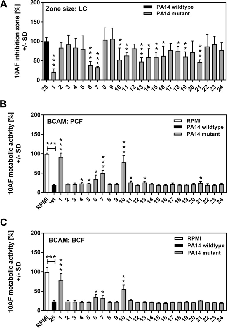
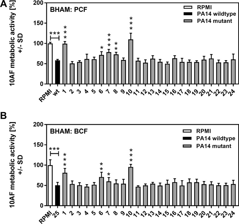
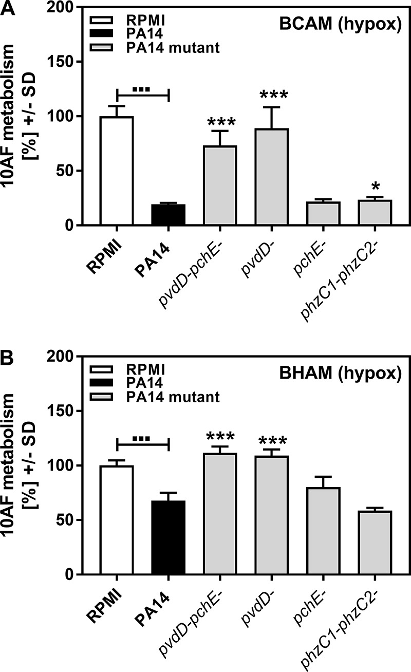
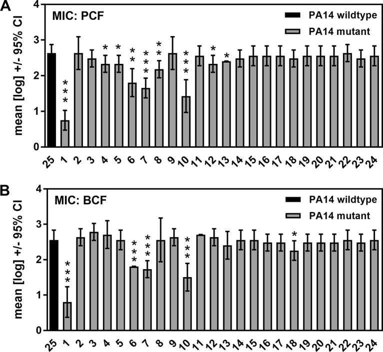
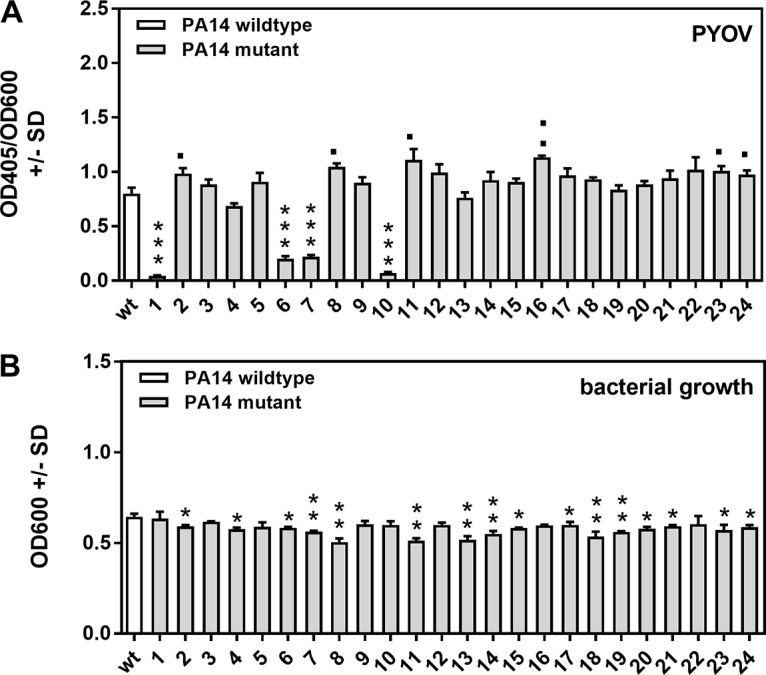
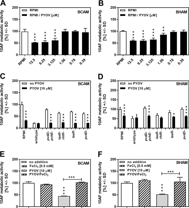
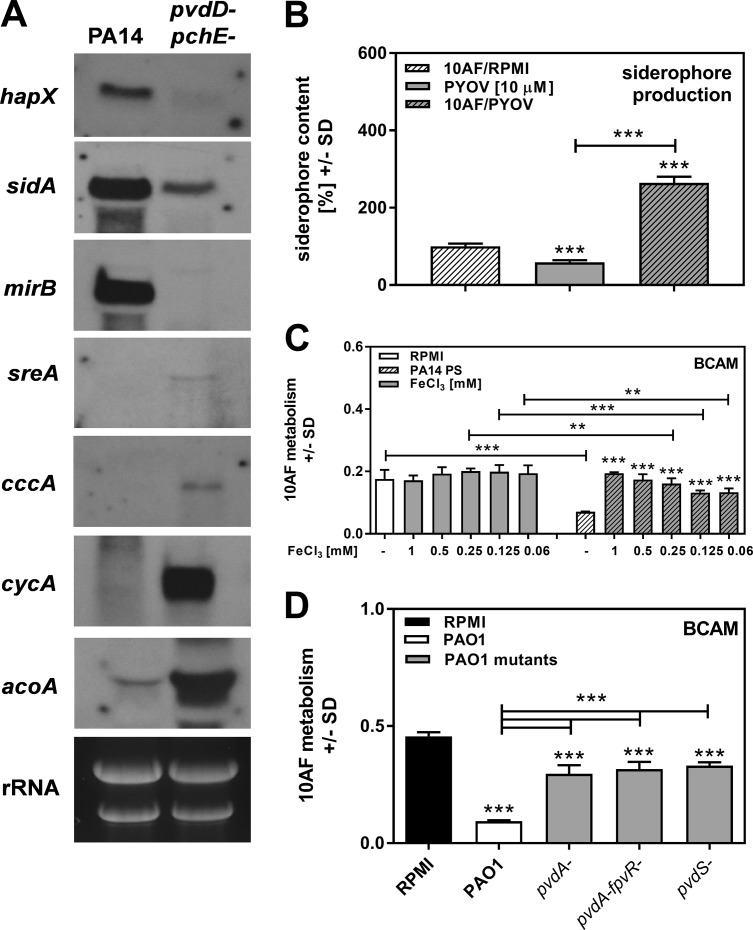
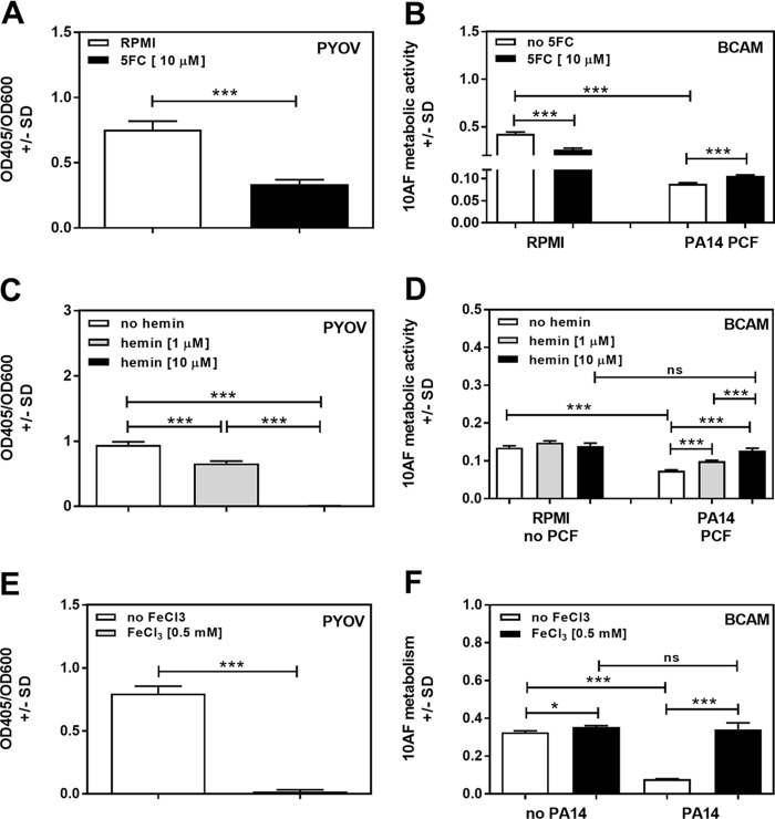
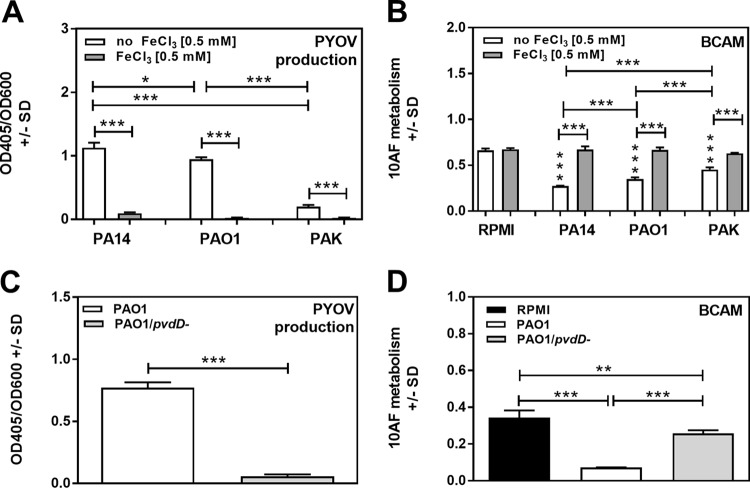
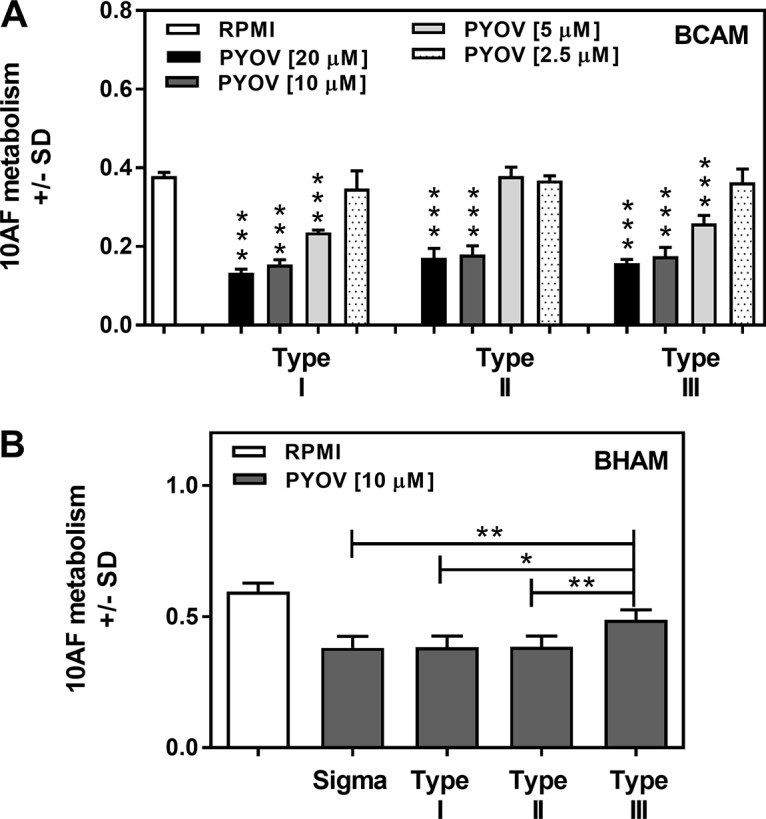
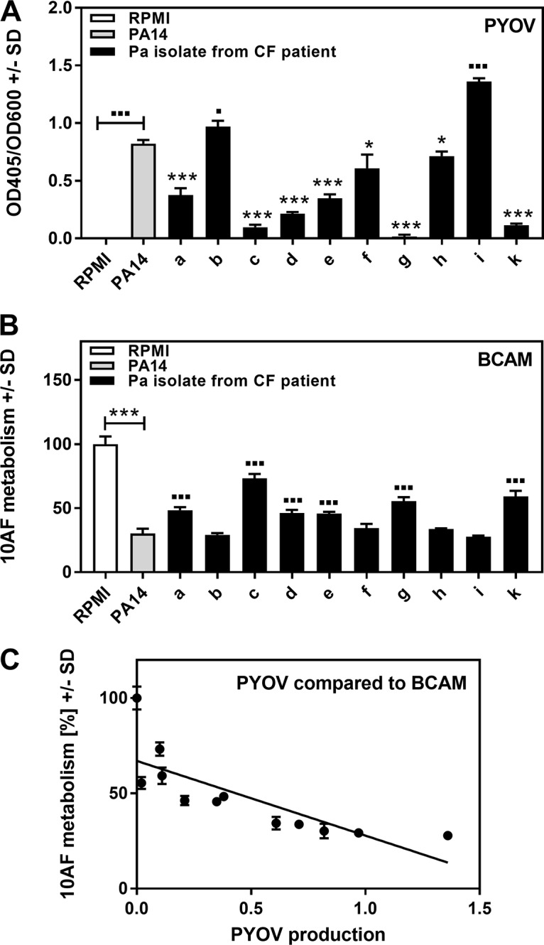
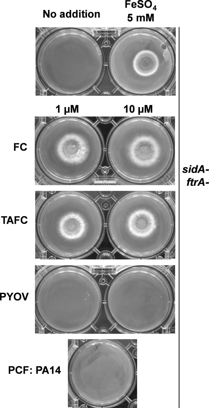
Pseudomonas aeruginosa and Aspergillus fumigatus are common opportunistic bacterial and fungal pathogens, respectively. They often coexist in airways of immunocompromised patients and individuals with cystic fibrosis, where they form biofilms and cause acute and chronic illnesses. Hence, the interactions between them have long been of interest and it is known that P. aeruginosa can inhibit A. fumigatusin vitro We have approached the definition of the inhibitory P. aeruginosa molecules by studying 24 P. aeruginosa mutants with various virulence genes deleted for the ability to inhibit A. fumigatus biofilms. The ability of P. aeruginosa cells or their extracellular products produced during planktonic or biofilm growth to affect A. fumigatus biofilm metabolism or planktonic A. fumigatus growth was studied in agar and liquid assays using conidia or hyphae. Four mutants, the pvdD pchE, pvdD, lasR rhlR, and lasR mutants, were shown to be defective in various assays. This suggested the P. aeruginosa siderophore pyoverdine as the key inhibitory molecule, although additional quorum sensing-regulated factors likely contribute to the deficiency of the latter two mutants. Studies of pure pyoverdine substantiated these conclusions and included the restoration of inhibition by the pyoverdine deletion mutants. A correlation between the concentration of pyoverdine produced and antifungal activity was also observed in clinical P. aeruginosa isolates derived from lungs of cystic fibrosis patients. The key inhibitory mechanism of pyoverdine was chelation of iron and denial of iron to A. fumigatusIMPORTANCE Interactions between human pathogens found in the same body locale are of vast interest. These interactions could result in exacerbation or amelioration of diseases. The bacterium Pseudomonas aeruginosa affects the growth of the fungus Aspergillus fumigatus Both pathogens form biofilms that are resistant to therapeutic drugs and host immunity. P. aeruginosa and A. fumigatus biofilms are found in vivo, e.g., in the lungs of cystic fibrosis patients. Studying 24 P. aeruginosa mutants, we identified pyoverdine as the major anti-A. fumigatus compound produced by P. aeruginosa Pyoverdine captures iron from the environment, thus depriving A. fumigatus of a nutrient essential for its growth and metabolism. We show how microbes of different kingdoms compete for essential resources. Iron deprivation could be a therapeutic approach to the control of pathogen growth.

Keywords: Aspergillus fumigatus; Pseudomonas aeruginosa; biofilms; intermicrobial interaction; iron; mutants; pyoverdine.


Figures

FIG 1
FIG 1
Effects of LC, PCF, and BCF on A. fumigatus strain 10AF biofilm formation and metabolism. (A) Wells in agar with A. fumigatus conidia were loaded with wild-type or mutant PA14 LC suspensions and incubated, and the area of the fungus-free (inhibition) zone around each well was measured. The inhibition zone created by wild-type PA14 LC was regarded as 100% inhibition, and inhibition zones created by mutants were compared to this. The results of four experiments are combined, with duplicates of each study article in each. *, P ≤ 0.05; **, P ≤ 0.01; ***, P ≤ 0.001 (mutant [no. 1 to 24] versus wild type [no. 25]). (B and C) Agar and A. fumigatus strain 10AF conidia were distributed into 96-well cell culture plates that were loaded with wild-type or mutant PA14 PCF (B) or wild-type or mutant PA14 BCF (C) and incubated at 37°C for 24 h. A. fumigatus strain 10AF metabolism was evaluated by XTT assay. Metabolism in the presence of RPMI medium alone was regarded as 100% A. fumigatus strain 10AF metabolic activity. The data shown are the mean ± SD of four independent experiments (with duplicates of each group studied in each experiment). *, P ≤ 0.05; **, P ≤ 0.01; ***, P ≤ 0.001. Vertical asterisks, mutants (no. 1 to 24) versus wild-type PA14 (no. 25); horizontal asterisks, RPMI medium versus wild-type PA14. Bars: 1, pvdD pchE mutant; 2, pqsE mutant; 3, mvfR mutant; 4, pqsA mutant; 5, pqsH mutant; 6, lasR rhlR mutant; 7, lasR mutant; 8, rsmA mutant; 9, pqsA pqsH not polar mutant; 10, pvdD mutant; 11, rhlR mutant; 12, ΔHSI-I ΔHSI-II mutant; 13, pvcA mutant; 14, rhlA mutant; 15, phzC1 phzC2 mutant; 16, pchE mutant; 17, exoU mutant; 18, rsmY rsmZ mutant; 19, ΔHSI-II ΔHSI-III mutant; 20, ΔHSI-I ΔHSI-III mutant; 21, pqsA pqsH polar mutant; 22, chiC mutant; 23, lecA mutant; 24, hcnA mutant.
FIG 2
FIG 2
Effects of PCF or BCF on A. fumigatus strain 10AF preformed biofilm growth and metabolism. A. fumigatus strain 10AF conidia in agar were distributed into 96-well cell culture plates. Plates were incubated at 37°C for 24 h to allow hyphal growth and then loaded with wild-type or mutant PA14 PCF (A) or wild-type or mutant PA14 BCF (B) and incubated at 37°C for 24 h. A. fumigatus strain 10AF metabolism was evaluated by XTT assay. Metabolism in the presence of RPMI medium alone was regarded as 100% A. fumigatus strain 10AF metabolic activity. The data shown are the mean ± SD of four independent experiments (with duplicates of each group studied in each experiment). *, P ≤ 0.05; **, P ≤ 0.01; ***, P ≤ 0.001 (mutant [no. 1 to 24] versus wild type [no. 25]). Horizontal asterisks, RPMI medium versus wild-type PA14. Bars: 1, pvdD pchE mutant; 2, pqsE mutant; 3, mvfR mutant; 4, pqsA mutant; 5, pqsH mutant; 6, lasR rhlR mutant; 7, lasR mutant; 8, rsmA mutant; 9, pqsA pqsH not polar mutant; 10, pvdD mutant; 11, rhlR mutant; 12, ΔHSI-I ΔHSI-II mutant; 13, pvcA mutant; 14, rhlA mutant; 15, phzC1 phzC2 mutant; 16, pchE mutant; 17, exoU mutant; 18, rsmY rsmZ mutant; 19, ΔHSI-II ΔHSI-III mutant; 20, ΔHSI-I ΔHSI-III mutant; 21, pqsA pqsH polar mutant; 22, chiC mutant; 23, lecA mutant; 24, hcnA mutant.
FIG 3
FIG 3
Effects of PCF on A. fumigatus strain 10AF forming and preformed biofilm metabolism under hypoxic conditions. A. fumigatus strain 10AF conidia in agar were distributed into 96-well cell culture plates. Plates were either loaded immediately after solidification (BCAM assay) (A) or incubated at 37°C for 24 h to allow hyphal growth (BHAM assay) (B). Plates were loaded with wild-type or mutant PA14 PCF and incubated under hypoxic conditions at 37°C for 24 h. A. fumigatus strain 10AF metabolism was evaluated by XTT assay. Metabolism in the presence of RPMI medium alone was regarded as 100% A. fumigatus strain 10AF metabolic activity. The data shown are the mean ± SD of four replicates. *, P ≤ 0.05; ***, P ≤ 0.001 (mutant versus wild-type PA14). Other comparisons are indicated by the ends of the brackets above the bars.
FIG 4
FIG 4
Effects of PCF or BCF on A. fumigatus strain 10AF planktonic growth (MIC). A. fumigatus strain 10AF conidia were incubated in the presence of RPMI medium and PCF dilutions (A) or RPMI medium and BCF dilutions (B) at 37°C for 48 h. Fungal growth was determined in 2-fold dilution steps for wild-type (no. 25) and mutant (no. 1 to 24) PA14 (dilutions, 1:2 to 1:1,024). For each supernatant, the last dilution showing fungal growth less than that in RPMI medium alone was determined. The means of four independent experiments were determined. The data shown are the mean ± SD. *, P ≤ 0.05; **, P ≤ 0.01; ***, P ≤ 0.001 (mutant [no. 1 to 24] versus wild type [no. 25]). Bars: 1, pvdD pchE mutant; 2, pqsE mutant; 3, mvfR mutant; 4, pqsA mutant; 5, pqsH mutant; 6, lasR rhlR mutant; 7, lasR mutant; 8, rsmA mutant; 9, pqsA pqsH not polar mutant; 10, pvdD mutant; 11, rhlR mutant; 12, ΔHSI-I ΔHSI-II mutant; 13, pvcA mutant; 14, rhlA mutant; 15, phzC1 phzC2 mutant; 16, pchE mutant; 17, exoU mutant; 18, rsmY rsmZ mutant; 19, ΔHSI-II ΔHSI-III mutant; 20, ΔHSI-I ΔHSI-III mutant; 21, pqsA pqsH polar mutant; 22, chiC mutant; 23, lecA mutant; 24, hcnA mutant. CI, confidence interval.
FIG 5
FIG 5
Pyoverdine production by wild-type and mutant PA14. Wild-type or mutant PA14 bacteria (5 × 10⁷/ml) were incubated in RPMI medium overnight. Bacterial growth was measured spectrophotometrically at 600 nm. PCF was harvested, and pyoverdine (PYOV) production was measured at 405 nm (A). Measurements were normalized to bacterial growth (B) with the formula relative pyoverdine expression = OD405/OD600. Experiments in both panels were performed three times (four replicates of each group in each experiment), and the results were combined. ***, P ≤ 0.001 (decrease in pyoverdine production or bacterial growth in mutants compared to wild-type PA14); ■, P ≤ 0.05; ■■, P ≤ 0.01 (significant increase in pyoverdine production compared to wild-type PA14). Bars: 1, pvdD pchE mutant; 2, pqsE mutant; 3, mvfR mutant; 4, pqsA mutant; 5, pqsH mutant; 6, lasR rhlR mutant; 7, lasR mutant; 8, rsmA mutant; 9, pqsA pqsH not polar mutant; 10, pvdD mutant; 11, rhlR mutant; 12, ΔHSI-I ΔHSI-II mutant; 13, pvcA mutant; 14, rhlA mutant; 15, phzC1 phzC2 mutant; 16, pchE mutant; 17, exoU mutant; 18, rsmY rsmZ mutant; 19, ΔHSI-II ΔHSI-III mutant; 20, ΔHSI-I ΔHSI-III mutant; 21, pqsA pqsH polar mutant; 22, chiC mutant; 23, lecA mutant; 24, hcnA mutant.
FIG 6
FIG 6
Effects of pyoverdine on A. fumigatus strain 10AF biofilm metabolism and antifungal activities of PA14 mutants. (A and B) Increasing concentrations of pyoverdine (PYOV) in RPMI 1640 (0.39 to 12.5 μM) were assayed for activity against A. fumigatus strain 10AF biofilm formation (A) or preformed A. fumigatus strain 10AF biofilm (B). Fungal metabolism was measured by XTT assay. Statistical analysis was performed by one-way ANOVA of the RPMI 1640 control versus pyoverdine concentrations: **, P ≤ 0.01; ***, P ≤ 0.001. (C and D) Antifungal activities of wild-type PA14 and pyoverdine-lacking mutants with or without supplementation with 10 μM pyoverdine were measured against forming A. fumigatus strain 10AF biofilm (C) or preformed A. fumigatus strain 10AF biofilm (D). No pyoverdine supplementation versus pyoverdine supplementation: *, P ≤ 0.05; ***, P ≤ 0.001. (E and F) A. fumigatus strain 10AF conidia were incubated in the presence of RPMI 1640 (unstriped bars) or RPMI 1640 supplemented with 0.5 μM FeCl3 (striped bars) with (gray bars) or without (white bars) 10 μM pyoverdine. A. fumigatus strain 10AF metabolism was evaluated by XTT assay. Metabolism in the presence of RPMI 1640 without FeCl3 or pyoverdine was regarded as 100% A. fumigatus strain 10AF metabolic activity. Vertical asterisks, comparison with RPMI 1640; horizontal asterisks, RPMI 1640 containing pyoverdine versus RPMI 1640 containing pyoverdine and FeCl3. ***, P ≤ 0.001. Experiments in panel A were performed twice, those in panel B were performed three times, those in panel D were performed six times, and representative results are shown. Every experiment had four replicates per group studied.
FIG 7
FIG 7
Effects of P. aeruginosa siderophores on A. fumigatus iron metabolism. (A) A. fumigatus strain AfS77 conidia were grown at 37°C in 15 ml of liquid 2YT medium for 15 h. Subsequently, 10 ml of the culture supernatant was replaced with 10 ml of wild-type or pvdA pvdE mutant PA14 PCF and incubated for 3 h. A Northern blot analysis was performed for genes inducible by iron starvation (hapX, sidA, and mirB) and genes repressed by iron starvation (sreA, cccA, cycA, and acoA). Measurement of rRNA served as a loading control. (B) A. fumigatus strain 10AF conidia were incubated in the presence of RPMI 1640 (striped bar) or RPMI 1640 supplemented with 10 μM pyoverdine (gray striped bar) for 24 h. Supernatants were sterile filtered. As a control, 10 μM pyoverdine (PYOV) solution in RPMI 1640 was prepared (solid gray bar). A CAS assay measuring siderophore content in A. fumigatus supernatants was performed, and the results were normalized to those of an XTT assay prepared in parallel with the CAS assay measuring fungal metabolism. Siderophore production by A. fumigatus was regarded as 100% and compared to all other bars. Other comparisons are indicated by the ends of the brackets above the bars. ***, P ≤ 0.001. Pyoverdine itself as a siderophore was measurable by CAS assay. (C) RPMI 1640 or PA14 PCF with or without increasing amounts of FeCl3 was subjected to a BCAM assay measuring effects on the metabolism of A. fumigatus strain 10AF forming biofilm. A. fumigatus strain 10AF metabolism was evaluated by XTT assay. Metabolism in the presence of RPMI 1640 alone was regarded as 100% A. fumigatus strain 10AF metabolic activity. The data shown are the mean ± SD from four replicates. **, P ≤ 0.01; ***, P ≤ 0.001 (RPMI 1640 versus RPMI 1640 containing FeCl3 [left side] or PCF versus PCF containing FeCl3 [right side]). Other comparisons are indicated by the ends of the brackets above the bars. (D) Agar and A. fumigatus strain 10AF conidia were distributed into 96-well cell culture plates that were loaded with wild-type or mutant PAO1 PCF and incubated at 37°C for 24 h. A. fumigatus strain 10AF metabolism was evaluated by XTT assay. Metabolism in the presence of RPMI medium alone was compared to metabolism in the presence of PCF. Other comparisons are indicated by the ends of the brackets above the bars. ***, P ≤ 0.001.
FIG 8
FIG 8
Effects of pyoverdine reduction on A. fumigatus strain 10AF biofilm metabolism. RPMI 1640 with or without 10 μM 5FC (A, B), 1 or 10 μM hemin (C, D), or 0.5 mM FeCl3 (E, F) was inoculated with 5 × 10⁷ PA14 bacteria/ml. Pyoverdine (PYOV) production was measured after 24 h of incubation (A, C, E). PCFs derived from panels A, C, and E were subjected to BCAM assays measuring effects on the metabolism of A. fumigatus strain 10AF forming biofilm (B, D, F). Groups being compared are indicated by the ends of the brackets above the bars. The experiments shown were performed three times (four replicates of each group in each experiment), and the results were combined. *, P ≤ 0.05; ***, P ≤ 0.001.
FIG 9
FIG 9
Pyoverdine production by P. aeruginosa PAO1, PAK, and PA14 and effects on A. fumigatus strain 10AF forming biofilm. (A) RPMI 1640 with or without 0.5 mM FeCl3 was inoculated with P. aeruginosa strains PAO1, PAK and PA14 at 5 × 10⁷ bacteria/ml. Pyoverdine (PYOV) production was measured after 24 h of incubation. (B) PCFs derived from panel A were studied in assays measuring effects on the metabolism of A. fumigatus strain 10AF forming biofilm (BCAM assay). Vertical asterisks, comparison with the negative control (RPMI 1640 alone). (C) Pyoverdine production by wild-type or pvdD mutant PAO1 bacteria (5 × 10⁷/ml) was measured after 24 h of incubation. (D) PCFs derived from panel C were studied in assays measuring effects on the metabolism of A. fumigatus strain 10AF forming biofilm (BCAM). Groups being compared are indicated by the ends of the brackets above the bars. Representative results are shown. Each group in each experiment contained at least four replicates. *, P ≤ 0.05; **, P ≤ 0.01; ***, P ≤ 0.001.
FIG 10
FIG 10
Titration of purified pyoverdines. (A) Pyoverdine types were titrated 2-fold from 20 to 2.5 μM against A. fumigatus strain 10AF biofilm formation. Mean results of four replicates of each group are shown. ***, P ≤ 0.001 (versus RPMI 1640 control). (B) Part of a titration series against preformed A. fumigatus strain 10AF biofilm (10 μM comparison) is shown. Sigma is the commercial pyoverdine (PYOV; type I) used in the experiments whose results are shown in the preceding figures. Mean results of four replicates of each group are shown. Groups being compared are indicated by the ends of the brackets above the bars. *, P ≤ 0.05; **, P ≤ 0.01. All three types of pyoverdine inhibited preformed biofilm metabolism compared to the RPMI 1640 control. Type III, P ≤ 0.01; type I or II, P ≤ 0.001.
FIG 11
FIG 11
Correlation between pyoverdine production and antifungal activity of clinical P. aeruginosa (Pa) isolates. Ten CF isolates designated a to i and k, as well as PA14, were tested for pyoverdine (PYOV) production (A), as well as for activity against the metabolism of A. fumigatus strain 10AF forming biofilm (BCAM assay) (B). RPMI medium served as a control. Results of panels A and B were compared by linear regression curve analysis (panel C). Symbols: ■ or *, P ≤ 0.01; ■■■ or ***, P ≤ 0.001. Each comparison without brackets in panels A and B is PA14 versus 1 of the 10 CF isolates. Comparisons with brackets are as indicated. *, significant decreases; ■, significant increases. n = 3 for pyoverdine production. n = 5 for A. fumigatus strain 10AF metabolic activity.
FIG 12
FIG 12
Measurement of xenosiderophores in PCF. A. fumigatus sidA ftrA mutant conidia (104) were point inoculated onto solid minimal medium agar with or without supplementation with FeSO4 to a final concentration of 5 mM, pyoverdine (PYOV) to final concentrations of 1 and 10 μM, TAFC or FC to a final concentration of 1 or 10 μM, or 600 μl of wild-type PA14 PCF. The plates were incubated for 48 h at 37°C.

Similar articles

Cited by

References

    1. Williams HD, Davies JC. 2012. Basic science for the chest physician: Pseudomonas aeruginosa and the cystic fibrosis airway. Thorax 67:465–467. doi:10.1136/thoraxjnl-2011-201498. - DOI - PubMed
    1. Smyth AR, Hurley MN. 2010. Targeting the Pseudomonas aeruginosa biofilm to combat infections in patients with cystic fibrosis. Drugs Future 35:1007–1014. doi:10.1358/dof.2010.035.012.1537937. - DOI
    1. Folkesson A, Jelsbak L, Yang L, Johansen HK, Ciofu O, Høiby N, Molin S. 2012. Adaptation of Pseudomonas aeruginosa to the cystic fibrosis airway: an evolutionary perspective. Nat Rev Microbiol 10:841–851. doi:10.1038/nrmicro2907. - DOI - PubMed
    1. Sabino R, Ferreira JA, Moss RB, Valente J, Veríssimo C, Carolino E, Clemons KV, Everson C, Banaei N, Penner J, Stevens DA. 2015. Molecular epidemiology of Aspergillus collected from cystic fibrosis patients. J Cyst Fibros 14:474–481. doi:10.1016/j.jcf.2014.10.005. - DOI - PubMed
    1. Fillaux J, Brémont F, Murris M, Cassaing S, Rittié JL, Tétu L, Segonds C, Abbal M, Bieth E, Berry A, Pipy B, Magnaval JF. 2012. Assessment of Aspergillus sensitization or persistent carriage as a factor in lung function impairment in cystic fibrosis patients. Scand J Infect Dis 44:842–847. doi:10.3109/00365548.2012.695454. - DOI - PubMed

MeSH terms

LinkOut - more resources