. 2017 Dec 1;292(48):19721-19732.
doi: 10.1074/jbc.M117.814392. Epub 2017 Oct 16.

Cystine uptake through the cystine/glutamate antiporter xCT triggers glioblastoma cell death under glucose deprivation

Affiliations

Cystine uptake through the cystine/glutamate antiporter xCT triggers glioblastoma cell death under glucose deprivation

Takeo Goji et al. J Biol Chem. .

Abstract

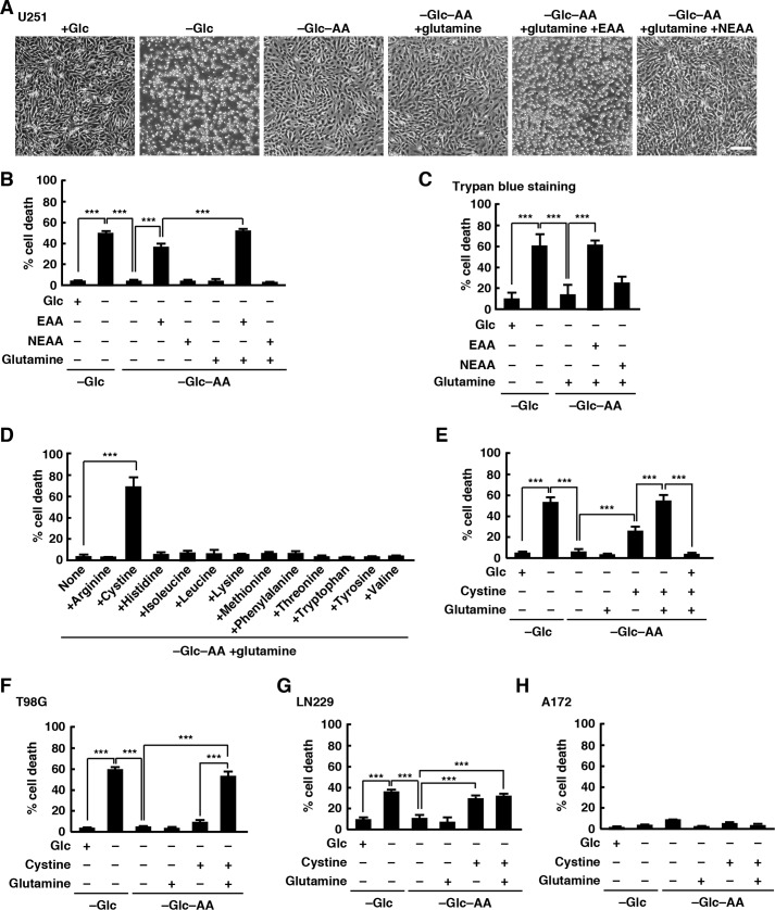
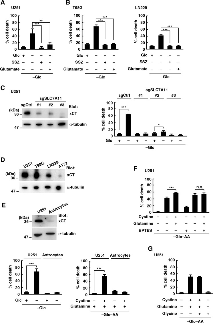
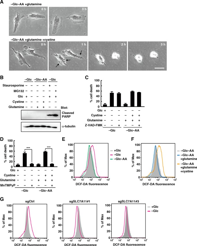
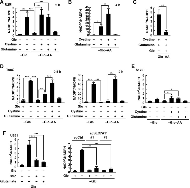
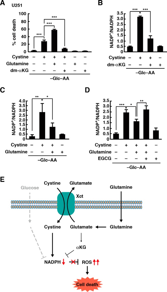
Oncogenic signaling in cancer cells alters glucose uptake and utilization to supply sufficient energy and biosynthetic intermediates for survival and sustained proliferation. Oncogenic signaling also prevents oxidative stress and cell death caused by increased production of reactive oxygen species. However, elevated glucose metabolism in cancer cells, especially in glioblastoma, results in the cells becoming sensitive to glucose deprivation (i.e. in high glucose dependence), which rapidly induces cell death. However, the precise mechanism of this type of cell death remains unknown. Here, we report that glucose deprivation alone does not trigger glioblastoma cell death. We found that, for cell death to occur in glucose-deprived glioblastoma cells, cystine and glutamine also need to be present in culture media. We observed that cystine uptake through the cystine/glutamate antiporter xCT under glucose deprivation rapidly induces NADPH depletion, reactive oxygen species accumulation, and cell death. We conclude that although cystine uptake is crucial for production of antioxidant glutathione in cancer cells its transport through xCT also induces oxidative stress and cell death in glucose-deprived glioblastoma cells. Combining inhibitors targeting cancer-specific glucose metabolism with cystine and glutamine treatment may offer a therapeutic approach for glioblastoma tumors exhibiting high xCT expression.

Keywords: amino acid transport; cell death; cystine; glioblastoma; glucose; reactive oxygen species (ROS).


Conflict of interest statement

The authors declare that they have no conflicts of interest with the contents of this article

Figures

Figure 1.
Figure 1.
Cystine and glutamine induce cell death during glucose deprivation. A and B, U251 cells were placed in glucose-free or glucose- and amino acid-free medium with or without glucose (5 mm), EAA, or NEAA solutions for 24 h. Scale bar, 200 μm (A). Quantification of cell death was performed using an LDH release assay. Cells treated with 0.1% Tween 20 were used for calculating 100% cell death (B). C, quantification of cell death was performed using a trypan blue exclusion assay. D–H, cells were placed in glucose-free or glucose- and amino acid-free medium with or without glucose, glutamine (2 mm), or cystine (0.2 mm) for 24 h (D, E, G, and H) or 12 h (F). Quantification of cell death was performed using an LDH release assay. Error bars represent S.D. (n = 3). ***, p < 0.001, calculated by one-way ANOVA with Tukey's post hoc test. Glc, glucose; AA, amino acids.
Figure 2.
Figure 2.
Glucose deprivation-induced cell death involves xCT. A and B, cells were placed in glucose-free medium with or without glucose (5 mm), SSZ (250 μm), or glutamic acid (10 mm) for 24 h. Quantification of cell death was performed using an LDH release assay. Error bars represent S.D. (n = 3). **, p < 0.01; ***, p < 0.001, calculated by one-way ANOVA with Tukey's post hoc test. C, immunoblot analysis of U251 cells after CRISPR-based gene editing with SLC7A11 sgRNAs (sgSLC7A11) or sgCtrl. Quantification of cell death was performed 24 h after glucose deprivation using an LDH release assay. Error bars represent S.D. (n = 3). *, p < 0.05; ***, p < 0.001, calculated by one-way ANOVA with Tukey's post hoc test. D, immunoblot analysis of glioblastoma cell lines. E, immunoblot analysis of U251 cells and rat astrocytes. Quantification of cell death was performed 24 h after glucose deprivation using an LDH release assay. Error bars represent S.D. (n = 3). *, p < 0.05; ***, p < 0.001, calculated by one-way ANOVA with Tukey's post hoc test. F and G, cells were placed in glucose- and amino acid-free medium with or without cystine (0.2 mm), glutamine (2 mm), glycine (0.4 mm), or BPTES (10 μm) for 24 h. Quantification of cell death was performed using an LDH release assay. Error bars represent S.D. (n = 3). **, p < 0.01; ***, p < 0.001, calculated by one-way ANOVA with Tukey's post hoc test. n.s., not significant; Glc, glucose; AA, amino acids.
Figure 3.
Figure 3.
Cystine uptake through xCT causes accumulation of ROS under glucose deprivation. A, after the medium was replaced with glucose- and amino acid-free medium containing glutamine (2 mm) or glutamine and cystine (0.2 mm), phase-contrast images of U251 cells were captured at the indicated times. Scale bar, 20 μm. B, immunoblot analysis of U251 cells after the medium was replaced with glucose-free or glucose- and amino acid-free medium with or without glucose (5 mm), glutamine (2 mm), cystine (0.2 mm), staurosporine (0.1 μm), or MG132 (10 μm) for 24 h. C and D, U251 cells were placed in glucose-free or glucose- and amino acid-free medium with or without glucose (5 mm), glutamine (2 mm), cystine (0.2 mm), Z-VAD-fmk (10 μm), or MnTMPyP (10 μm) for 24 h. Quantification of cell death was performed using an LDH release assay. Error bars represent S.D. (n = 3). ***, p < 0.001, calculated by one-way ANOVA with Tukey's post hoc test. E–G, FACS analysis of ROS generation in U251 cells. Cells were placed in glucose-free or glucose- and amino acid-free medium with or without glucose or cystine for 3 h. Dark traces, cells placed in the medium with glucose (E and G) or in the medium without glucose and amino acids (F). Similar results were obtained in three independent experiments. Glc, glucose; AA, amino acids; DCF-DA, 2′,7′-dichlorofluorescein diacetate.
Figure 4.
Figure 4.
Cystine uptake through xCT induces NADPH depletion during glucose deprivation. After the medium was replaced with glucose-free or glucose- and amino acid-free medium containing glucose (5 mm), glutamine (2 mm), cystine (0.2 mm), SSZ (250 μm), or glutamic acid (10 mm) for 2 (A, C, E, and F), 0.5 or 2 (D), or 4 h (B), the NADP+/NADPH ratio was measured. Error bars represent S.D. (n = 3). *, p < 0.05; **, p < 0.01; ***, p < 0.001, calculated by one-way ANOVA with Tukey's post hoc test. Glc, glucose; AA, amino acids.
Figure 5.
Figure 5.
Glutamine and α-ketoglutarate inhibit cystine-induced NADPH depletion. A, cells were placed in glucose- and amino acid-free medium with or without glutamine (2 mm), cystine (0.2 mm), or dm-αKG (4 mm) for 24 h. Quantification of cell death was performed using an LDH release assay. B–D, after the medium was replaced with glucose- and amino acid-free medium containing glutamine (2 mm), cystine (0.2 mm), dm-αKG (4 mm), or EGCG (20 μm) for 2 (B) or 1 h (C and D), the NADP+/NADPH ratio was measured. Error bars represent S.D. (n = 3). *, p < 0.05; **, p < 0.01; ***, p < 0.001, calculated by one-way ANOVA with Tukey's post hoc test. E, model of xCT-mediated cell death under glucose deprivation conditions. Glc, glucose; AA, amino acids.

Similar articles

Cited by

References

    1. Hanahan D., and Weinberg R. A. (2011) Hallmarks of cancer: the next generation. Cell 144, 646–674 - PubMed
    1. Ward P. S., and Thompson C. B. (2012) Metabolic reprogramming: a cancer hallmarks even Warburg did not anticipate. Cancer Cell 21, 297–308 - PMC - PubMed
    1. Furnari F. B., Fenton T., Bachoo R. M., Mukasa A., Stommel J. M., Stegh A., Hahn W. C., Ligon K. L., Louis D. N., Brennan C., Chin L., DePinho R. A., and Cavenee W. K. (2007) Malignant astrocytic glioma: genetics, biology, and paths to treatment. Genes Dev. 21, 2683–2710 - PubMed
    1. Elstrom R. L., Bauer D. E., Buzzai M., Karnauskas R., Harris M. H., Plas D. R., Zhuang H., Cinalli R. M., Alavi A., Rudin C. M., and Thompson C. B. (2004) Akt stimulates aerobic glycolysis in cancer cells. Cancer Res. 64, 3892–3899 - PubMed
    1. Jelluma N., Yang X., Stokoe D., Evan G. I., Dansen T. B., and Haas-Kogan D. A. (2006) Glucose withdrawal induces oxidative stress followed by apoptosis in glioblastoma cells but not in normal astrocytes. Mol. Cancer Res. 4, 319–330 - PubMed

Publication types