. 2018 Jan 2;217(1):413-427.
doi: 10.1083/jcb.201604048. Epub 2017 Oct 16.

The small GTPase RSG1 controls a final step in primary cilia initiation

Affiliations

The small GTPase RSG1 controls a final step in primary cilia initiation

Stephanie O Agbu et al. J Cell Biol. .

Abstract

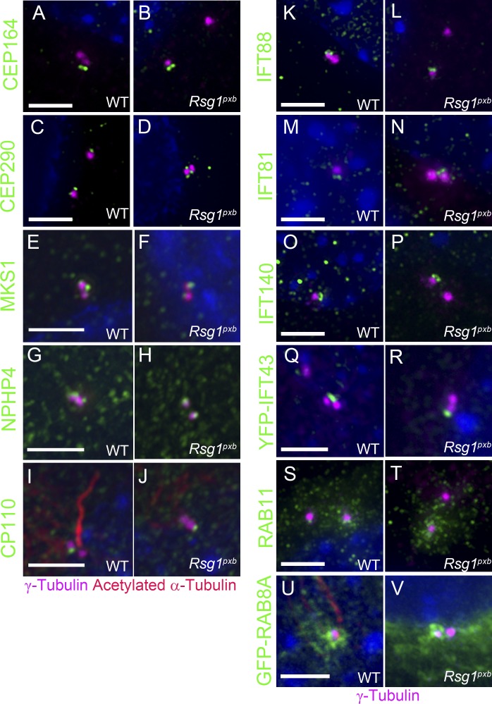
Primary cilia, which are essential for normal development and tissue homeostasis, are extensions of the mother centriole, but the mechanisms that remodel the centriole to promote cilia initiation are poorly understood. Here we show that mouse embryos that lack the small guanosine triphosphatase RSG1 die at embryonic day 12.5, with developmental abnormalities characteristic of decreased cilia-dependent Hedgehog signaling. Rsg1 mutant embryos have fewer primary cilia than wild-type embryos, but the cilia that form are of normal length and traffic Hedgehog pathway proteins within the cilium correctly. Rsg1 mother centrioles recruit proteins required for cilia initiation and dock onto ciliary vesicles, but axonemal microtubules fail to elongate normally. RSG1 localizes to the mother centriole in a process that depends on tau tubulin kinase 2 (TTBK2), the CPLANE complex protein Inturned (INTU), and its own GTPase activity. The data suggest a specific role for RSG1 in the final maturation of the mother centriole and ciliary vesicle that allows extension of the ciliary axoneme.


Figures

Figure 1.
Figure 1.
Shh signaling phenotypes of Rsg1 embryos. (A and B) Whole-mount images of WT (A) and Rsg1pxb mutant (B) embryonic limbs at E12.5. Note extra preaxial digits in the mutant (arrows). Bar, 500 µm. (C and D) Whole-mount RNA in situ hybridization for Gli1 transcripts in E10.5 WT (C) and Rsg1pxb (D) limbs (n = 6 embryos). Bar, 250 µm. (E) Western blot analysis of GLI3 from extracts of E10.5 limbs, showing the defect in processing of the full-length protein (FL) to the repressor form (R). α-Tubulin was the loading control (n = 3 independent experiments). (F–M) Confocal images of WT, Rsg1pxb, Rsg1pxb/CRISPR, and Rsg1CRISPR lumbar neural tube sections stained for SHH-dependent ventral cell populations marked by FOXA2 (floor plate), NKX2.2 (V3 interneuron progenitors), OLIG2 (motor neuron progenitors), and PAX6 (lateral neural progenitors; n = 3 embryos per genotype). Bars, 70 µm. In contrast to the results shown here, ventral cell fates were specified correctly in the rostral neural tube (not depicted), consistent with a milder defect in SHH response than seen in mutants that completely lack primary cilia.
Figure 2.
Figure 2.
Reduction of cilia number in Rsg1 mutants. (A–C) Primary cilia in WT (A), Rsg1pxb (B), and Rsg1CRISPR (C) mesenchymal cells in the E10.5 limb. Bar, 3 µm (n = 3 embryos). (D–F) Primary cilia in WT (D), Rsg1pxb (E), and Rsg1CRISPR (F) MEFs. Bar, 3 µm. (G) Quantification of primary cilia formation in limb mesenchyme. Mean ± SD. WT, n = 870 cells; Rsg1pxb, n = 645 cells; Rsg1CRISPR, n = 589 cells. (H) Quantification of primary cilia formation in WT, Rsg1pxb, and Rsg1CRISPR MEFs. Mean ± SD. WT, n = 220 cells; Rsg1pxb, n = 239 cells; Rsg1CRISPR, n = 803 cells. (I and J) Higher-resolution images showing the length of primary cilia in WT (I) and Rsg1pxb (J) MEFs. Bar, 3 µm. (K) Quantification of cilia length in WT and Rsg1pxb MEFs. Mean ± SD. WT, n = 227 cilia; Rsg1pxb, n = 122 cilia. Error bars are SDs. (L and M) Scanning electron micrographs of primary cilia in the WT (L) and Rsg1pxb (M) brachial neural tube. Bar, 1 µm. t test compared with WT, ****, P < 0.0001; ns, no significant difference.
Figure 3.
Figure 3.
Trafficking of Hedgehog pathway proteins is normal in Rsg1 mutant cilia. (A–D) SMO staining in WT (A and B) and Rsg1pxb (C and D) MEF cilia, in the absence of pathway activation (–SAG) or when the pathway was activated by the small molecule SAG (+SAG). (E–H) GLI2 at cilia tips in WT (E and F) and Rsg1pxb (G and H) MEFs, in the presence or absence of SAG. (I–L) KIF7 staining in WT (I and J) and Rsg1pxb (K and L) MEFs, in the presence or absence of SAG. Bars, 1 µm (n = 3 independent experiments). (M and N) Expression of Gli1 and Ptch1 mRNAs in WT, Rsg1pxb, and Rsg1CRISPR MEFs in the absence of pathway activation (–SAG) or when the pathway was activated by SAG (+SAG). Fold change is normalized to WT cells (=1) without SAG treatment; data are presented as mean ± SD, n = 3 experiments. t test *, P < 0.05; **, P < 0.01; ***, P < 0.001; ns, no significant difference.
Figure 4.
Figure 4.
RSG1 localizes to the mother centriole and the transition zone of the primary cilium. (A and B) Immunolocalization of endogenous RSG1 in human RPE1 cells. (A) In cilia-promoting conditions (absence of serum), RSG1 localizes to the ciliary transition zone, between the γ-tubulin+ mother basal body (pink) and the acetylated α-tubulin+ ciliary axoneme (red). (B) When grown in the presence of serum, the majority of cells lack cilia, and RSG1 was detected at one of the two centrioles in ∼30% of the unciliated cells. (C and D) GFP-RSG1 localization in transiently transfected ciliated (C) and unciliated (D) WT cells (n = 3 independent experiments). (E and F) GFP-RSG1 partially colocalized with CEP164 in transiently transfected ciliated (E) and unciliated (F) WT cells. (G and H) Rescue of cilia formation in Rsg1pxb MEFs by GFP-RSG1. Untransfected Rsg1pxb MEFs (G) and Rsg1pxb MEFs (H) transfected with GFP-RSG1 (n = 3 independent experiments). (I–K) GTPase activity is required for RSG1 localization to the mother centriole. (I) Transfected GFP-RSG1V169D localizes to the transition zone of transfected Rsg1pxb MEFs. (J) In the rare cilia that form after transfection of Rsg1pxb MEFs with GFP-RSG1T69N, GFP was not detected at the base of the cilium (n = 3 independent experiments). (K) Quantification of cilia frequency in MEFs transfected with GFP-RSG1, GFP-RSG1V169D, and GFP-RSG1T69N constructs. WT, n = 70 cells; Rsg1pxb, n = 67 cells; GFP-RSG1, n = 65 cells; GFP-RSG1V169D, n = 63 cells; and GFP-RSG1T69N, n = 55 cells. Mean ± SD. Cilia frequency in GFP-RSG1V169D and GFP-RSG1T69N was significantly reduced compared with WT cells and Rsg1pxb MEFs transfected with WT GFP-RSG1 (t test, *, P < 0.05; ****, P < 0.0001; n = 3 independent experiments). Bars, 2 µm.
Figure 5.
Figure 5.
Recruitment of appendage, transition zone, IFT, and cilia-associated RAB proteins to centrioles is normal in Rsg1pxb MEFs. (A and B) CEP164 staining in WT and Rsg1pxb MEFs. As in WT, CEP164 associated with Rsg1pxb mother centrioles (100 ± 0.0% in WT [n = 92 cells] vs. 98.5 ± 1.5% in Rsg1pxb [n = 91 cells]). (C and D) CEP290 localized to mother centrioles in both genotypes (99.5 ± 0.5% in WT [n = 47 cells] vs. 98.6 ± 0.9% in Rsg1pxb [n = 113 cells]). (E and F) MKS1, a transition zone protein, localized normally to Rsg1pxb centrioles (100 ± 0.0% in WT [n = 77 cells] vs. 93.0% ± 2.5% in Rsg1pxb [n = 87 cells]). (G and H) NPHP4, a component of the nephronophthisis (NPHP) complex, was recruited normally to Rsg1pxb centrioles (98.9 ± 1.1% in WT [n = 59 cells] vs. 94.9 ± 1.8% in Rsg1pxb [n = 111 cells]). (I and J) CP110 in WT (I) and Rsg1pxb (J) MEFs (88.8 ± 3.3% in WT [n = 293 cells] vs. 81.1 ± 4.2% in Rsg1pxb [n = 351 cells]). IFT88 (K and L), IFT81 (M and N), IFT140 (O and P), and YFP-IFT43 (Q and R) localization in WT and Rsg1pxb MEFs (IFT88, 98.7 ± 1.0% in WT [n = 134 cells] vs. 94.4 ± 2.1% in Rsg1pxb [n = 381 cells]; IFT81, 99.3 ± 0.4% in WT [n = 133 cells] vs. 96.7% ± 1.2% in Rsg1pxb [n = 178 cells]; IFT43-YFP, 83.6 ± 5.05% in WT [n = 64 cells] vs. 71.4 ± 5.0% in Rsg1pxb [n = 66 cells]). (S and T) RAB11 localization at WT and Rsg1pxb centrosomes after 1 h of serum starvation (46.6 ± 9.2% in WT [n = 34 centrosomes] vs. 56.7 ± 7.8% in Rsg1pxb [n = 57 centrosomes]). n = 3 independent experiments. (U and V) GFP-RAB8A is enriched near the mother centriole in both WT and Rsg1pxb cells. For all experiments, each genotype was examined in three independent experiments. Bars, 2 µm.
Figure 6.
Figure 6.
Mother centrioles associate with ciliary vesicles in the absence of RSG1. (A) SMO is not detected at centrioles of serum-fed unciliated WT MEFs. (B and C) SMO localizes to WT (B) and Rsg1pxb (C) mother centrioles after serum starvation. (D and E) SMO localization to WT (D) and Rsg1CRISPR (E) mother centrioles (n = 3 independent experiments). (A–E) Bars, 3 µm. (F and G) Transmission electron micrographs of ciliary vesicles associated with WT (F) and Rsg1CRISPR (G) mother centrioles from serial sections of the E11.5 neural tube. Bar, 250 nm.
Figure 7.
Figure 7.
RSG1 localization to the mother centriole depends on TTBK2. (A–C) 3D-SIM image of GFP-RSG1 (A) and TTBK2 (B) localization at the ciliary base of a transfected WT MEF. Merged image is shown in C. Bar, 0.1 µm. (D and E) TTBK2 localization in serum-starved WT (D) and Rsg1pxb (E) MEFs. (F and G) GFP-RSG1 localization in serum-starved WT (F) and Ttbk2bby (G) MEFs. GFP-RSG1 never localized to the mother centriole in Ttbk2bby mutant MEFs (n = 60). n = 3 independent experiments. (H and I) TTBK2 localization in serum-starved WT (H) and Intu−/− (I) MEFs. (J and K) GFP-INTU localization in serum-starved WT (J) and Ttbk2bby (K) MEFs. GFP-INTU never localized to the mother centriole in Ttbk2bby mutant MEFs (n = 75). (D–K) Bars, 3 µm. n = 3 independent experiments.
Figure 8.
Figure 8.
RSG1 localization to the mother centriole depends on INTU. (A and B) Transfected GFP-INTU localizes to the mother centriole (A) of unciliated Intu−/− cells and the ciliary transition zone of Intu−/− ciliated MEFs (B). (C and D) GFP-INTU localization to the centriole (C) and cilium (D) was normal in Rsg1pxb mutant MEFs. (E and F) In contrast, GFP-RSG1 failed to localize to the centriole (E) and cilium (F) of Intu null MEFs. Bars, 2 µm. (G) Expression of GFP-RSG1 partially rescued the ciliogenesis defect of Intu−/− cells. GFP-RSG1 rescues the cilia ratio of Intu−/− cells to 20.0%, whereas expression of GFP-INTU does not increase the fraction of ciliated Rsg1pxb cells. (H) Expression of GFP-RSG1 does not increase the length of Intu−/− cilia, whereas expression of GFP-INTU rescues cilium length in Intu−/− cells; ****, P < 0.0001; ns, no significant difference. Error bars are the SD. (I) Working model for the network of interactions that controls cilia initiation.

Similar articles

Cited by

References

    1. Adler P.N., and Wallingford J.B.. 2017. From planar cell polarity to ciliogenesis and back: The curious tale of the PPE and CPLANE proteins. Trends Cell Biol. 27:379–390. 10.1016/j.tcb.2016.12.001 - DOI - PMC - PubMed
    1. Aoto K., Nishimura T., Eto K., and Motoyama J.. 2002. Mouse GLI3 regulates Fgf8 expression and apoptosis in the developing neural tube, face, and limb bud. Dev. Biol. 251:320–332. 10.1006/dbio.2002.0811 - DOI - PubMed
    1. Arts H.H., Bongers E.M., Mans D.A., van Beersum S.E., Oud M.M., Bolat E., Spruijt L., Cornelissen E.A., Schuurs-Hoeijmakers J.H., de Leeuw N., et al. . 2011. C14ORF179 encoding IFT43 is mutated in Sensenbrenner syndrome. J. Med. Genet. 48:390–395. 10.1136/jmg.2011.088864 - DOI - PubMed
    1. Bangs F.K., Schrode N., Hadjantonakis A.K., and Anderson K.V.. 2015. Lineage specificity of primary cilia in the mouse embryo. Nat. Cell Biol. 17:113–122. 10.1038/ncb3091 - DOI - PMC - PubMed
    1. Belo J.A., Bouwmeester T., Leyns L., Kertesz N., Gallo M., Follettie M., and De Robertis E.M.. 1997. Cerberus-like is a secreted factor with neutralizing activity expressed in the anterior primitive endoderm of the mouse gastrula. Mech. Dev. 68:45–57. 10.1016/S0925-4773(97)00125-1 - DOI - PubMed

Publication types

MeSH terms