

GATA transcription factors in testicular adrenal rest tumours
- PMID: 29038332
- PMCID: PMC5682415
- DOI: 10.1530/EC-17-0215
GATA transcription factors in testicular adrenal rest tumours
Abstract
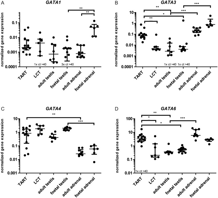
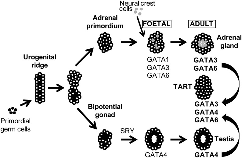
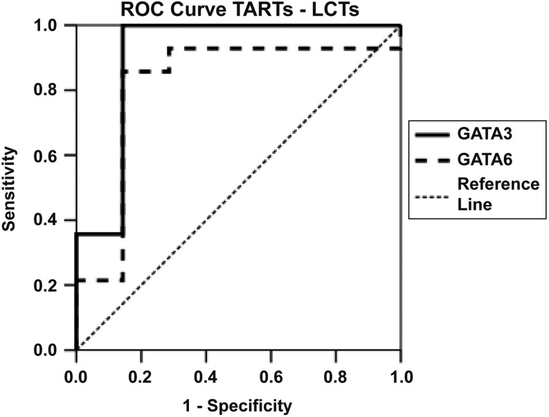
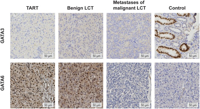
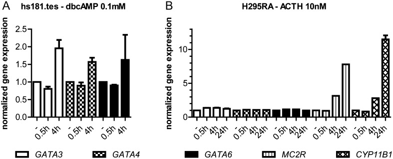
Testicular adrenal rest tumours (TARTs) are benign adrenal-like testicular tumours that frequently occur in male patients with congenital adrenal hyperplasia. Recently, GATA transcription factors have been linked to the development of TARTs in mice. The aim of our study was to determine GATA expression in human TARTs and other steroidogenic tissues. We determined GATA expression in TARTs (n = 16), Leydig cell tumours (LCTs; n = 7), adrenal (foetal (n = 6) + adult (n = 10)) and testis (foetal (n = 13) + adult (n = 8)). We found testis-like GATA4, and adrenal-like GATA3 and GATA6 gene expressions by qPCR in human TARTs, indicating mixed testicular and adrenal characteristics of TARTs. Currently, no marker is available to discriminate TARTs from LCTs, leading to misdiagnosis and incorrect treatment. GATA3 and GATA6 mRNAs exhibited excellent discriminative power (area under the curve of 0.908 and 0.816, respectively), while immunohistochemistry did not. GATA genes contain several CREB-binding sites and incubation with 0.1 mM dibutyryl cAMP for 4 h stimulated GATA3, GATA4 and GATA6 expressions in a human foetal testis cell line (hs181.tes). Incubation of adrenocortical cells (H295RA) with ACTH, however, did not induce GATA expression in vitro Although ACTH did not dysregulate GATA expression in the only human ACTH-sensitive in vitro model available, our results do suggest that aberrant expression of GATA transcription factors in human TARTs might be involved in TART formation.
Keywords: GATA transcription factors; Leydig cell tumour; congenital adrenal hyperplasia; testicular adrenal rest tumour.
© 2017 The authors.
Figures
Similar articles
-
Abundance of DLK1, differential expression of CYP11B1, CYP21A2 and MC2R, and lack of INSL3 distinguish testicular adrenal rest tumours from Leydig cell tumours.Eur J Endocrinol. 2015 Apr;172(4):491-9. doi: 10.1530/EJE-14-0810. Epub 2015 Jan 21. Eur J Endocrinol. 2015. PMID: 25609776
-
Bilateral testicular masses and adrenal insufficiency: is congenital adrenal hyperplasia the only possible diagnosis? First two cases of TARTS described in Addison-only X-linked adrenoleukodystrophy and a brief review of literature.J Endocrinol Invest. 2021 Mar;44(3):391-402. doi: 10.1007/s40618-020-01362-x. Epub 2020 Jul 20. J Endocrinol Invest. 2021. PMID: 32691371 Review.
-
Leydig Cell Tumor Associated with Testicular Adrenal Rest Tumors in a Patient with Congenital Adrenal Hyperplasia due to 11β-Hydroxylase Deficiency.Case Rep Urol. 2012;2012:648643. doi: 10.1155/2012/648643. Epub 2012 Feb 12. Case Rep Urol. 2012. PMID: 22606635 Free PMC article.
-
Risk factors for testicular adrenal rest tumors in pediatric patients with congenital adrenal hyperplasia.J Pediatr Urol. 2023 Aug;19(4):398.e1-398.e7. doi: 10.1016/j.jpurol.2023.03.028. Epub 2023 Mar 24. J Pediatr Urol. 2023. PMID: 37029011
-
Testicular Adrenal Rest Tumors: Current Insights on Prevalence, Characteristics, Origin, and Treatment.Endocr Rev. 2019 Aug 1;40(4):973-987. doi: 10.1210/er.2018-00258. Endocr Rev. 2019. PMID: 30882882 Review.
Cited by
-
Interpretation of Steroid Biomarkers in 21-Hydroxylase Deficiency and Their Use in Disease Management.J Clin Endocrinol Metab. 2023 Aug 18;108(9):2154-2175. doi: 10.1210/clinem/dgad134. J Clin Endocrinol Metab. 2023. PMID: 36950738 Free PMC article. Review.
-
Extensive Bilateral Adrenal Rest Testicular Tumors in a Patient With 3β-Hydroxysteroid Dehydrogenase Type 2 Deficiency.J Endocr Soc. 2018 May 1;2(6):513-517. doi: 10.1210/js.2018-00082. eCollection 2018 Jun 1. J Endocr Soc. 2018. PMID: 29850650 Free PMC article.
-
Case Report:clinical experience of bilateral giant pediatric Testicular adrenal rest tumors with 3 Beta-Hydroxysteroid Dehydrogenase-2 family history.BMC Pediatr. 2021 Sep 15;21(1):405. doi: 10.1186/s12887-021-02883-x. BMC Pediatr. 2021. PMID: 34526000 Free PMC article.
-
Transcriptional comparison of testicular adrenal rest tumors with fetal and adult tissues.Eur J Endocrinol. 2022 Sep 29;187(5):607-615. doi: 10.1530/EJE-22-0143. Print 2022 Nov 1. Eur J Endocrinol. 2022. PMID: 36047744 Free PMC article.
-
Morphologic and Molecular Characterization of Adrenals and Adrenal Rest Affected by Congenital Adrenal Hyperplasia.Front Endocrinol (Lausanne). 2021 Sep 20;12:730947. doi: 10.3389/fendo.2021.730947. eCollection 2021. Front Endocrinol (Lausanne). 2021. PMID: 34616364 Free PMC article.
References
-
- Stikkelbroeck NM, Otten BJ, Pasic A, Jager GJ, Sweep CG, Noordam K, Hermus AR. High prevalence of testicular adrenal rest tumors, impaired spermatogenesis, and Leydig cell failure in adolescent and adult males with congenital adrenal hyperplasia. Journal of Clinical Endocrinology and Metabolism 2001. 86 5721–5728. (10.1210/jcem.86.12.8090) - DOI - PubMed
-
- Claahsen-van der Grinten HL, Otten BJ, Takahashi S, Meuleman EJ, Hulsbergen-van de Kaa C, Sweep FC, Hermus AR. Testicular adrenal rest tumors in adult males with congenital adrenal hyperplasia: evaluation of pituitary-gonadal function before and after successful testis-sparing surgery in eight patients. Journal of Clinical Endocrinology and Metabolism 2007. 92 612–615. (10.1210/jc.2006-1311) - DOI - PubMed
-
- Clark RV, Albertson BD, Munabi A, Cassorla F, Aguilera G, Warren DW, Sherins RJ, Loriaux DL. Steroidogenic enzyme activities, morphology, and receptor studies of a testicular adrenal rest in a patient with congenital adrenal hyperplasia. Journal of Clinical Endocrinology and Metabolism 1990. 70 1408–1413. (10.1210/jcem-70-5-1408) - DOI - PubMed
-
- Claahsen-van der Grinten HL, Otten BJ, Sweep FC, Span PN, Ross HA, Meuleman EJ, Hermus AR. Testicular tumors in patients with congenital adrenal hyperplasia due to 21-hydroxylase deficiency show functional features of adrenocortical tissue. Journal of Clinical Endocrinology and Metabolism 2007. 92 3674–3680. (10.1210/jc.2007-0337) - DOI - PubMed
LinkOut - more resources
Full Text Sources
Other Literature Sources
