. 2017 Oct;5(19):e13439.
doi: 10.14814/phy2.13439. Epub 2017 Oct 16.

Local production of neurostradiol affects gonadotropin-releasing hormone (GnRH) secretion at mid-gestation in Lagostomus maximus (Rodentia, Caviomorpha)

Affiliations

Local production of neurostradiol affects gonadotropin-releasing hormone (GnRH) secretion at mid-gestation in Lagostomus maximus (Rodentia, Caviomorpha)

Santiago E Charif et al. Physiol Rep. 2017 Oct.

Abstract

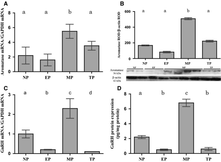
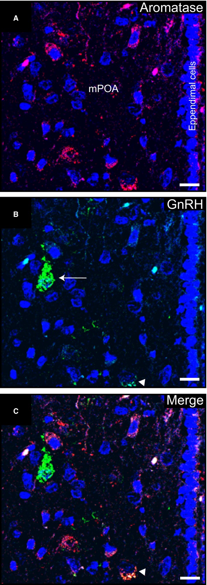
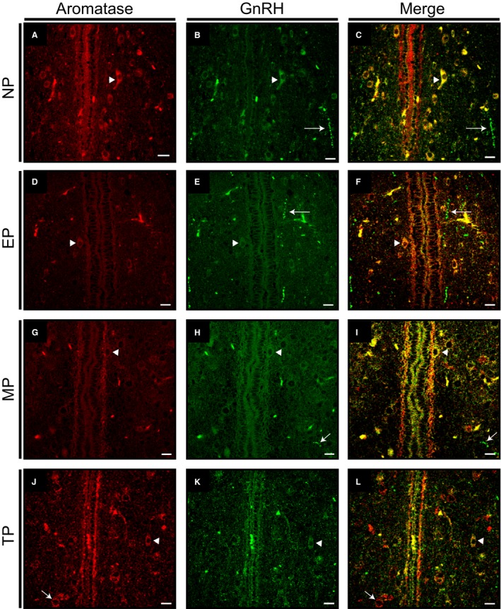
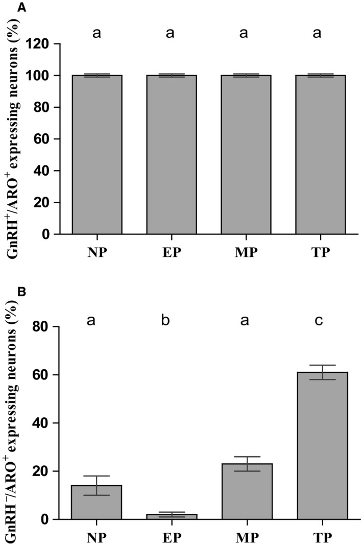
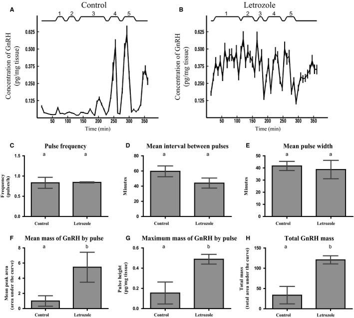
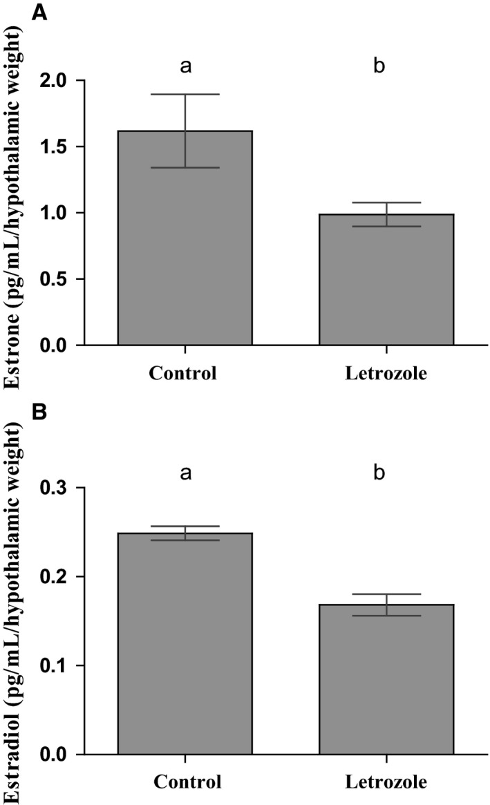
Females of the South American plains vizcacha, Lagostomus maximus, show peculiar reproductive features such as massive polyovulation up to 800 oocytes per estrous cycle and an ovulatory process around mid-gestation arising from the reactivation of the hypothalamic-hypophyseal-ovary (H.H.O.) axis. Estradiol (E2) regulates gonadotropin-releasing hormone (GnRH) expression. Biosynthesis of estrogens results from the aromatization of androgens by aromatase, which mainly occurs in the gonads, but has also been described in the hypothalamus. The recently described correlation between GnRH and ERα expression patterns in the hypothalamus of the vizcacha during pregnancy, with coexpression in the same neurons of the medial preoptic area, suggests that hypothalamic synthesis of E2 may affect GnRH neurons and contribute with systemic E2 to modulate GnRH delivery during the gestation. To elucidate this hypothesis, hypothalamic expression and the action of aromatase on GnRH release were evaluated in female vizcachas throughout pregnancy. Aromatase and GnRH expression was increased significantly in mid-pregnant and term-pregnant vizcachas compared to early-pregnant and nonpregnant females. In addition, aromatase and GnRH were colocalized in neurons of the medial preoptic area of the hypothalamus throughout gestation. The blockage of the negative feedback of E2 induced by the inhibition of aromatase resulted in a significant increment of GnRH-secreted mass by hypothalamic explants. E2 produced in the same neurons as GnRH may drive intracellular E2 to higher levels than those obtained from systemic circulation alone. This may trigger for a prompt GnRH availability enabling H.H.O. activity at mid-gestation with ovulation and formation of accessory corpora lutea with steroidogenic activity that produce the necessary progesterone to maintain gestation to term and guarantee the reproductive success.

Keywords: LH; Lagostomus maximus; Estradiol; GnRH; reproduction.


Figures

Figure 1
Figure 1
Hypothalamic aromatase expression during gestation is correlated with GnRH expression. Aromatase mRNA and protein expressions normalized to GAPDH and β‐actin, respectively, show a significant increase in MP with respect to the other groups (A and B). Representative images of aromatase and β‐actin blots obtained by Western blot are shown (B). GnRH mRNA and protein expression, with GAPDH or total protein expression normalization, respectively, show a significant increase in MP when compared to the other groups (C and D). NP: nonpregnant, EP: early‐pregnant, MP: mid‐pregnant, TP: term‐pregnant females, ROD: relative optical density. Different letters in bars indicate significant differences (P < 0.05) among groups. N = 5 or 6 per group and per technique.
Figure 2
Figure 2
Immunolocalization of aromatase and GnRH in the medial preoptic area of a nonpregnant vizcacha. (A) Representative images of the medial preoptic area (mPOA) showing cytoplasmic aromatase immunoreactive neurons (red). (B) GnRH immunoreactive neurons (green) in the same optic area shown in A with low (arrowhead) or high (arrow) GnRH labeling. (C) Merged image showing expression of aromatase in a GnRH low‐expressing neuron (arrowhead), and a GnRH high‐expressing neuron (green) surrounded by aromatase‐expressing neurons (red). Nuclei were counterstained with DAPI (blue). Scale bar: 10 μm. N = 5.
Figure 3
Figure 3
Aromatase and GnRH colocalization in the medial preoptic area of the hypothalamus throughout different reproductive stages. (A, D, G, J) Representative images of the medial preoptic area (mPOA) showing cytoplasmic aromatase immunoreactive neurons (red, arrowhead). (B, E, H, K) Low labeling GnRH immunoreactive neurons (green, arrowhead) in the same optic area shown in A, D, G, and J, respectively. (C, F, I, L) Merged images showing expression of aromatase in GnRH low‐expressing neurons (yellow, arrowhead) and in neurons lacking GnRH immunoreactivity (red). (L) Neurons showing only aromatase expression could be observed at term pregnancy (red, arrow). GnRH immunoreactive axonal varicosities distributed in the mPOA could be observed in all groups (arrows). NP, nonpregnant; EP, early‐pregnant; MD, mid‐pregnant; TP, term‐pregnant vizcachas. N = 5 per group. Scale bars: 10 μm
Figure 4
Figure 4
Quantification of cells with aromatase and GnRH colocalization. (A) Quantification of aromatase and GnRH colocalization in cells of POA showing no differences in the percentage of GnRH cells with aromatase immunoreactivity throughout the reproductive cycle (GnRH+/ARO+). (B) Significant increase in the percentage of aromatase cells without GnRH immunoreactivity with pregnancy progression (GnRH /ARO+). Different letters indicate significant differences among groups with P < 0.05. NP, nonpregnant; EP, early‐pregnant; MP, mid‐pregnant; TP, term‐pregnant females. N = 5 per group.
Figure 5
Figure 5
Neuroestradiol is involved in GnRH hypothalamic release. Representative GnRH pulsatile profiles from female hypothalamic explants control (A) and supplemented with letrozole (B), determined by radioimmunoassay (RIA). Five pulses were detected in both conditions during 6 h of treatment. The number of each pulse is indicated above the graph, whereas the bottom line indicates the time lapse of the GnRH release study. Several pulsatil parameters were evaluated: Pulse frequency (C), mean interval between pulses (D), mean pulse width (E), mean mass of GnRH delivered by pulse (mean area of the peaks (F), the maximum mass of GnRH secreted by pulse (mean pulse height), (G), and the total GnRH mass secreted during the 6 h of the experiment (under curve area), (H). Significant increase was determined in maximum mass of GnRH delivered by pulse, the mass of GnRH secreted by pulse, and total GnRH delivered in letrozole‐supplemented hypothalamus related to control. Different letters indicate significant differences between groups with P < 0.05. N = 5 per group.
Figure 6
Figure 6
Hypothalamic synthesis and delivery of estrone and estradiol is decreased by letrozole. Hypothalamic synthesis and delivery of E1 (A) and E2 (B) was significantly decreased by inhibition of aromatase activity as a result of treatment with the specific inhibitor letrozole. E1 and E2 content was expressed as pg/mL/hypothalamic weight in order to normalize steroid content to the weight of the corresponding hypothalamic explant. For each evaluated group, values represent the average of E1 or E2 in the incubation media corresponding to the aliquots where each GnRH pulse showed its maximum height. Different letters indicate significant differences between groups with P < 0.05. N = 5 per group.

Similar articles

Cited by

References

    1. Abraham, G. E. , Odell W. D., Swerdloff R. S., and Hopper K.. 1972. Simultaneous radioimmunoassay of plasma FSH, LH, progesterone, 17‐hydroxyprogesterone and estradiol‐17β during the menstrual cycle. J. Clin. Endocrinol. Metab. 34:312–318. - PubMed
    1. Abrahám, I. M. , Todman M. G., Korach K. S., and Herbison A. E.. 2004. Critical in vivo roles for classical estrogen receptors in rapid estrogen actions on intracellular signaling in mouse brain. Endocrinol. 145:3055–3061. - PubMed
    1. Balthazart, J. , Baillien M., Charlier T. D., and Ball G. F.. 2003. Calcium dependent phosphorylation processes control brain aromatase in quail. Eur. J. Neurosci. 17:1591–1606. - PubMed
    1. Boukhliq, R. , Goodman R. L., Berriman S. J., Adrian B., and Lehman M. N.. 1999. A subset of gonadotropin‐releasing hormone neurons in the ovine medial basal hypothalamus is activated during increased pulsatile luteinizing hormone secretion. Endocrinol. 140:5929–5936. - PubMed
    1. Bradford, M. M. 1976. Rapid and sensitive method for the quantitation of microgram quantities of protein utilizing the principle of protein‐dye binding. Anal. Biochem. 72:248–254. - PubMed

LinkOut - more resources