

Non-steroidal anti-inflammatory drug delays corneal wound healing by reducing production of 12-hydroxyheptadecatrienoic acid, a ligand for leukotriene B4 receptor 2
- PMID: 29038497
- PMCID: PMC5643301
- DOI: 10.1038/s41598-017-13122-8
Non-steroidal anti-inflammatory drug delays corneal wound healing by reducing production of 12-hydroxyheptadecatrienoic acid, a ligand for leukotriene B4 receptor 2
Abstract
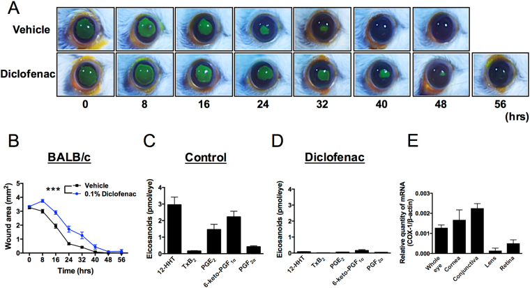
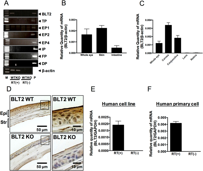
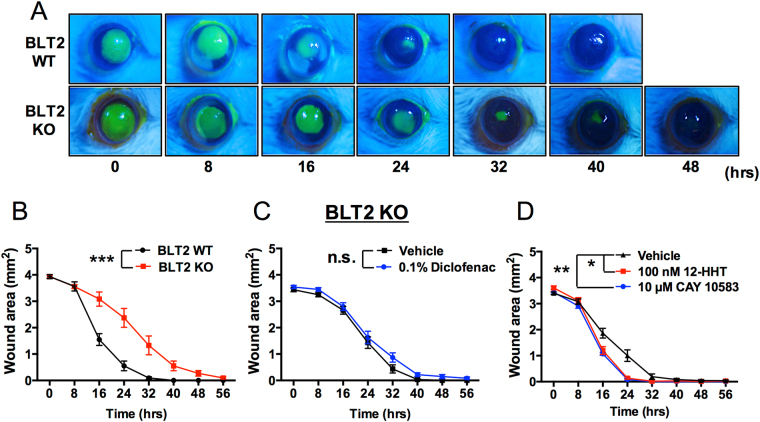
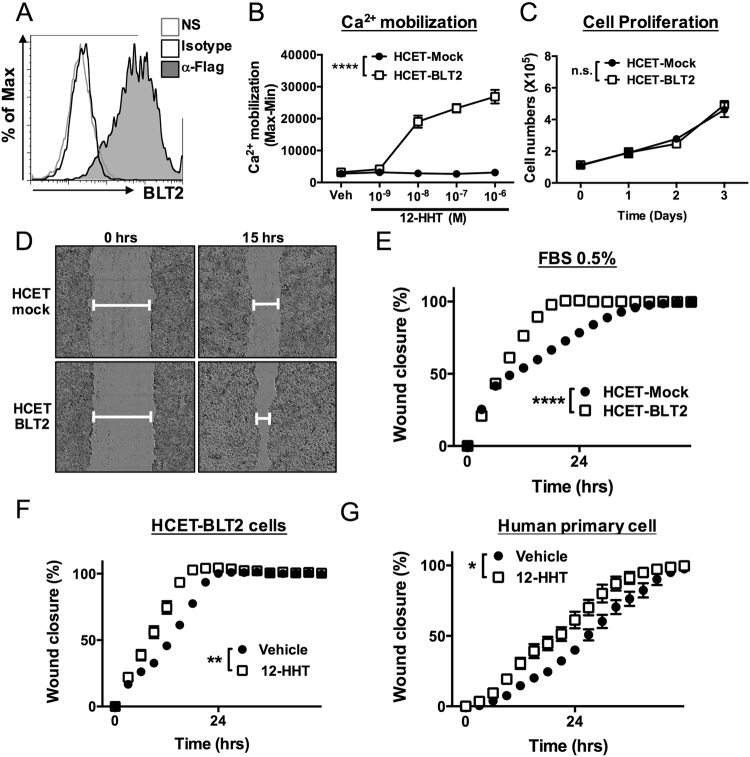
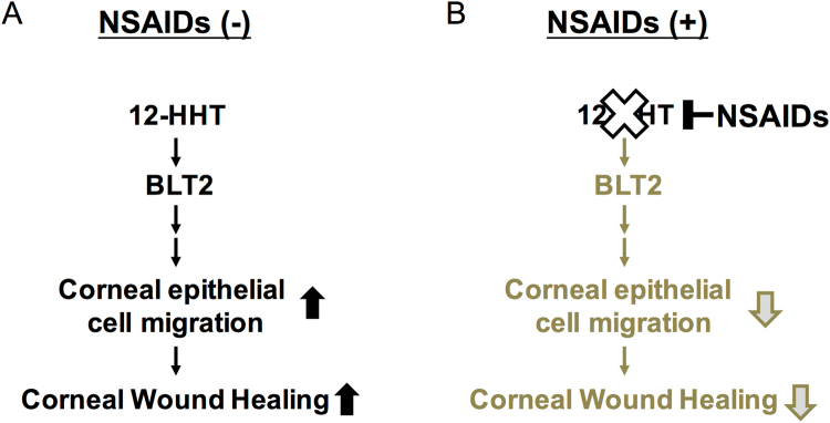
Non-steroidal anti-inflammatory drugs (NSAIDs) are widely used to reduce inflammation by suppressing cyclooxygenases (COXs). NSAID eye drops are frequently prescribed after ocular surgery to reduce inflammation and pain, but this treatment has clinically significant side effects, including corneal ulcer and perforation. The molecular mechanisms underlying these side effects remain unknown. Recently, the COX product 12(S)-hydroxyheptadeca-5Z,8E,10E-trienoic acid (12-HHT) was identified as an endogenous ligand for leukotriene B4 receptor 2 (BLT2), which is important in maintenance of epithelial homeostasis. We hypothesized that NSAID-dependent corneal damage is caused by reduced production of 12-HHT. Diclofenac eye drops decreased the abundance of downstream products of COX and delayed corneal wound healing in BALB/c mice. Expression of BLT2 was observed in murine ocular tissues including cornea, and in human corneal epithelial cell line and human primary corneal epithelial cells. In BLT2-knockout mice, corneal wound healing was delayed, but the diclofenac-dependent delay in corneal wound healing disappeared. 12-HHT accelerated wound closure both in BLT2-transfected corneal cell line and human primary corneal epithelial cells. Thus, our results reveal that NSAIDs delay corneal wound healing by inhibiting 12-HHT production, and suggest that stimulation of the 12-HHT/BLT2 axis represents a novel therapeutic approach to corneal wound healing.
Conflict of interest statement
The authors declare that they have no competing interests.
Figures
Similar articles
-
12-Hydroxyheptadecatrienoic acid promotes epidermal wound healing by accelerating keratinocyte migration via the BLT2 receptor.J Exp Med. 2014 Jun 2;211(6):1063-78. doi: 10.1084/jem.20132063. Epub 2014 May 12. J Exp Med. 2014. PMID: 24821912 Free PMC article.
-
Biological functions of 12(S)-hydroxyheptadecatrienoic acid as a ligand of leukotriene B4 receptor 2.Inflamm Regen. 2018 Oct 29;38:29. doi: 10.1186/s41232-018-0087-4. eCollection 2018. Inflamm Regen. 2018. PMID: 30397418 Free PMC article. Review.
-
12(S)-hydroxyheptadeca-5Z,8E,10E-trienoic acid (12-HHT) induces cell growth and improves barrier function through BLT2 interaction in intestinal epithelial Caco-2 cell cultures.Biochem Pharmacol. 2021 Aug;190:114663. doi: 10.1016/j.bcp.2021.114663. Epub 2021 Jun 20. Biochem Pharmacol. 2021. PMID: 34161796
-
Metabolism and biological functions of 12(S)-hydroxyheptadeca-5Z,8E,10E-trienoic acid.Prostaglandins Other Lipid Mediat. 2021 Feb;152:106502. doi: 10.1016/j.prostaglandins.2020.106502. Epub 2020 Oct 16. Prostaglandins Other Lipid Mediat. 2021. PMID: 33075476 Review.
-
Leukotriene B4 receptor BLT2 negatively regulates allergic airway eosinophilia.FASEB J. 2013 Aug;27(8):3306-14. doi: 10.1096/fj.12-217000. Epub 2013 Apr 19. FASEB J. 2013. PMID: 23603839
Cited by
-
Two distinct forms of human BLT2: long-form and short-form BLT2.Front Cell Dev Biol. 2023 Oct 26;11:1288373. doi: 10.3389/fcell.2023.1288373. eCollection 2023. Front Cell Dev Biol. 2023. PMID: 37954206 Free PMC article. Review.
-
Regenerative Therapy for Corneal Scarring Disorders.Biomedicines. 2024 Mar 14;12(3):649. doi: 10.3390/biomedicines12030649. Biomedicines. 2024. PMID: 38540264 Free PMC article. Review.
-
A Method for Real-Time Assessment of Mitochondrial Respiration Using Murine Corneal Biopsy.Invest Ophthalmol Vis Sci. 2023 Aug 1;64(11):33. doi: 10.1167/iovs.64.11.33. Invest Ophthalmol Vis Sci. 2023. PMID: 37642632 Free PMC article.
-
Combined Therapy Using Human Corneal Stromal Stem Cells and Quiescent Keratocytes to Prevent Corneal Scarring after Injury.Int J Mol Sci. 2022 Jun 23;23(13):6980. doi: 10.3390/ijms23136980. Int J Mol Sci. 2022. PMID: 35805991 Free PMC article.
-
Efficacy of low-level laser therapy in pain management after root canal treatment or retreatment: a systematic review.Lasers Med Sci. 2019 Sep;34(7):1305-1316. doi: 10.1007/s10103-019-02793-6. Epub 2019 May 1. Lasers Med Sci. 2019. PMID: 31044364
References
Publication types
MeSH terms
Substances
LinkOut - more resources
Full Text Sources
Other Literature Sources
Medical
Molecular Biology Databases
