. 2017 Oct 16;7(1):13250.
doi: 10.1038/s41598-017-13753-x.

Punctate White Matter Lesions Associated With Altered Brain Development And Adverse Motor Outcome In Preterm Infants

Affiliations

Punctate White Matter Lesions Associated With Altered Brain Development And Adverse Motor Outcome In Preterm Infants

Nora Tusor et al. Sci Rep. .

Abstract

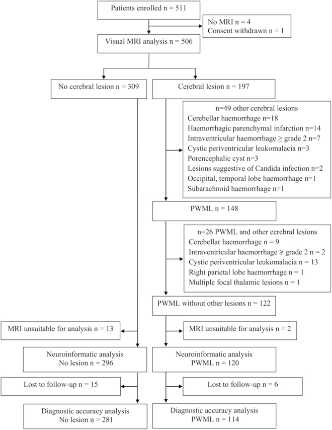
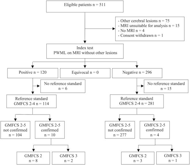
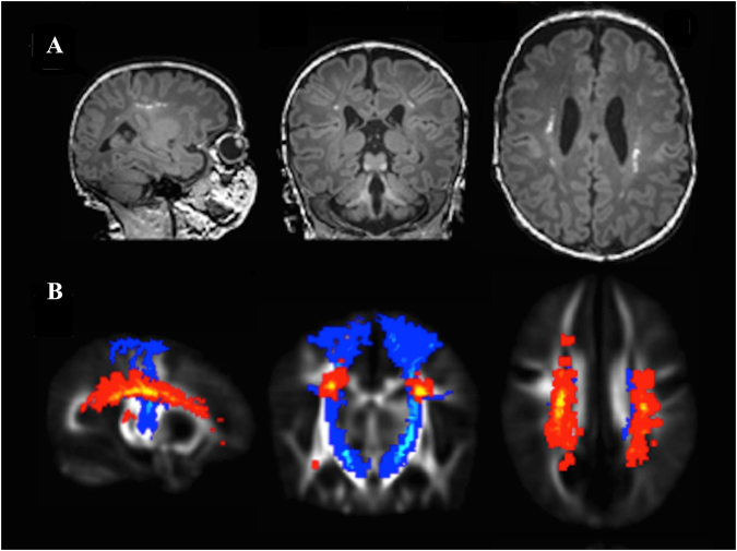
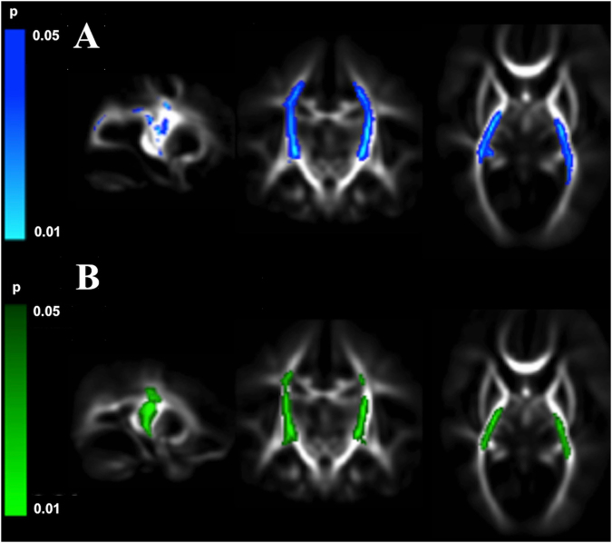
Preterm infants who develop neurodevelopmental impairment do not always have recognized abnormalities on cerebral ultrasound, a modality routinely used to assess prognosis. In a high proportion of infants, MRI detects punctate white matter lesions that are not seen on ultrasonography. To determine the relation of punctate lesions to brain development and early neurodevelopmental outcome we used multimodal brain MRI to study a large cohort of preterm infants. Punctate lesions without other focal cerebral or cerebellar lesions were detected at term equivalent age in 123 (24.3%) (59 male) of the 506 infants, predominantly in the centrum semiovale and corona radiata. Infants with lesions had higher gestational age, birth weight, and less chronic lung disease. Punctate lesions showed a dose dependent relation to abnormalities in white matter microstructure, assessed with tract-based spatial statistics, and reduced thalamic volume (p < 0.0001), and predicted unfavourable motor outcome at a median (range) corrected age of 20.2 (18.4-26.3) months with sensitivity (95% confidence intervals) 71 (43-88) and specificity 72 (69-77). Punctate white matter lesions without associated cerebral lesions are common in preterm infants currently not regarded as at highest risk for cerebral injury, and are associated with widespread neuroanatomical abnormalities and adverse early neurodevelopmental outcome.


Conflict of interest statement

The authors declare that they have no competing interests.

Figures

Figure 1
Figure 1
CONSORT diagram. PWML, punctate white matter lesion.
Figure 2
Figure 2
STARD diagram. GMFCS, Gross Motor Classification System; PWML, punctate white matter lesion.
Figure 3
Figure 3
Typical location and distribution of punctate white matter lesions. (A) Punctate white matter lesions apparent as high signal focal lesions on magnetization-prepared rapid gradient echo image in a single infant. (B) Group-level probabilistic lesion map (yellow-red) in relation to the corticospinal tracts (blue) overlaid on a 40-week T2-weighted template. Images are displayed in sagittal, coronal and transverse views.
Figure 4
Figure 4
Punctate white matter lesions are associated with altered white matter microstructure. Mean fractional anisotropy maps in sagittal, coronal, and transverse views showing white matter regions where infants with punctate lesions had significantly (A). higher radial diffusivity (blue) and (B). lower fractional anisotropy (green) as assessed with tract-based spatial statistics.

Similar articles

Cited by

References

    1. Ancel PY, et al. Cerebral palsy among very preterm children in relation to gestational age and neonatal ultrasound abnormalities: the EPIPAGE cohort study. Pediatrics. 2006;117:828–835. doi: 10.1542/peds.2005-0091. - DOI - PubMed
    1. Hintz SR, et al. Neuroimaging and neurodevelopmental outcome in extremely preterm infants. Pediatrics. 2015;135:e32–42. doi: 10.1542/peds.2014-0898. - DOI - PMC - PubMed
    1. Wagenaar N, et al. Clinical Risk Factors for Punctate White Matter Lesions on Early Magnetic Resonance Imaging in Preterm Newborns. The Journal of pediatrics. 2017;182:34–40 e31. doi: 10.1016/j.jpeds.2016.11.073. - DOI - PubMed
    1. Leijser LM, et al. Brain imaging findings in very preterm infants throughout the neonatal period: part I. Incidences and evolution of lesions, comparison between ultrasound and MRI. Early human development. 2009;85:101–109. doi: 10.1016/j.earlhumdev.2008.11.010. - DOI - PubMed
    1. Miller SP, et al. Comparing the diagnosis of white matter injury in premature newborns with serial MR imaging and transfontanel ultrasonography findings. AJNR. American journal of neuroradiology. 2003;24:1661–1669. - PMC - PubMed

Publication types

LinkOut - more resources