. 2017 Oct 16;8(1):960.
doi: 10.1038/s41467-017-01075-5.

A structural model of flagellar filament switching across multiple bacterial species

Affiliations

A structural model of flagellar filament switching across multiple bacterial species

Fengbin Wang et al. Nat Commun. .

Abstract

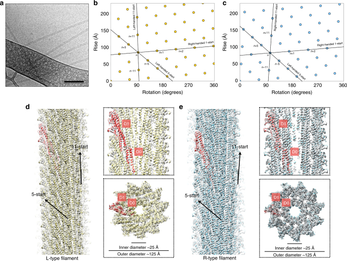
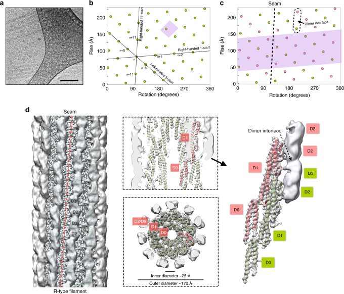
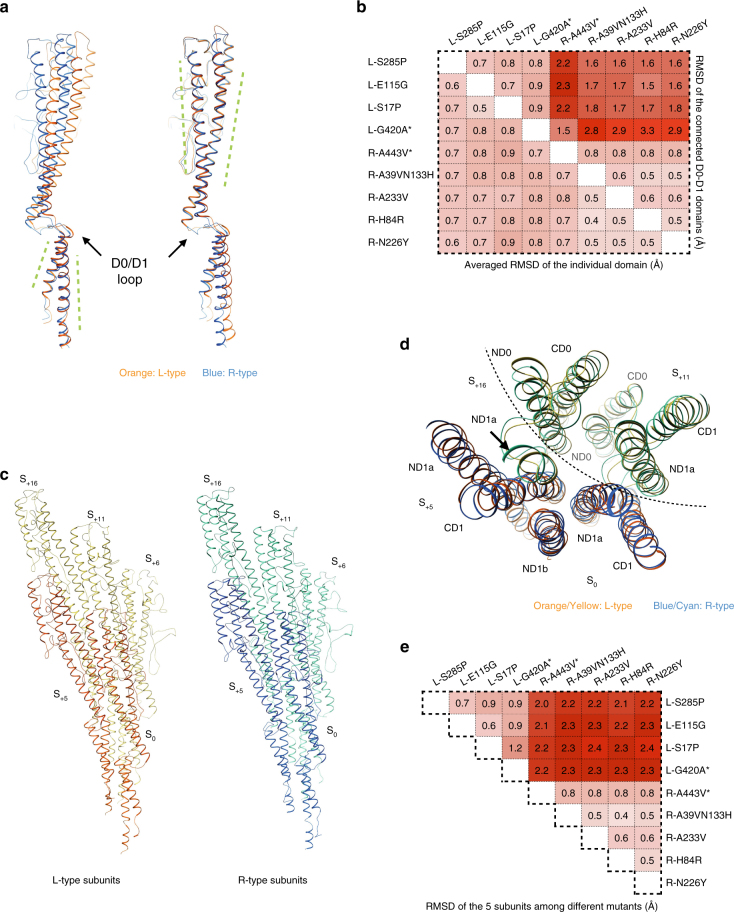
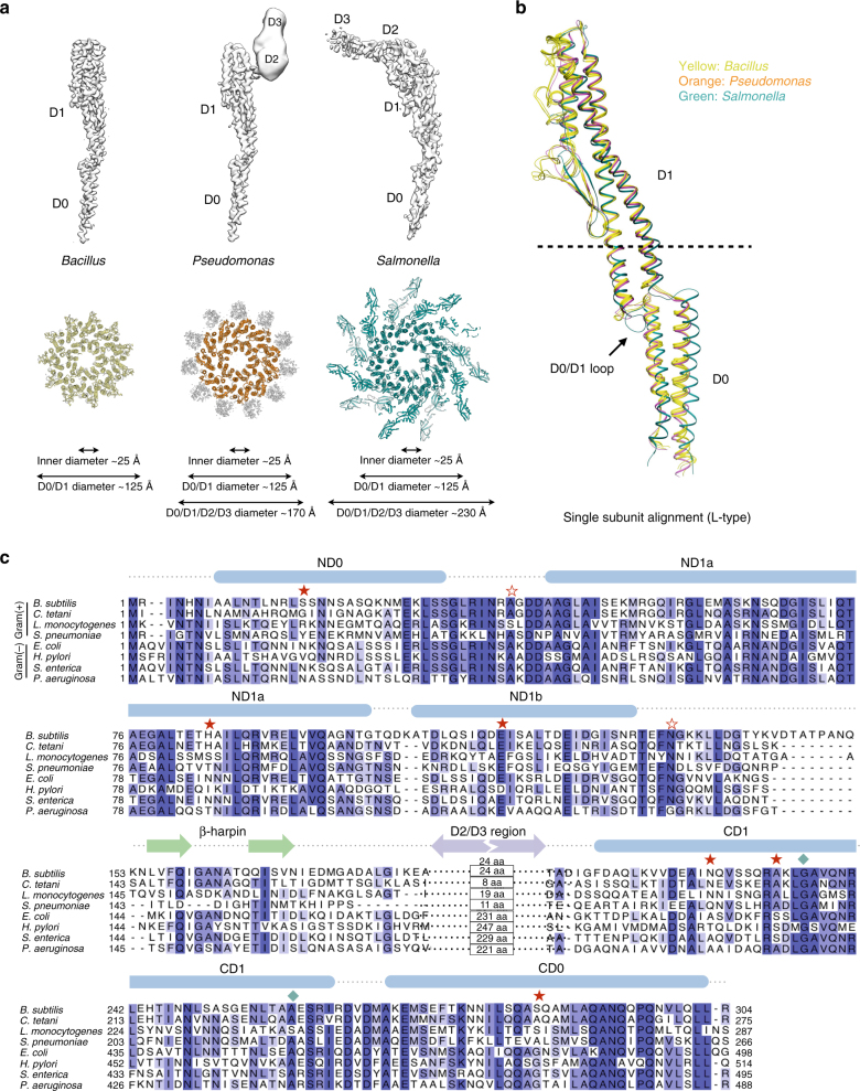
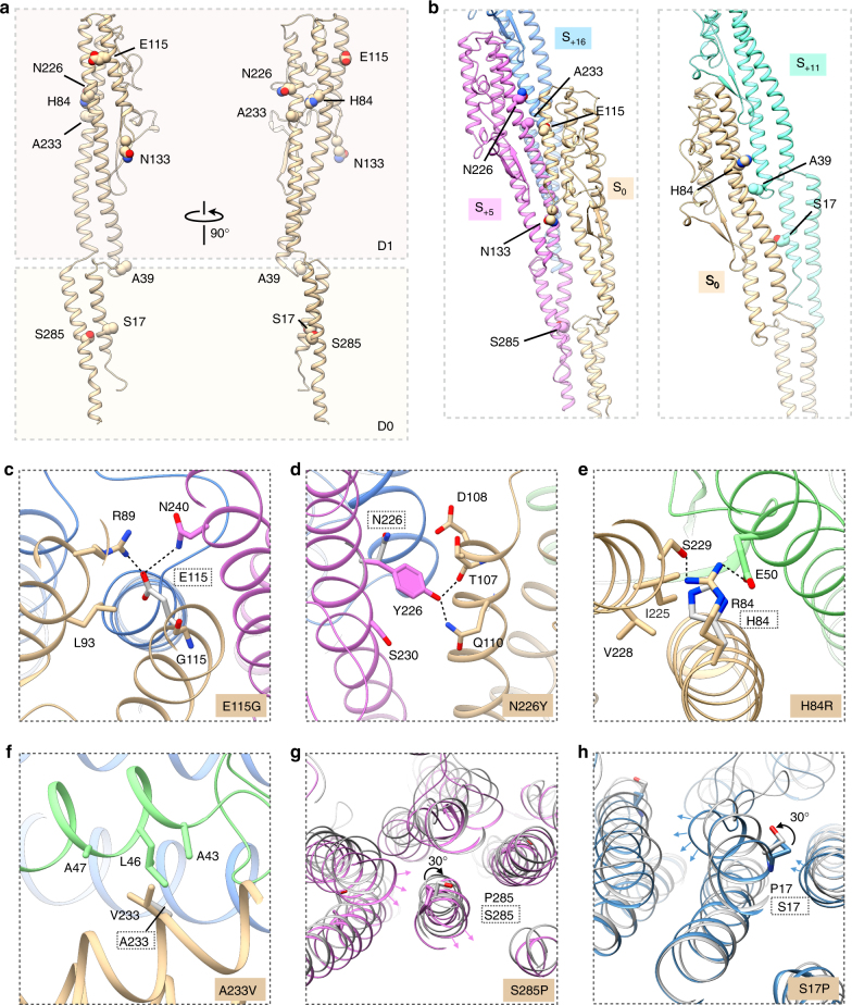
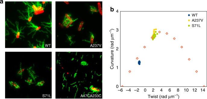
The bacterial flagellar filament has long been studied to understand how a polymer composed of a single protein can switch between different supercoiled states with high cooperativity. Here we present near-atomic resolution cryo-EM structures for flagellar filaments from both Gram-positive Bacillus subtilis and Gram-negative Pseudomonas aeruginosa. Seven mutant flagellar filaments in B. subtilis and two in P. aeruginosa capture two different states of the filament. These reliable atomic models of both states reveal conserved molecular interactions in the interior of the filament among B. subtilis, P. aeruginosa and Salmonella enterica. Using the detailed information about the molecular interactions in two filament states, we successfully predict point mutations that shift the equilibrium between those two states. Further, we observe the dimerization of P. aeruginosa outer domains without any perturbation of the conserved interior of the filament. Our results give new insights into how the flagellin sequence has been "tuned" over evolution.Bacterial flagellar filaments are composed almost entirely of a single protein-flagellin-which can switch between different supercoiled states in a highly cooperative manner. Here the authors present near-atomic resolution cryo-EM structures of nine flagellar filaments, and begin to shed light on the molecular basis of filament switching.


Conflict of interest statement

The authors declare no competing financial interests.

Figures

Fig. 1
Fig. 1
Cryo-EM reconstruction and flagellar filament model of B. subtilis. a Cryo-electron micrograph of B. subtilis flagellar filaments. The scale bar represents 100 nm. b and c The helical net of the B. subtilis flagellar filament using the convention that the surface is unrolled and we are looking from the outside. The L-type filaments are shown in b and the R-type filaments are shown in c. d and e The surfaces of the side view, the central slice through the lumen, and the top view of the cryo-EM reconstructions (L-type straight filament S285P shown in d, R-type straight filament N226Y shown in e)
Fig. 2
Fig. 2
Cryo-EM reconstruction and flagellar filament model of P. aeruginosa. a Cryo-electron micrograph of P. aeruginosa flagellar filament. The scale bar represents 100 nm. b The helical net of R-type flagellar filament core (D0 and D1 domain) of P. aeruginosa. c The helical net of D2/D3 domain in the R-type flagellar filament of P. aeruginosa. d The side view, the central slice through the lumen, the top view and the segmented dimer view of the cryo-EM reconstructions of the R-type flagellar filament A443V of P. aeruginosa
Fig. 3
Fig. 3
Comparison of the Cα backbones of L- and R- type structures. a Superposition of a single L-type (orange, S285P) and R-type (blue, N226Y) subunit of B. subtilis, by D0 domain (left) and D1 domain (right). b RMSD calculations of individual domains (bottom left) and RMSD calculations of the combined D0 plus D1 domains (upper right). Two P. aeruginosa mutants are labeled with an asterisk. The intensity of red corresponds to the RMSD level. c Five subunits of L-type (yellow and orange, S285P) and R-type (blue and cyan, N226Y) subunits, containing all unique contacts within the filaments. d Comparison of the relative arrangement of these five subunits from the horizontal plane (yellow and orange: L-type S285, blue and cyan: R-type N226Y). e RMSD calculations of the complex of these five subunits among all the nine filament mutants
Fig. 4
Fig. 4
Comparison with flagellar filaments of other bacterial species. a A comparison of L-type flagellar structures in B. subtilis (S285P), P. aeruginosa (G420A) and S. enterica (PDB: 3A5X, EMD-1641). A comparison of the segmented maps corresponding to a single subunit is shown on top. A diameter comparison from the top view of the filaments is shown on the bottom. b Superimposition of the single L-type subunits: three from B. subtilis (yellow, S285P, S17P, E115G), one from P. aeruginosa (orange, G420A) and one from S. enterica (dark green, 3A5X). The dashed line indicates where the crystal structure of S. enterica ends. c Alignments of the flagellin amino acid sequence from four gram-positive bacteria and four gram-negative bacteria. Single mutants in B. subtilis are marked with filled red stars, the double mutants are marked with empty red stars, and the mutants in P. aeruginosa are marked with filled blue squares
Fig. 5
Fig. 5
Mutation sites of B. subtilis and their molecular basis that lead to straight filaments. a Locating the mutation sites on the single flagellin subunit. b Locating the mutation sites on the 5-start interface (left) and the 11-start interface (right). cf Subunits S0, S+5, S+11 and S+16 are colored in dark yellow, purple, green and blue, respectively. Un-mutated residues are shown in gray, and adapted from the other mutants in the same hand. Mutation sites for: left-handed mutation E115G c, right-handed mutation N226Y d, right-handed mutation H84R e, and right-handed mutation A233V f. g Top view of two mutant structures N226Y (gray) and S285P (purple) aligned by upper part of D0 domain (amino acids 18–32 and 268–284). h Top view of two mutant structures N226Y (gray) and S17P (blue) aligned by the same upper part of D0 domain
Fig. 6
Fig. 6
Predicted mutations of B. subtilis flagella filaments. a Fluorescence images of wildtype, curly form mutations A237V and S71L, and relatively straight form disulfide double mutant A47CA233C. b Plot of curvature κ against twist τ for measured helical forms of those B. subtilis mutants. The 12 discrete points corresponding to different theoretical waveforms in S. enterica are shown by empty red squares

References

    1. Macnab RM. How bacteria assemble flagella. Annual Reviews in Microbiology. 2003;57:77–100. doi: 10.1146/annurev.micro.57.030502.090832. - DOI - PubMed
    1. Namba K, Vonderviszt F. Molecular architecture of bacterial flagellum. Q. Rev. Biophys. 1997;30:1–65. doi: 10.1017/S0033583596003319. - DOI - PubMed
    1. Kearns DB. A field guide to bacterial swarming motility. Nat. Rev. Microbiol. 2010;8:634–644. doi: 10.1038/nrmicro2405. - DOI - PMC - PubMed
    1. Ramos HC, Rumbo M, Sirard JC. Bacterial flagellins: mediators of pathogenicity and host immune responses in mucosa. Trends. Microbiol. 2004;12:509–517. doi: 10.1016/j.tim.2004.09.002. - DOI - PubMed
    1. Postel S, et al. Bacterial flagellar capping proteins adopt diverse oligomeric states. Elife. 2016;5:e18857. doi: 10.7554/eLife.18857. - DOI - PMC - PubMed

Publication types

LinkOut - more resources