. 2017 Nov;27(11):1309-1326.
doi: 10.1038/cr.2017.126. Epub 2017 Oct 17.

Intermittent fasting promotes adipose thermogenesis and metabolic homeostasis via VEGF-mediated alternative activation of macrophage

Affiliations

Intermittent fasting promotes adipose thermogenesis and metabolic homeostasis via VEGF-mediated alternative activation of macrophage

Kyoung-Han Kim et al. Cell Res. 2017 Nov.

Abstract

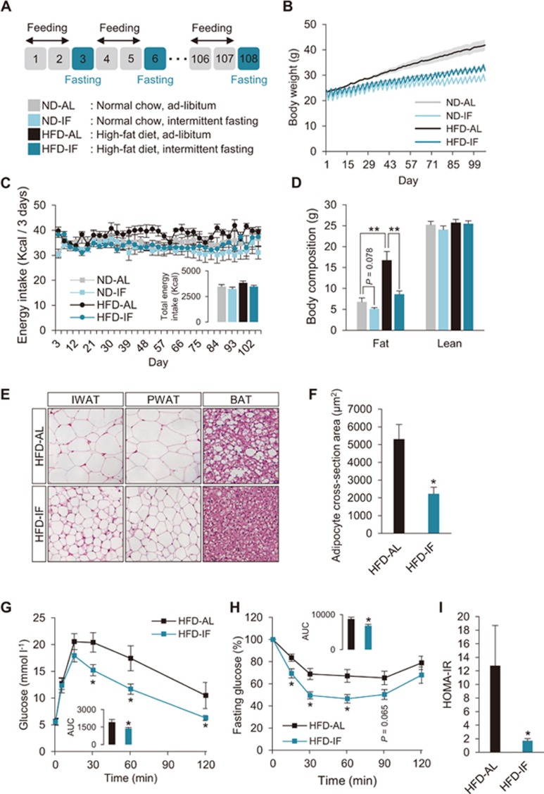
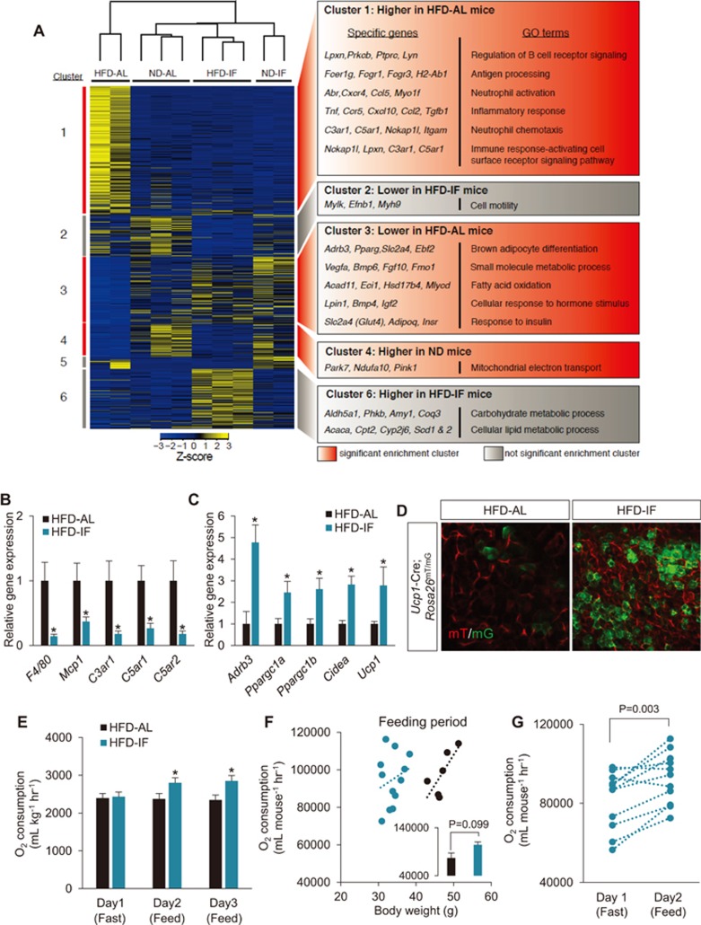
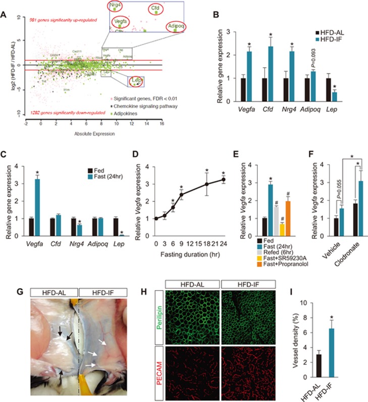
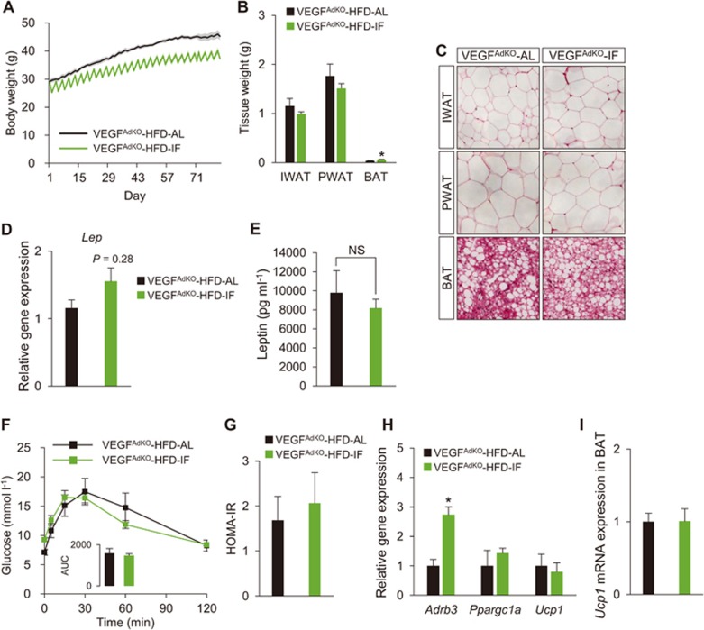
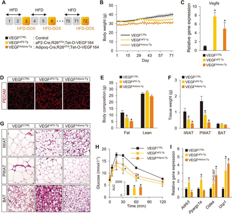
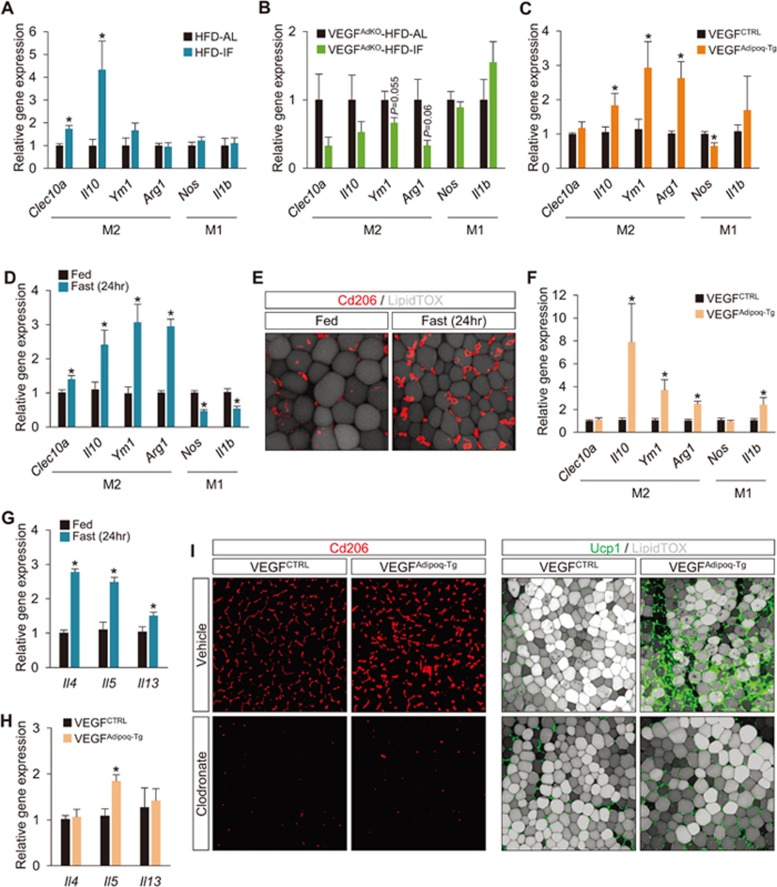
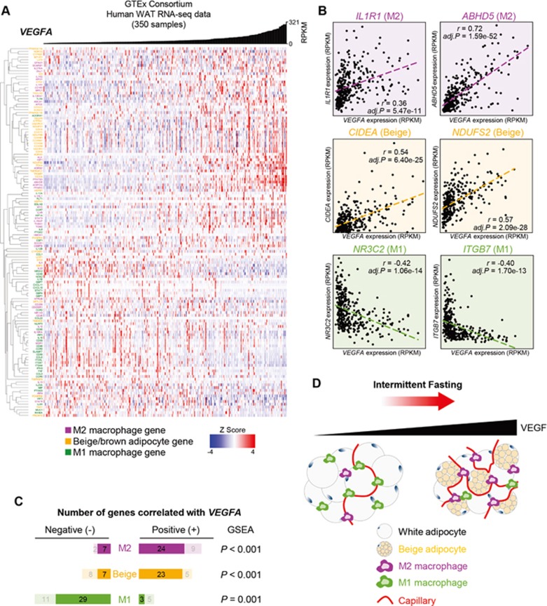
Intermittent fasting (IF), a periodic energy restriction, has been shown to provide health benefits equivalent to prolonged fasting or caloric restriction. However, our understanding of the underlying mechanisms of IF-mediated metabolic benefits is limited. Here we show that isocaloric IF improves metabolic homeostasis against diet-induced obesity and metabolic dysfunction primarily through adipose thermogenesis in mice. IF-induced metabolic benefits require fasting-mediated increases of vascular endothelial growth factor (VEGF) expression in white adipose tissue (WAT). Furthermore, periodic adipose-VEGF overexpression could recapitulate the metabolic improvement of IF in non-fasted animals. Importantly, fasting and adipose-VEGF induce alternative activation of adipose macrophage, which is critical for thermogenesis. Human adipose gene analysis further revealed a positive correlation of adipose VEGF-M2 macrophage-WAT browning axis. The present study uncovers the molecular mechanism of IF-mediated metabolic benefit and suggests that isocaloric IF can be a preventive and therapeutic approach against obesity and metabolic disorders.


Figures

Figure 1
Figure 1
IF protects mice from diet-induced obesity and metabolic dysfunction. (A) Schematic illustration of the 2:1 IF regimen. (B) Body weight measurement during 16 weeks of IF. (C) Changes of energy intake during 16 weeks of IF cycles. The inset shows total energy intake during 16 weeks of IF cycles. (D) Body composition showing fat and lean mass. (E) H&E-stained sections of adipose tissues; IWAT (subcutaneous), PWAT (visceral), and BAT. (F) Average of cross-sectioned area of subcutaneous white adipocytes, revealing reduced white adipocyte size for HFD-IF mice compared to HFD-AL mice. (G) GTT in HFD-AL and HFD-IF mice. The inset shows AUC. (H) ITT. The inset shows AUC. (I) HOMA-IR. Data are expressed as mean ± SEM (ND-AL: n = 7; ND-IF: n = 8; HFD-AL: n = 7; and HFD-IF: n = 8); one or two-way ANOVA with Student-Newman-Keuls post hoc analysis and two-tailed unpaired Student's t-test; *P < 0.05 and **P < 0.01 vs HFD-AL. AL, ad libitum; AUC, area under the curve; HFD, high-fat diet; ND, normal diet.
Figure 2
Figure 2
Transcriptome analysis upon IF reveals browning of WAT. (A) Heatmap displaying 3 644 differentially expressed genes among ND-AL, ND-IF, HFD-AL, and HFD-IF groups that were clustered into six distinct gene groups. Enriched GO terms and representative genes are shown at the right for each cluster. Significantly enriched clusters: red boxes. Not significantly enriched clusters: gray boxes. (B) Gene expression analysis on inflammation-related genes. (C) Marker gene expression of sympathetic activation (i.e., Adrb3) and brown/beige adipocyte (i.e., Ppargc1a, Ppargc1b, Cidea and Ucp1) (HFD, AL/IF: n = 6/8). (D) Representative images of whole-mount WAT of the brown/beige chaser (Ucp1-Cre;Rosa26mT/mG) mice subjected to AL and IF. Beige adipocytes are visualized by membrane-targeted GFP (mG; green). (E) O2 consumption during fasting and feeding condition. (F) Linear regression analysis of O2 consumption as a function of body weight during feeding period. The inset shows O2 consumption during feeding condition adjusted with body weight at 38.54 g using ANCOVA (HFD, AL/IF: n = 6/12). (G) Significant changes in O2 consumption between fasting and feeding periods in HFD-IF mice. Values are mean ± SEM; two-tailed unpaired and paired Student's t-test; *P < 0.05 vs HFD-AL.
Figure 3
Figure 3
Fasting induces adipose-VEGF expression. (A) MA plot highlighting significantly altered mRNA expression of adipose-derived factors in PWAT of HFD-IF mice, compared to that of HFD-AL mice. Vegfa, Cfd, Nrg4, Adipoq, and Lep encode vascular endothelial growth factor, adipsin, neuregulin 4, adiponectin, and leptin, respectively. (B) qPCR validation of adipose-derived factors in PWAT (HFD, AL/IF: n = 6/8). (C) mRNA expression levels of Vegfa, Cfd, Nrg4, Adipoq, and Lep in PWAT at feeding and 24 h of fasting (n = 5 per group). (D) Vegfa mRNA expression in PWAT at different fasting durations (n = 5-6 per group). (E) Vegfa mRNA expression in PWAT at feeding, 24 h of fasting, 6 h of refeeding, fasting with β3-AR antagonist, SR59230A (5 mg/kg, i.p.), and fasting with non-specific β-AR antagonist, Propranolol (5 mg/kg, i.p.; n = 5 per group). (F) Vegfa mRNA expression in PWAT at feeding and 24 h of fasting with treatments of vehicle or clodronate (n = 5). (G) A representative macroscopic image illustrating increased vascularization in IWAT of HFD-IF mice, compared to HFD-AL mice. Black and white arrows indicate IWAT of HFD-AL and HFD-IF mice, respectively. (H) Representative microscopic images of adipocytes and blood vessels, visualized with perilipin and PECAM-1 antibodies, respectively, in whole-mount PWAT. (I) Quantification of vessel densities in PWAT. Data are mean ± SEM; one- or two-way ANOVA with Student-Newman-Keuls post hoc analysis and two-tailed unpaired Student's t-test; *P < 0.05 vs HFD-AL or Fed. #P < 0.05 vs Fast (24 h).
Figure 4
Figure 4
Adipose-VEGF is required for IF-mediated metabolic benefits. (A) Body weight measurements of aP2-Cre;Vegfaflox/flox mice (VEGFAdKO) subjected to AL and IF under HFD feeding (VEGFAdKO-HFD, AL/IF: n = 5/6). (B) Tissue weight of IWAT, PWAT, and BAT in VEGFAdKO-HFD-AL and -IF mice. (C) H&E-stained sections of IWAT, PWAT, and BAT show no noticeable differences between VEGFAdKO-HFD-AL and -IF mice. (D) Lep mRNA expression of PWAT in VEGFAdKO-HFD mice. (E) Plasma leptin levels. (F) GTT in VEGFAdKO-HFD mice. (G) HOMA-IR in VEGFAdKO-HFD mice. (H) Gene expression analysis revealed that IF increased sympathetic activation (Adrb3), but did not affect brown/beige adipocyte maker expression (i.e., Ucp1) in PWAT of VEGFAdKO mice. (I) No changes in Ucp1 expression in BAT of VEGFAdKO mice upon IF. Data are mean ± SEM; two-tailed unpaired Student's t-test; *P < 0.05 vs VEGFAdKO-HFD-AL. Lep, leptin.
Figure 5
Figure 5
Intermittent adipose-VEGF overexpression is sufficient to mimic the IF-mediated metabolic benefits. (A) Schematic illustration of the 2:1 intermittent VEGF induction regimen and two different VEGF overexpression mouse models. (B) Body weight measurements during 10 weeks of adipose VEGF cyclic inductions. (C) Vegfa mRNA expression in PWAT at different fasting durations (n = 5-6 per group). (D) Representative microscopic images of PECAM-stained blood vessels in whole-mount PWAT. (E) Body composition showing fat and lean mass. (F) Tissue weight of IWAT, PWAT, and BAT. (G) H&E-stained sections of IWAT, PWAT, and BAT. (H) GTT. An insert graph shows AUC. (I) Gene expression analysis on WAT browning markers in PWAT. Data are expressed as mean ± SEM (VEGFCTRL: n = 10; VEGFaP2-Tg: n = 6; and VEGFAdipoq-Tg: n = 5); one- or two-way ANOVA with Student-Newman-Keuls post hoc analysis; *P < 0.05 vs VEGFCTRL.
Figure 6
Figure 6
Fasting and adipose-VEGF induce alternative activation of macrophage. (A) M1/M2 macrophage marker gene expression analysis in HFD-AL and -IF mice. (B) M1/M2 macrophage marker gene expression in VEGFAdKO-HFD-AL and -IF mice. (C) M1/M2 macrophage marker gene expression after intermittent adipose-VEGF upregulation in VEGFAdipoq-Tg mice. (D) M1/M2 macrophage marker gene expression analysis in fed and fasted (24 h) mice. (E) Representative images of M2 marker Cd206-stained cells in whole-mount PWAT of fed and fasted (24 h) mice. (F) M1/M2 macrophage marker gene expression analysis after acute adipose-VEGF upregulation in VEGFAdipoq-Tg mice. (G) Type 2 cytokine gene expression in fed and fasted (24 h) mice. (H) Type 2 cytokine gene expression after acute adipose-VEGF upregulation (48 h) in VEGFAdipoq-Tg mice. (I) Representative images of M2 macrophages and Ucp1 expression in WAT after acute adipose-VEGF upregulation in VEGFAdipoq-Tg mice with treatments of vehicle or clodronate. Values are mean ± SEM (post HFD-AL: n = 7 and post HFD-IF: n = 9); two-tailed unpaired Student's t-test; *P < 0.05 vs HFD-AL, VEGFAdKO-HFD-AL, VEGFCTRL or fed mice. See also Supplementary information, Figure S7.
Figure 7
Figure 7
VEGF expression in human WAT correlates with M2 macrophage and WAT browning. (A) A correlation heatmap of VEGFA gene with unsupervised hierarchical clustering of M1/M2 macrophages- and beige/brown adipocyte-associated genes in human WAT. A histogram of VEGFA gene expression level (RPKM) is shown on the top of the heatmap. (B) Representative scatter plots showing correlation of VEGFA with IL1R1 & ABHD5 (M2), CIDEA & NUDFS2 (beige), and NR3C2 & ITGB7 (M1) genes. (C) Summary of VEGFA correlation with M2, beige, and M1-associated genes. Permutation P-values with the GSEA are shown. (D) Schematic model of IF-mediated VEGF expression underlying adipose thermogenesis and M2 macrophage polarization. GSEA, gene set enrichment analysis.

Comment in

References

    1. Rutkowski JM, Stern JH, Scherer PE. The cell biology of fat expansion. J Cell Biol 2015; 208:501–512. - PMC - PubMed
    1. Stern JH, Scherer PE. Adipose tissue biology in 2014: advances in our understanding of adipose tissue homeostasis. Nat Rev Endocrinol 2015; 11:71–72. - PMC - PubMed
    1. Kajimura S, Spiegelman BM, Seale P. Brown and beige fat: physiological roles beyond heat generation. Cell Metab 2015; 22:546–559. - PMC - PubMed
    1. Wang W, Seale P. Control of brown and beige fat development. Nat Rev Mol Cell Biol 2016; 17:691–702. - PMC - PubMed
    1. Odegaard JI, Chawla A. Type 2 responses at the interface between immunity and fat metabolism. Curr Opin Immunol 2015; 36:67–72. - PMC - PubMed

Substances