. 2017 Dec;40(6):1869-1880.
doi: 10.3892/ijmm.2017.3166. Epub 2017 Sep 29.

Rivaroxaban attenuates thrombosis by targeting the NF-κB signaling pathway in a rat model of deep venous thrombus

Affiliations

Rivaroxaban attenuates thrombosis by targeting the NF-κB signaling pathway in a rat model of deep venous thrombus

Junhao Ma et al. Int J Mol Med. 2017 Dec.

Retraction in

Abstract

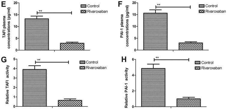
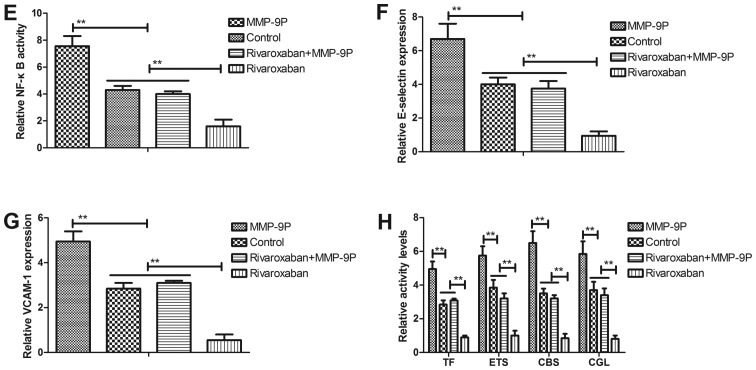
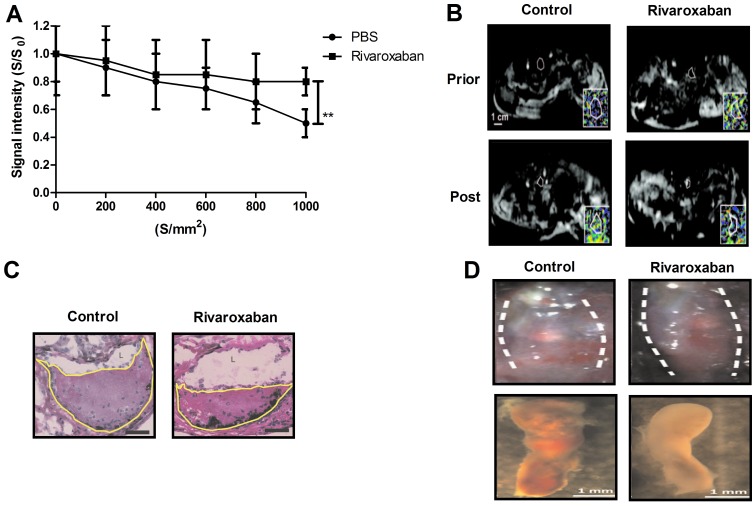
Anticoagulant therapy is commonly used for the prevention and treatment of patients with deep venous thrombus. Evidence has shown that rivaroxaban is a potential oral anticoagulant drug for the acute treatment of venous thromboembolism. However, the rivaroxaban-mediated molecular mechanism involved in the progression of deep venous thrombosis has not been investigated. In the present study, we investigated the efficacy of rivaroxaban and the underlying signaling pathways in the prevention and treatment of rats with deep venous thrombosis. A rat model with deep vein thrombus formation was established and received treatment with rivaroxaban or PBS as control. The thrombin-activatable fibrinolysis inhibitor (TAFI) and plasminogen activator inhibitor-1 (PAI-1) were analyzed both in vitro and in vivo. The progression of thrombosis and stroke was evaluated after treatment with rivaroxaban or PBS. Nuclear factor-κB (NF-κB) signaling pathway in venous endothelial cells and in the rat model of deep venous thrombus was assessed. The therapeutic effects of rivaroxaban were evaluated as determined by changes in deep venous thrombosis in the rat model. Our results showed that rivaroxaban markedly inhibited TAFI and PAI-1 expression levels, neutrophils, tissue factor, neutrophil extracellular traps (NETs), myeloperoxidase and macrophages in venous endothelial cells and in the rat model of deep venous thrombus. Expression levels of ADP, PAIs, von Willebrand factor (vWF) and thromboxane were downregulated in vein endothelial cells and in serum from the experimental rats. Importantly, the incidences of inferior vena cava filter thrombus were protected by rivaroxaban during heparin-induced thrombolysis deep venous thrombosis in the rat model. We observed that activity of the NF-κB signaling pathway was inhibited by rivaroxaban in vein endothelial cells both in vitro and in vivo. Notably, immunohistology indicated that rivaroxaban attenuated deep venous thrombosis and the accumulation of inflammatory factors in the lesions in venous thrombus. Matrix metalloproteinase (MMP) expression and activity were downregulated in rivaroxaban-treated rats with deep venous thrombus. Rivaroxaban inhibited the elasticity of the extracellular matrix and collagen-elastin fibers. On the whole, these results indicate that rivaroxaban attenuates deep venous thrombus through MMP-9-mediated NF-κB signaling pathway.


Figures

Figure 1
Figure 1
Expression and activity of thrombin-activatable fibrinolysis inhibitor (TAFI) and plasminogen activator inhibitor-1 (PAI-1) in vein endothelial cells and a rat model of deep venous thrombosis. (A and B) TAFI (A) and PAI-1 (B) protein expression levels after treatment with rivaroxaban in vein endothelial cells. (C and D) Activity of TAFI (C) and PAI-1 (D) was detected in vein endothelial cells after treatment with rivaroxaban or PBS. (E and F) Plasma concentration levels of TAFI (E) and PAI-1 (F) were decreased in serum in rivaroxaban-treated rat. (G and H) Activity of TAFI (G) and PAI-1 (H) was also decreased in rivaroxaban-treated rat. (I and J) Activity of fibrinolysis (I) and plasma concentration of fibrinolysis (J) in a rat model of deep venous thrombosis. All data are represented as means ± SEM of triplicate samples. One-way ANOVA revealed a significant effect. *P<0.05 and **P<0.01 vs. the control.
Figure 1
Figure 1
Expression and activity of thrombin-activatable fibrinolysis inhibitor (TAFI) and plasminogen activator inhibitor-1 (PAI-1) in vein endothelial cells and a rat model of deep venous thrombosis. (A and B) TAFI (A) and PAI-1 (B) protein expression levels after treatment with rivaroxaban in vein endothelial cells. (C and D) Activity of TAFI (C) and PAI-1 (D) was detected in vein endothelial cells after treatment with rivaroxaban or PBS. (E and F) Plasma concentration levels of TAFI (E) and PAI-1 (F) were decreased in serum in rivaroxaban-treated rat. (G and H) Activity of TAFI (G) and PAI-1 (H) was also decreased in rivaroxaban-treated rat. (I and J) Activity of fibrinolysis (I) and plasma concentration of fibrinolysis (J) in a rat model of deep venous thrombosis. All data are represented as means ± SEM of triplicate samples. One-way ANOVA revealed a significant effect. *P<0.05 and **P<0.01 vs. the control.
Figure 2
Figure 2
Expression levels of inflammatory factors in endothelial cells and a rat model of deep venous thrombosis. (A) Representative multi-wavelength IVM of a rat with heparin-induced deep venous thrombus in the thigh. (B) Analysis of inflammatory signals in thrombus samples from a rat with deep venous thrombus. (C and D) Inflammatory factor expression levels in endothelial cells (C) and tissues (D) in rats with deep venous thrombosis after treatment with rivaroxaban. (E and F) Analysis of venous inflammatory leukocytes distributed in clusters (E) or layers adjacent to the intact endothelium (F). (G) Inflammatory responses of rats with example of images of vein thrombus induced by heparin. (H) Analysis of the efficacy of rivaroxaban for inflammatory cells in lesions in deep venous thrombosis. (I and J) Analysis of the number of neutrophils and monocytes in lesions as determined by immunostaining (I) and intravital two-photon microscopy (J). All data are represented as means ± SEM of triplicate samples. One-way ANOVA revealed a significant effect. **P<0.01 vs. the control.
Figure 2
Figure 2
Expression levels of inflammatory factors in endothelial cells and a rat model of deep venous thrombosis. (A) Representative multi-wavelength IVM of a rat with heparin-induced deep venous thrombus in the thigh. (B) Analysis of inflammatory signals in thrombus samples from a rat with deep venous thrombus. (C and D) Inflammatory factor expression levels in endothelial cells (C) and tissues (D) in rats with deep venous thrombosis after treatment with rivaroxaban. (E and F) Analysis of venous inflammatory leukocytes distributed in clusters (E) or layers adjacent to the intact endothelium (F). (G) Inflammatory responses of rats with example of images of vein thrombus induced by heparin. (H) Analysis of the efficacy of rivaroxaban for inflammatory cells in lesions in deep venous thrombosis. (I and J) Analysis of the number of neutrophils and monocytes in lesions as determined by immunostaining (I) and intravital two-photon microscopy (J). All data are represented as means ± SEM of triplicate samples. One-way ANOVA revealed a significant effect. **P<0.01 vs. the control.
Figure 3
Figure 3
Evaluation of the effects of rivaroxaban on clotting and anti-clotting factors in vein endothelial cells. (A–C) Expression levels of ADP (A), von Will-ebrand factor (vWF) (B) and thromboxane (C) in vein endothelial cells. (D–F) The activity of ETS (D), CBS (E) and CGL (F) was detected in vein endothelial cells. (G and H) Plasma concentration levels of monounsaturated and saturated fatty acids (G) and the ratio MUFA:SFA (H) in rats with deep venous thrombosis. (I) Expression levels of TF, fibrinogen, and tissue-type plasminogen activator (t-PA). All data are represented as means ± SEM of triplicate samples. One-way ANOVA revealed a significant effect. **P<0.01 vs. the control.
Figure 3
Figure 3
Evaluation of the effects of rivaroxaban on clotting and anti-clotting factors in vein endothelial cells. (A–C) Expression levels of ADP (A), von Will-ebrand factor (vWF) (B) and thromboxane (C) in vein endothelial cells. (D–F) The activity of ETS (D), CBS (E) and CGL (F) was detected in vein endothelial cells. (G and H) Plasma concentration levels of monounsaturated and saturated fatty acids (G) and the ratio MUFA:SFA (H) in rats with deep venous thrombosis. (I) Expression levels of TF, fibrinogen, and tissue-type plasminogen activator (t-PA). All data are represented as means ± SEM of triplicate samples. One-way ANOVA revealed a significant effect. **P<0.01 vs. the control.
Figure 4
Figure 4
Analysis of matrix metalloproteinase-9 (MMP-9) expression and inflammatory cell recruitment and collagen metabolism in thrombus resolution. (A and B) Expression level (A) and activity (B) of MMP-9 were analyzed in vein endothelial cells in rats with heparin-induced deep venous thrombosis. (C) The number of macrophages in vein endothelial cells after treatment with rivaroxaban. (D) Expression of von Willebrand factor (vWF) in endothelial cells from rivaroxaban-treated rats with deep venous thrombosis. (E) Analysis of the effects of MMP-9 on collagen metabolism in vein endothelial cells. (F) Viability of vein endothelial cells after treatment with rivaroxaban. (G and H) Improvement in elastin fibers (G) and the stiffness of collagen (H) were analyzed after thrombus resolution by rivaroxaban. All data are represented as means ± SEM of triplicate samples. One-way ANOVA revealed a significant effect. **P<0.01 vs. the control.
Figure 4
Figure 4
Analysis of matrix metalloproteinase-9 (MMP-9) expression and inflammatory cell recruitment and collagen metabolism in thrombus resolution. (A and B) Expression level (A) and activity (B) of MMP-9 were analyzed in vein endothelial cells in rats with heparin-induced deep venous thrombosis. (C) The number of macrophages in vein endothelial cells after treatment with rivaroxaban. (D) Expression of von Willebrand factor (vWF) in endothelial cells from rivaroxaban-treated rats with deep venous thrombosis. (E) Analysis of the effects of MMP-9 on collagen metabolism in vein endothelial cells. (F) Viability of vein endothelial cells after treatment with rivaroxaban. (G and H) Improvement in elastin fibers (G) and the stiffness of collagen (H) were analyzed after thrombus resolution by rivaroxaban. All data are represented as means ± SEM of triplicate samples. One-way ANOVA revealed a significant effect. **P<0.01 vs. the control.
Figure 5
Figure 5
Rivaroxaban improves deep venous thrombosis through matrix metalloproteinase-9 (MMP-9)-induced nuclear factor-κB (NF-κB) signaling pathway. (A) Expression levels of NF-κB target genes in vein endothelial cells from the experimental rat. (B) Expression levels of IκBα, IκBβ and IκBε in vein endothelial cells from experimental rats. (C and D) The activity of thrombin-activatable fibrinolysis inhibitor (TAFI) (C) and plasminogen activator inhibitor-1 (PAI-1) (D) was analysis in vein endothelial cells after treatment with MMP-9P and/or rivaroxaban. (E) NF-κB activity in vein endothelial cells from experimental rats. (F and G) Expression levels of E-selectin (F) and VCAM-1 (G) in vein endothelial cells from experimental rats. (H) TF and ETS, CBS and CGL activities in vein endothelial cells from experimental rats. All data are represented as means ± SEM of triplicate samples. One-way ANOVA revealed a significant effect. *P<0.05 and **P<0.01 vs. the control.
Figure 5
Figure 5
Rivaroxaban improves deep venous thrombosis through matrix metalloproteinase-9 (MMP-9)-induced nuclear factor-κB (NF-κB) signaling pathway. (A) Expression levels of NF-κB target genes in vein endothelial cells from the experimental rat. (B) Expression levels of IκBα, IκBβ and IκBε in vein endothelial cells from experimental rats. (C and D) The activity of thrombin-activatable fibrinolysis inhibitor (TAFI) (C) and plasminogen activator inhibitor-1 (PAI-1) (D) was analysis in vein endothelial cells after treatment with MMP-9P and/or rivaroxaban. (E) NF-κB activity in vein endothelial cells from experimental rats. (F and G) Expression levels of E-selectin (F) and VCAM-1 (G) in vein endothelial cells from experimental rats. (H) TF and ETS, CBS and CGL activities in vein endothelial cells from experimental rats. All data are represented as means ± SEM of triplicate samples. One-way ANOVA revealed a significant effect. *P<0.05 and **P<0.01 vs. the control.
Figure 6
Figure 6
In vivo effects of rivaroxaban on rats with heparin-induced deep venous thrombus. (A) Signal intensity(S/S0) changes in deep venous thrombus after treatment with rivaroxaban or PBS. (B) Representative venograms and ADC maps at thrombus organization after treatment with rivaroxaban or PBS. (C) Thrombus outlined after treatment with rivaroxaban or PBS. (D) Representative in situ images of the inferior deep venous thrombus after treatment with rivaroxaban or PBS. (E) Fibrin and collagen plasma levels in the rivaroxaban-treated rats compared to PBS. (F) Matrix metalloproteinase-9 (MMP-9) and nuclear factor-κB (NF-κB) expression in the presence of endothelium-lined channels within the thrombi. (G) H&E and Masson trichrome solution were used to analyze the thrombus samples after treatment with rivaroxaban or PBS. (H) Expression levels of apolipoprotein and thrombomodulin in thrombus samples after treatment with rivaroxaban or PBS. All data are represented as means ± SEM of triplicate samples. One-way ANOVA revealed a significant effect. **P<0.01 vs. the control.
Figure 6
Figure 6
In vivo effects of rivaroxaban on rats with heparin-induced deep venous thrombus. (A) Signal intensity(S/S0) changes in deep venous thrombus after treatment with rivaroxaban or PBS. (B) Representative venograms and ADC maps at thrombus organization after treatment with rivaroxaban or PBS. (C) Thrombus outlined after treatment with rivaroxaban or PBS. (D) Representative in situ images of the inferior deep venous thrombus after treatment with rivaroxaban or PBS. (E) Fibrin and collagen plasma levels in the rivaroxaban-treated rats compared to PBS. (F) Matrix metalloproteinase-9 (MMP-9) and nuclear factor-κB (NF-κB) expression in the presence of endothelium-lined channels within the thrombi. (G) H&E and Masson trichrome solution were used to analyze the thrombus samples after treatment with rivaroxaban or PBS. (H) Expression levels of apolipoprotein and thrombomodulin in thrombus samples after treatment with rivaroxaban or PBS. All data are represented as means ± SEM of triplicate samples. One-way ANOVA revealed a significant effect. **P<0.01 vs. the control.

Similar articles

Cited by

References

    1. Sharifi M, Bay C, Mehdipour M, Sharifi J, TORPEDO Investigators Thrombus obliteration by rapid percutaneous endovenous intervention in deep venous occlusion (TORPEDO) trial: Midterm results. J Endovasc Ther. 2012;19:273–280. doi: 10.1583/11-3674MR.1. - DOI - PubMed
    1. Meissner MH, Gloviczki P, Comerota AJ, Dalsing MC, Eklof BG, Gillespie DL, Lohr JM, McLafferty RB, Murad MH, Padberg F, et al. Society for Vascular Surgery. American Venous Forum Early thrombus removal strategies for acute deep venous thrombosis: Clinical practice guidelines of the Society for Vascular Surgery and the American Venous Forum. J Vasc Surg. 2012;55:1449–1462. doi: 10.1016/j.jvs.2011.12.081. - DOI - PubMed
    1. Santin BJ, Lohr JM, Panke TW, Neville PM, Felinski MM, Kuhn BA, Recht MH, Muck PE. Venous duplex and pathologic differences in thrombus characteristics between de novo deep vein thrombi and endovenous heat-induced thrombi. J Vasc Surg Venous Lymphat Disord. 2015;3:184–189. doi: 10.1016/j.jvsv.2014.08.004. - DOI - PubMed
    1. Sevuk U, Altindag R, Bahadir MV, Ay N, Demirtas E, Ayaz F. Value of platelet indices in identifying complete resolution of thrombus in deep venous thrombosis patients. Indian J Hematol Blood Transfus. 2015;31:71–76. doi: 10.1007/s12288-014-0365-5. - DOI - PMC - PubMed
    1. Comerota AJ, Paolini D. Treatment of acute iliofemoral deep venous thrombosis: a strategy of thrombus removal. Eur J Vasc Endovasc Surg. 2007;33:351–362. doi: 10.1016/j.ejvs.2006.11.013. - DOI - PubMed

Publication types