

Gene expression profile identifies potential biomarkers for human intervertebral disc degeneration
- PMID: 29039500
- PMCID: PMC5779940
- DOI: 10.3892/mmr.2017.7741
Gene expression profile identifies potential biomarkers for human intervertebral disc degeneration
Abstract
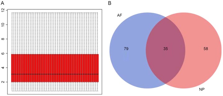
The present study aimed to reveal the potential genes associated with the pathogenesis of intervertebral disc degeneration (IDD) by analyzing microarray data using bioinformatics. Gene expression profiles of two regions of the intervertebral disc were compared between patients with IDD and controls. GSE70362 containing two groups of gene expression profiles, 16 nucleus pulposus (NP) samples from patients with IDD and 8 from controls, and 16 annulus fibrosus (AF) samples from patients with IDD and 8 from controls, was downloaded from the Gene Expression Omnibus database. A total of 93 and 114 differentially expressed genes (DEGs) were identified in NP and AF samples, respectively, using a limma software package for the R programming environment. Gene Ontology (GO) function enrichment analysis was performed to identify the associated biological functions of DEGs in IDD, which indicated that the DEGs may be involved in various processes, including cell adhesion, biological adhesion and extracellular matrix organization. Pathway enrichment analysis using the Kyoto Encyclopedia of Genes and Genomes (KEGG) demonstrated that the identified DEGs were potentially involved in focal adhesion and the p53 signaling pathway. Further analysis revealed that there were 35 common DEGs observed between the two regions (NP and AF), which may be further regulated by 6 clusters of microRNAs (miRNAs) retrieved with WebGestalt. The genes in the DEG‑miRNA regulatory network were annotated using GO function and KEGG pathway enrichment analysis, among which extracellular matrix organization was the most significant disrupted biological process and focal adhesion was the most significant dysregulated pathway. In addition, the result of protein‑protein interaction network modules demonstrated the involvement of inflammatory cytokine interferon signaling in IDD. These findings may not only advance the understanding of the pathogenesis of IDD, but also identify novel potential biomarkers for this disease.
Figures


Similar articles
-
Identification of Key Genes Potentially Related to Intervertebral Disk Degeneration by Microarray Analysis.Genet Test Mol Biomarkers. 2019 Sep;23(9):610-617. doi: 10.1089/gtmb.2019.0043. Epub 2019 Aug 1. Genet Test Mol Biomarkers. 2019. PMID: 31368816
-
Identification of key potential targets for TNF-α/TNFR1-related intervertebral disc degeneration by bioinformatics analysis.Connect Tissue Res. 2021 Sep;62(5):531-541. doi: 10.1080/03008207.2020.1797709. Epub 2020 Aug 26. Connect Tissue Res. 2021. PMID: 32686499
-
Bioinformatics analysis of molecular mechanisms involved in intervertebral disc degeneration induced by TNF-α and IL-1β.Mol Med Rep. 2016 Mar;13(3):2925-31. doi: 10.3892/mmr.2016.4861. Epub 2016 Feb 4. Mol Med Rep. 2016. PMID: 26847698
-
MicroRNA Expression Profiles, Target Genes, and Pathways in Intervertebral Disk Degeneration: A Meta-Analysis of 3 Microarray Studies.World Neurosurg. 2019 Jun;126:389-397. doi: 10.1016/j.wneu.2019.03.120. Epub 2019 Mar 20. World Neurosurg. 2019. PMID: 30904808 Review.
-
A Bioinformatic Analysis of MicroRNAs' Role in Human Intervertebral Disc Degeneration.Pain Med. 2019 Dec 1;20(12):2459-2471. doi: 10.1093/pm/pnz015. Pain Med. 2019. PMID: 30953590 Review.
Cited by
-
Single Cell RNA-Sequence Analyses Reveal Uniquely Expressed Genes and Heterogeneous Immune Cell Involvement in the Rat Model of Intervertebral Disc Degeneration.Appl Sci (Basel). 2022 Aug 2;12(16):8244. doi: 10.3390/app12168244. Epub 2022 Aug 18. Appl Sci (Basel). 2022. PMID: 36451894 Free PMC article.
-
Integrating multiple microarray dataset analysis and machine learning methods to reveal the key genes and regulatory mechanisms underlying human intervertebral disc degeneration.PeerJ. 2020 Oct 13;8:e10120. doi: 10.7717/peerj.10120. eCollection 2020. PeerJ. 2020. PMID: 33083145 Free PMC article.
-
Significance of Immune-Related Genes in the Diagnosis and Classification of Intervertebral Disc Degeneration.J Immunol Res. 2022 Aug 30;2022:2616260. doi: 10.1155/2022/2616260. eCollection 2022. J Immunol Res. 2022. PMID: 36081453 Free PMC article.
-
Transcriptome signatures reveal candidate key genes in the whole blood of patients with lumbar disc prolapse.Exp Ther Med. 2019 Dec;18(6):4591-4602. doi: 10.3892/etm.2019.8137. Epub 2019 Oct 25. Exp Ther Med. 2019. PMID: 31777557 Free PMC article.
-
Quality Assessment of Surgical Disc Samples Discriminates Human Annulus Fibrosus and Nucleus Pulposus on Tissue and Molecular Level.Int J Mol Sci. 2018 Jun 13;19(6):1761. doi: 10.3390/ijms19061761. Int J Mol Sci. 2018. PMID: 29899321 Free PMC article.
References
-
- Costi JJ, Stokes IA, Gardner-Morse MG, Iatridis JC. Frequency-dependent behavior of the intervertebral disc in response to each of six degree of freedom dynamic loading: Solid phase and fluid phase contributions. Spine (Phila Pa 1976) 2008;33:1731–1738. doi: 10.1097/BRS.0b013e31817bb116. - DOI - PMC - PubMed
-
- Friedman BW, O'Mahony S, Mulvey L, Davitt M, Choi H, Xia S, Esses D, Bijur PE, Gallagher EJ. One-week and 3-month outcomes following an emergency department visit for undifferentiated musculoskeletal low back pain. Ann Emerg Med. 2012;59:128–133. doi: 10.1016/j.annemergmed.2011.09.012. - DOI - PubMed
MeSH terms
Substances
LinkOut - more resources
Full Text Sources
Other Literature Sources
Research Materials
Miscellaneous
