Antitumor effects of baicalin on ovarian cancer cells through induction of cell apoptosis and inhibition of cell migration in vitro
- PMID: 29039573
- PMCID: PMC5779949
- DOI: 10.3892/mmr.2017.7757
Antitumor effects of baicalin on ovarian cancer cells through induction of cell apoptosis and inhibition of cell migration in vitro
Abstract
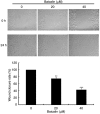
Baicalin, an active flavone isolated from Scutellaria baicalensis Georgi, has been demonstrated to induce various beneficial biochemical effects such as anti‑inflammatory, anti‑viral, and antitumor effects. However, the antitumor mechanism of baicalin is not well understood. In the present study, baicalin was demonstrated to inhibit the viability and migration of a widely used ovarian cancer cell line, A2780, in a dose‑dependent manner. MTT assays revealed that cell viability significantly decreased in ovarian cancer cells treated with baicalin compared with untreated cells, without effect on normal ovarian cells. Flow cytometric analysis indicated that baicalin suppressed cell proliferation by inducing apoptosis. The underlying mechanisms involved were indicated to be downregulation of the anti‑apoptotic protein B‑cell lymphoma 2 apoptosis regulator and activation of caspase‑3 and ‑9. In addition, wound healing and transwell assays revealed that cell migratory potential and expression of matrix metallopeptidase (MMP)‑2 and MMP‑9 were significantly inhibited when cells were exposed to baicalin, compared with untreated cells. The present study therefore suggested that baicalin has the potential to be used in novel anti‑cancer therapeutic formulations for treatment of ovarian cancer.
Figures


Similar articles
-
The anti-tumor effect and bioactive phytochemicals of Hedyotis diffusa willd on ovarian cancer cells.J Ethnopharmacol. 2016 Nov 4;192:132-139. doi: 10.1016/j.jep.2016.07.027. Epub 2016 Jul 15. J Ethnopharmacol. 2016. PMID: 27426510
-
Anti-tumor effects of osthole on ovarian cancer cells in vitro.J Ethnopharmacol. 2016 Dec 4;193:368-376. doi: 10.1016/j.jep.2016.08.045. Epub 2016 Aug 24. J Ethnopharmacol. 2016. PMID: 27566206
-
Formononetin, an isoflavone from Astragalus membranaceus inhibits proliferation and metastasis of ovarian cancer cells.J Ethnopharmacol. 2018 Jul 15;221:91-99. doi: 10.1016/j.jep.2018.04.014. Epub 2018 Apr 13. J Ethnopharmacol. 2018. PMID: 29660466
-
Anticancer Properties of Baicalin against Breast Cancer and other Gynecological Cancers: Therapeutic Opportunities based on Underlying Mechanisms.Curr Mol Pharmacol. 2024;17:e18761429263063. doi: 10.2174/0118761429263063231204095516. Curr Mol Pharmacol. 2024. PMID: 38284731 Review.
-
Antiviral Properties of Baicalin: a Concise Review.Rev Bras Farmacogn. 2021;31(4):408-419. doi: 10.1007/s43450-021-00182-1. Epub 2021 Oct 6. Rev Bras Farmacogn. 2021. PMID: 34642508 Free PMC article. Review.
Cited by
-
Baicalin alleviates bleomycin‑induced pulmonary fibrosis and fibroblast proliferation in rats via the PI3K/AKT signaling pathway.Mol Med Rep. 2020 Jun;21(6):2321-2334. doi: 10.3892/mmr.2020.11046. Epub 2020 Apr 1. Mol Med Rep. 2020. PMID: 32323806 Free PMC article.
-
Detarium microcarpum, Guiera senegalensis, and Cassia siamea Induce Apoptosis and Cell Cycle Arrest and Inhibit Metastasis on MCF7 Breast Cancer Cells.Evid Based Complement Alternat Med. 2019 May 23;2019:6104574. doi: 10.1155/2019/6104574. eCollection 2019. Evid Based Complement Alternat Med. 2019. PMID: 31239861 Free PMC article.
-
Dual effects of baicalin on osteoclast differentiation and bone resorption.J Cell Mol Med. 2018 Oct;22(10):5029-5039. doi: 10.1111/jcmm.13785. Epub 2018 Jul 16. J Cell Mol Med. 2018. PMID: 30010244 Free PMC article.
-
In vitro evaluation of the pogostone effects on the expression of PTEN and DACT1 tumor suppressor genes, cell cycle, and apoptosis in ovarian cancer cell line.Res Pharm Sci. 2022 Jan 15;17(2):164-175. doi: 10.4103/1735-5362.335175. eCollection 2022 Apr. Res Pharm Sci. 2022. PMID: 35280836 Free PMC article.
-
5,7,2',6'- Tetrahydroxyflavone affects the progression of ovarian cancer via hsa-miR-495-3p-ACTB/HSP90AA1 pathway.Discov Oncol. 2025 May 19;16(1):817. doi: 10.1007/s12672-025-02570-8. Discov Oncol. 2025. PMID: 40389695 Free PMC article.
References
-
- Markman M. Current status and future directions of platinum/paclitaxel-based chemotherapy of ovarian cancer. Semin Oncol. 1997;24(4 Suppl 11):S11–S27. - PubMed
MeSH terms
Substances
LinkOut - more resources
Full Text Sources
Other Literature Sources
Medical
Research Materials
Miscellaneous

