. 2017 Dec;16(6):9347-9354.
doi: 10.3892/mmr.2017.7795. Epub 2017 Oct 17.

MicroRNA‑218 promotes prostaglandin E2 to inhibit osteogenic differentiation in synovial mesenchymal stem cells by targeting 15‑hydroxyprostaglandin dehydrogenase [NAD(+)]

Affiliations

MicroRNA‑218 promotes prostaglandin E2 to inhibit osteogenic differentiation in synovial mesenchymal stem cells by targeting 15‑hydroxyprostaglandin dehydrogenase [NAD(+)]

Ruijun Cong et al. Mol Med Rep. 2017 Dec.

Abstract

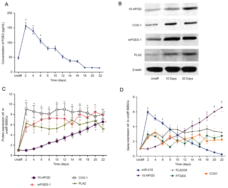
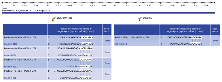
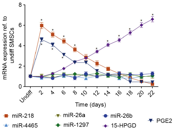
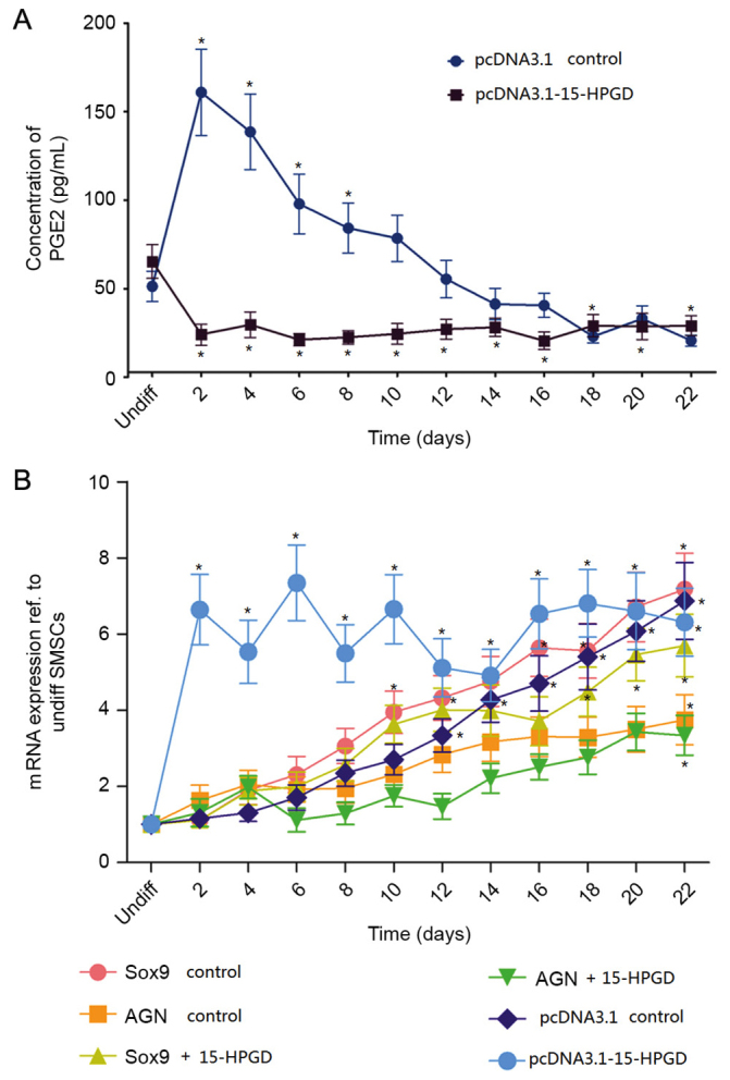
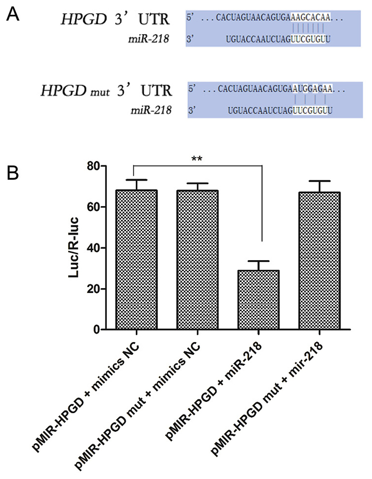
The chondrogenic differentiation of synovial mesenchymal stem cells (SMSCs) is regulated by essential transcription factors and signaling cascades. However, the precise mechanisms involved in this process remain unclear. MicroRNAs (miRs/miRNAs) are undersized non‑coding RNAs responsible for the post‑transcriptional regulation of gene expression, by binding to the 3'‑untranslated regions (3'‑UTRs) of their target mRNAs. miRNAs may constitute a promising tool to regulate SMSC differentiation and to advance the controlled differentiation of SMSCs in therapeutic applications. The aim of the present study was to examine the role of miR‑218 in SMSC differentiation towards chondrocytes. The present study comparatively analyzed the expression profile of known miRNAs and specific target genes in SMSCs between early and late differentiation stages. Western blotting and reverse transcription‑quantitative polymerase chain reaction analysis of gene expression demonstrated the upregulation of 15‑hydroxyprostaglandin dehydrogenase [NAD(+)] (15‑HPGD), prostaglandin E2 (PGE2) and rate limiting enzymes responsible for the synthesis of PGE2 precursors throughout chondrogenesis. Through correlation analysis, it was observed that there was a significant association between miR‑128, 15‑HPGD gene expression, 15‑HPGD protein expression and microsomal prostaglandin E synthase 1. Further experiments demonstrated that miR‑218 decreased PGE2 concentration by binding to the 3'‑UTR of 15‑HPGD. Using an immunofluorescence reporting system, it was observed that miR‑218 regulated the expression of 15‑HPGD during the differentiation of SMSCs into cartilage, and subsequently inhibited osteogenesis during chondrogenesis by acting on the 3'UTR of 15‑HPGD. Therefore, miR‑218 may be an important regulator targeting osteogenic factors and modulating cartilage formation and differentiation. The results of the present study provided a novel insight beneficial to cellular manipulation methods during cartilage regeneration, and in cartilage tissue engineering research.


Figures

Figure 1.
Figure 1.
15-HPGD is the rate limited degradation enzyme of PGE2, and 15-HPGD expression is negatively associated with PGE2. (A) PGE2 concentration during SMSC differentiation. In the early differentiation period (10 days ago), PGE2 expression was significantly different compared with the undifferentiated group. (B) Representative western blot images. (C) Expression of proteins at different time points during SMSC differentiation, determined by western blot analysis. (D) Gene expression at different time points during SMSC differentiation determined by reverse transcription-quantitative polymerase chain reaction. *P<0.05 vs. undifferentiated cells. 15-HPGD, 15-hydroxyprostaglandin dehydrogenase [NAD(+)]; PGE2, prostaglandin E2; COX-1, cyclooxygenase-1; mPGES-1, microsomal prostaglandin E synthase 1; PLA2, phospholipase A2; SMSC, synovial mesenchymal stem cells; miR, microRNA; undiff, undifferentiated.
Figure 2.
Figure 2.
Identification of miRs targeting 15-HPGD using TargetScan. miR, microRNA; UTR, untranslated region; 15-HPGD, 15-hydroxyprostaglandin dehydrogenase [NAD(+)]; UTR, untranslated region.
Figure 3.
Figure 3.
mRNA expression of 15-HPGD, PGE2 and miRs targeting 15-HPGD. Targeting miRs were identified using TargetScan and analyzed via reverse transcription-quantitative polymerase chain reaction. miRNA218 and PGE2 expression were significantly higher compared with undifferentiated cells at baseline. Following 10 days of differentiation, 15-HPGD was significantly increased compared with undifferentiated cells. *P<0.05 vs. Undifferentiated cells. miR, microRNA; 15-HPGD, 15-hydroxyprostaglandin dehydrogenase [NAD(+)]; PGE2, prostaglandin E2; undiff, undifferentiated.
Figure 4.
Figure 4.
15-HPGD decreases PGE2 concentration. (A) Comparison of the concentration of PGE2 in SMSCs transfected with the wide type 15-HPGD coding sequence (pcDNA3.1-15-HPGD) or normal SMSCs (empty control vector). (B) mRNA expression of differentiation-associated genes and 15-HPGD in SMSCs transfected with the 15-HPGD coding sequence (pcDNA3.1-15-HPGD) or empty control vector SMSCs. *P<0.05 vs. Undifferentiated cells. PGE2, prostaglandin E2; 15-HPGD, 15-hydroxyprostaglandin dehydrogenase [NAD(+)]; SMSCs, synovial mesenchymal stem cells; UTR, untranslated region; nom, normal; Sox9, transcription factor Sox9; AGN, aggrecan.
Figure 5.
Figure 5.
miR-218 targets the 15-HPGD 3′-UTR. (A) The three point mutations induced in the 3′UTR of 15-HPGD. (B) Overexpression of exogenous miR-218 suppressed the 15-HPGD expression, with the opposite effect observed with mutant 15-HPGD. **P<0.01. miR, microRNA; 15-HPGD, 15-hydroxyprostaglandin dehydrogenase [NAD(+)]; UTR, untranslated region; SMSCs, synovial mesenchymal stem cells; mut, mutant.

References

    1. Sakaguchi Y, Sekiya I, Yagishita K, Muneta T. Comparison of human stem cells derived from various mesenchymal tissues: Superiority of synovium as a cell source. Arthritis Rheum. 2005;52:2521–2529. doi: 10.1002/art.21212. - DOI - PubMed
    1. Shirasawa S, Sekiya I, Sakaguchi Y, Yagishita K, Ichinose S, Muneta T. In vitro chondrogenesis of human synovium-derived mesenchymal stem cells: Optimal condition and comparison with bone marrow-derived cells. J Cell Biochem. 2006;97:84–97. doi: 10.1002/jcb.20546. - DOI - PubMed
    1. Mochizuki T, Muneta T, Sakaguchi Y, Nimura A, Yokoyama A, Koga H, Sekiya I. Higher chondrogenic potential of fibrous synovium- and adipose synovium-derived cells compared with subcutaneous fat-derived cells: Distinguishing properties of mesenchymal stem cells in humans. Arthritis Rheum. 2006;54:843–853. doi: 10.1002/art.21651. - DOI - PubMed
    1. Yoshimura H, Muneta T, Nimura A, Yokoyama A, Koga H, Sekiya I. Comparison of rat mesenchymal stem cells derived from bone marrow, synovium, periosteum, adipose tissue and muscle. Cell Tissue Res. 2007;327:449–462. doi: 10.1007/s00441-006-0308-z. - DOI - PubMed
    1. Alm JJ, Heino TJ, Hentunen TA, Väänänen HK, Aro HT. Transient 100 nM dexamethasone treatment reduces inter- and intraindividual variations in osteoblastic differentiation of bone marrow-derived human mesenchymal stem cells. Tissue Eng Part C Methods. 2012;18:658–666. doi: 10.1089/ten.tec.2011.0675. - DOI - PubMed

MeSH terms