. 2018 Sep;38(3):292-298.
doi: 10.1097/WNO.0000000000000580.

Altered Macular Microvasculature in Mild Cognitive Impairment and Alzheimer Disease

Affiliations

Altered Macular Microvasculature in Mild Cognitive Impairment and Alzheimer Disease

Hong Jiang et al. J Neuroophthalmol. 2018 Sep.

Abstract

Background: The goal of the present study was to analyze the macular microvacular network in mild cognitive impirment (MCI) and Alzheimer disease (AD).

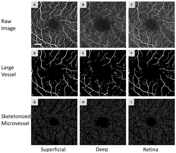
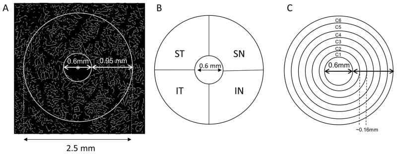
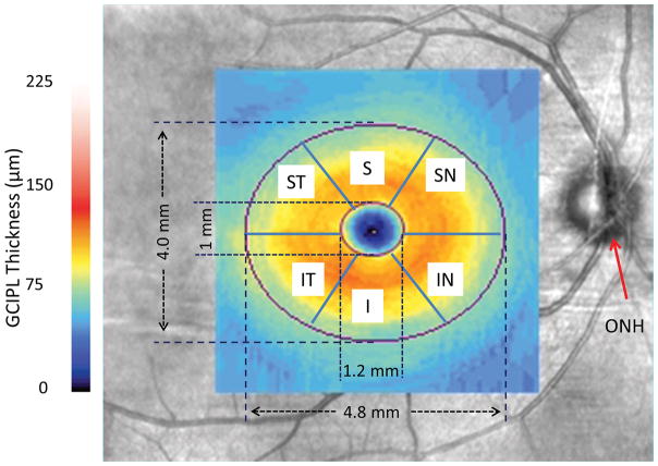
Methods: Twelve patients with AD and 19 patients with MCI were recruited together with 21 cognitively normal controls with a similar range of ages. Optical coherence tomography angiography was used to image the retinal microvascular network at the macular region, including retinal vascular network (RVN), superficial vascular plexus (SVP), and deep vascular plexus (DVP). Fractal analysis (box counting, Dbox) representing the microvascular density was performed in different annular zones and quadrantal sectors. The macular ganglion cell-inner plexiform layer (GC-IPL) thickness was measured using Zeiss OCT. The relationship between the retinal microvasculature and clinical manifestations was analyzed.

Results: Patients with AD had lower densities of RVN, SVP, and DVP in the annulus, from 0.6 to 2.5 mm in diameter (P < 0.05) in comparison with controls. Patients with MCI had lower density of DVP in the superior nasal quadrant (P < 0.05) than that of the controls. There were no significant differences of GC-IPL thickness among groups (P > 0.05). There was a trend of vascular density loss from control to MCI then AD (P < 0.05). Retinal microvascular density of DVP was correlated with GC-IPL thickness (P < 0.05) in patients with AD, but not in patients with MCI and controls.

Conclusions: Patients with AD had less density of retinal microvascular networks than controls. Our findings suggest the presence of retinal microvascular dysfunction in AD.


Figures

Figure 1
Figure 1. Segmentation of OCTA images
The superficial (A) and deep (B) vascular plexuses and retinal vascular network (C) from a patient with mild cognitive impairment. The segmentation software removed the large vessel (D, E and F) from the vascular network and extracted the skeletonized microvascular network using processing procedures, such as inverting, equalizing and removing background noise and non-vessel structures to create a binary image (G, H and I). Any vessels with a diameter ≥ 25 μm (D, E and F) were defined as large vessels and were separated from the remaining vessels, which were defined as microvessels. This procedure was also used to eliminate the shadow graphic projection artifact of the large vessels (E) in the superficial vascular plexus on the deep vascular plexus (B). Bar = 0.5 mm.
Figure 2
Figure 2. Partitions of the microvascular network
The center (marked as red asterisk*) of the foveal avascular zone (FAZ) was detected in the skeletonized microvasculature image (A) and the center of the FAZ was used to partition the quadrantal and annular zones. The annulus from 0.6 mm to 2.5 mm in diameter was defined as the annular zone with a width of 0.95 mm after removing the avascular zone (diameter = 0.6 mm) centered on the fovea (A). Using the hemispheric partition, the annular zone was then partitioned into four quadrantal sectors (B), named the superior temporal (ST), inferior temporal (IT), superior nasal (SN), and inferior nasal (IN). To analyze the changes in the thin annular zones from the center to the periphery, the annular zone (A) was partitioned into 6 thin annuli named C1 to C6 (D) with a width of ~0.16 mm.
Figure 3
Figure 3. GC-IPL imaging and partition
GC-IPL of the right eye from a patient with mild cognitive impairment was acquired in the macula using Zeiss OCTA and a thickness map was processed in the elliptic annulus with partitions of 6 sectors of the annulus after removal of the central elliptic area. ONH: optic nerve head; ST: superior temporal; S: superior; SN: superior nasal; IN: inferior nasal; I: inferior; and IT: inferior temporal.
Figure 4
Figure 4. Representative images of the retinal microvascular networks imaged using optical coherence tomography angiography (OCTA)
Patients with Alzheimer disease (AD) (A, B and C) and mild cognitive impairment (MCI) (D, E and F) as well as a control subject (G, H and I) were imaged. Compared to the normal control (G and I), the large vessels in AD and MCI patients had similar densities in the superficial vascular plexus (A and D) and retinal vascular network (C and F) but showed some degrees of tortuosity. The microvessels in the deep vascular plexus in AD (B) and MCI (E) patients appeared to be less dense compared to the normal control (H). Note: the deep vascular plexus images are raw images, showing the graphic projection artifact of the large vessels. Bar = 0.5 mm.

References

    1. de la Torre JC. Vascular risk factors: a ticking time bomb to Alzheimer’s disease. Am J Alzheimers Dis Other Demen. 2013;28:551–559. - PMC - PubMed
    1. Ott A, Stolk RP, van HF, Pols HA, Hofman A, Breteler MM. Diabetes mellitus and the risk of dementia: The Rotterdam Study. Neurology. 1999;53:1937–1942. - PubMed
    1. Hofman A, Ott A, Breteler MM, Bots ML, Slooter AJ, van HF, van Duijn CN, Van BC, Grobbee DE. Atherosclerosis, apolipoprotein E, and prevalence of dementia and Alzheimer’s disease in the Rotterdam Study. Lancet. 1997;349:151–154. - PubMed
    1. de la Torre JC. Vascular risk factor detection and control may prevent Alzheimer’s disease. Ageing Res Rev. 2010;9:218–225. - PubMed
    1. de la Torre JC. Cerebral hemodynamics and vascular risk factors: setting the stage for Alzheimer’s disease. J Alzheimers Dis. 2012;32:553–567. - PubMed

Publication types