

Dexamethasone-mediated inhibition of Glioblastoma neurosphere dispersal in an ex vivo organotypic neural assay
- PMID: 29040322
- PMCID: PMC5645119
- DOI: 10.1371/journal.pone.0186483
Dexamethasone-mediated inhibition of Glioblastoma neurosphere dispersal in an ex vivo organotypic neural assay
Abstract
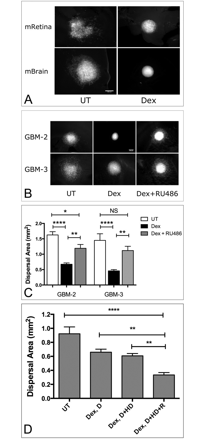
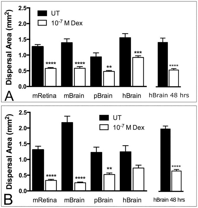
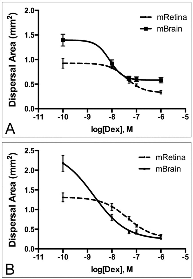
Glioblastoma is highly aggressive. Early dispersal of the primary tumor renders localized therapy ineffective. Recurrence always occurs and leads to patient death. Prior studies have shown that dispersal of Glioblastoma can be significantly reduced by Dexamethasone (Dex), a drug currently used to control brain tumor related edema. However, due to high doses and significant side effects, treatment is tapered and discontinued as soon as edema has resolved. Prior analyses of the dispersal inhibitory effects of Dex were performed on tissue culture plastic, or polystyrene filters seeded with normal human astrocytes, conditions which inherently differ from the parenchymal architecture of neuronal tissue. The aim of this study was to utilize an ex-vivo model to examine Dex-mediated inhibition of tumor cell migration from low-passage, human Glioblastoma neurospheres on multiple substrates including mouse retina, and slices of mouse, pig, and human brain. We also determined the lowest possible Dex dose that can inhibit dispersal. Analysis by Two-Factor ANOVA shows that for GBM-2 and GBM-3, Dex treatment significantly reduces dispersal on all tissue types. However, the magnitude of the effect appears to be tissue-type specific. Moreover, there does not appear to be a difference in Dex-mediated inhibition of dispersal between mouse retina, mouse brain and human brain. To estimate the lowest possible dose at which Dex can inhibit dispersal, LogEC50 values were compared by Extra Sum-of-Squares F-test. We show that it is possible to achieve 50% reduction in dispersal with Dex doses ranging from 3.8 x10⁻⁸M to 8.0x10⁻⁹M for GBM-2, and 4.3x10⁻⁸M to 1.8x10⁻⁹M for GBM-3, on mouse retina and brain slices, respectively. These doses are 3-30-fold lower than those used to control edema. This study extends our previous in vitro data and identifies the mouse retina as a potential substrate for in vivo studies of GBM dispersal.
Conflict of interest statement
Figures
References
-
- Brandes AA, Tosoni A, Franceschi E, Reni M, Gatta G, Vecht C. Glioblastoma in adults. Critical reviews in oncology/hematology. 2008;67(2):139–52. Epub 2008/04/09. doi: 10.1016/j.critrevonc.2008.02.005 . - DOI - PubMed
-
- Stupp R, Hegi ME, Mason WP, van den Bent MJ, Taphoorn MJ, Janzer RC, et al. Effects of radiotherapy with concomitant and adjuvant temozolomide versus radiotherapy alone on survival in glioblastoma in a randomised phase III study: 5-year analysis of the EORTC-NCIC trial. The Lancet Oncology. 2009;10(5):459–66. doi: 10.1016/S1470-2045(09)70025-7 . - DOI - PubMed
-
- Clarke J, Butowski N, Chang S. Recent advances in therapy for glioblastoma. Archives of neurology. 2010;67(3):279–83. Epub 2010/03/10. doi: 10.1001/archneurol.2010.5 . - DOI - PubMed
-
- Sabari J, Lax D, Connors D, Brotman I, Mindrebo E, Butler C, et al. Fibronectin matrix assembly suppresses dispersal of glioblastoma cells. PLoS One. 2011;6(9):e24810 doi: 10.1371/journal.pone.0024810 . - DOI - PMC - PubMed
-
- Shannon S, Vaca C, Jia D, Entersz I, Schaer A, Carcione J, et al. Dexamethasone-Mediated Activation of Fibronectin Matrix Assembly Reduces Dispersal of Primary Human Glioblastoma Cells. PLoS One. 2015;10(8):e0135951 doi: 10.1371/journal.pone.0135951 . - DOI - PMC - PubMed
MeSH terms
Substances
LinkOut - more resources
Full Text Sources
Other Literature Sources
