. 2017 Dec 1;158(12):4300-4316.
doi: 10.1210/en.2017-00660.

Sperm Release at Spermiation Is Regulated by Changes in the Organization of Actin- and Microtubule-Based Cytoskeletons at the Apical Ectoplasmic Specialization-A Study Using the Adjudin Model

Affiliations

Sperm Release at Spermiation Is Regulated by Changes in the Organization of Actin- and Microtubule-Based Cytoskeletons at the Apical Ectoplasmic Specialization-A Study Using the Adjudin Model

Linxi Li et al. Endocrinology. .

Abstract

The mechanism that regulates sperm release at spermiation is unknown. Herein, we used an animal model wherein rats were treated with adjudin, 1-(2,4-dichlorobenzyl)-1H-indazole-3-carbohydrazide, via oral gavage to induce premature release of elongating/elongated spermatids, followed by round spermatids and spermatocytes. Spermatid release mimicking spermiation occurred within 6 to 12 hours following adjudin treatment and, by 96 hours, virtually all tubules were devoid of elongating/elongated spermatids. Using this model, we tracked the organization of F-actin and microtubules (MTs) by immunofluorescence microscopy, and the association of actin or MT regulatory proteins that either promote or demolish cytoskeletal integrity through changes in the organization of actin microfilaments or MTs by coimmunoprecipitation. Adjudin treatment induced an increase in the association of (1) epidermal growth factor receptor pathway substrate 8 (an actin barbed-end capping and bundling protein) or formin 1 (an actin nucleator) with actin and (2) end-binding protein 1 (an MT stabilizing protein) with MT shortly after adjudin exposure (at 6 hours), in an attempt to maintain spermatid adhesion to the Sertoli cell at the apical ectoplasmic specialization (ES). However, this was followed by a considerable decline of their steady-state protein levels, replacing with an increase in association of (1) actin-related protein 3 (a branched actin nucleator that converts actin filaments into a branched/unbundled network) with actin and (2) MT affinity-regulating kinase 4 (an MT destabilizing protein kinase) with MTs by 12 hours after adjudin treatment. These latter changes thus promoted actin and MT disorganization, leading to apical ES disruption and the release of elongating/elongated spermatids, mimicking spermiation. In summary, spermiation is a cytoskeletal-dependent event, involving regulatory proteins that modify cytoskeletal organization.


Figures

Figure 1.
Figure 1.
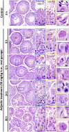
Adjudin treatment in adult rats rapidly induces defects in spermatogenesis prior to the occurrence of germ cell release from the seminiferous epithelium that mimics spermiation. Adult rats treated with a single dose of adjudin at 50 mg/kg b.w. by oral gavage at time 0 (controls; n = 5 rats). Thereafter, rats (n = 5) were terminated at specified time points at 6, 12, 24, and 96 hours vs 0 hours (control) for histological analysis using paraffin cross-sections of testes and H&E staining. By 6 and 12 hours, defects in spermatogenesis were noted in the testis. For instance, many elongated spermatids were trapped deep inside the epithelium (annotated by green arrowheads) when “unwanted spermiation” had taken place in nonstage VIII tubules but also stage VIII tubules; and phagosomes were found in a XIII tubule, which appeared to be derived from multinucleated round spermatids (annotated by red arrowheads; see also enlarged image in inset at 6 hours). In an apparently stage I to III tubule, a meiotic spermatocyte (annotated by a yellow arrowhead), phagosomes (annotated by red arrowheads), and a pachytene spermatocyte (annotated by a blue arrowhead) were detected in the tubule lumen when steps 15 and 16 spermatids (see green square box and the corresponding inset) were also depleting to the tubule lumen. By 24 hours, few elongating/elongated spermatids were found in the epithelium across the testis sections except those that were trapped deep inside the epithelium (annotated by green arrowheads and the corresponding boxed areas shown in insets), and both round spermatids, spermatocytes, and even some phagosomes were also emptied into the tubule lumen (see inset). At 96 hours (i.e., day 4), obvious defects in spermatogenesis were detected in the testis. For instance, virtually all elongated/elongated spermatids from 98% of tubules examined were not found in the seminiferous epithelium but depleted from the testis, yet some elongated spermatids remain trapped deep inside the epithelium, and this extensive germ cell loss also led to a reduction in tubule diameters. Scale bars = 180 µm in left panel at 0 hours, 70 µm in the blue boxed rectangle, and 30 µm in green boxed square, which apply to corresponding images in the same column or row.
Figure 2.
Figure 2.
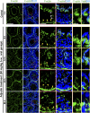
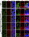
Adjudin treatment induces defects in F-actin organization prior to germ cell release from the seminiferous epithelium that mimics spermiation. Adult rats (∼300 g b.w.) were treated with a single dose of adjudin at 50 mg/kg b.w. by oral gavage at 0 (controls; n = 5 rats). Thereafter, rats (n = 5) were terminated at specified time points at 6, 12, 24, and 96 h (i.e., day 4) for IF microscopy to visualize the organization of F-actin network across the seminiferous epithelium using FITC-conjugated phalloidin (green fluorescence; Invitrogen). Cell nuclei were visualized by DAPI (Invitrogen). A section of the images (boxed in red) on the second column was magnified and shown on the third and fourth columns; and a section of the images (boxed in yellow) on the fourth column was also magnified and shown on the fifth and sixth columns. The relative location of the base of the tunica propria was annotated by a dashed white line in the third column, and the relative location of the BTB was annotated by yellow arrowheads. The track-like structures (annotated by white arrowheads) conferred by F-actin were also shown in control testes. After adjudin treatment, F-actin that supported the BTB was no longer tightly aligned at the BTB (see white bracket in control testes on the third column), but diffusely localized in particular by 12 to 96 hours (see yellow brackets). These changes in F-actin distribution were semiquantified and shown in Supplemental Fig. 1. Also, the track-like structures conferred by F-actin were virtually nondetectable by 12 hours following adjudin treatment. Moreover, F-actin that appeared as bulb-like structures located at the concave side of spermatid heads was extensively mislocalized by 6 hours after adjudin treatment, moving to the concave side of spermatid heads and considerably diminished by 24 and 96 hours. This time-dependent F-actin disorganization at the apical ES induced by adjudin thus led to germ cell release from the epithelium, mimicking spermiation. Scale bars = 180, 70, and 30 µm in the micrograph in the first, third, and fifth column, which apply to corresponding images.
Figure 3.
Figure 3.
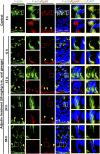
Adjudin treatment induces considerable changes in the spatiotemporal expression of Eps8 and its colocalization with F-actin at the ES prior to germ cell release from the seminiferous epithelium that mimics spermiation. Adult rats (∼300 g b.w.) were treated with a single dose of adjudin at 50 mg/kg b.w. by oral gavage at time 0 (controls; n = 3 rats). Thereafter, rats (n = 5) were terminated at specified time points for IF microscopy to visualize changes in the spatial expression of Eps8 (red fluorescence) and its colocalization with F-actin (green fluorescence) using frozen sections of testes. Cell nuclei were visualized by DAPI. Eps8 is an actin barbed-end capping and bundling protein capable of assembling actin microfilaments to form bundles to support ES function at the Sertoli-spermatid interface (apical ES) and at the Sertoli cell-cell interface (basal ES/BTB). Eps8 (similar to F-actin) appeared as bulb-like ultrastructures at the apical ES, colocalizing with F-actin at the concave (ventral) side of spermatid heads. Following adjudin treatment, a considerable decline in Eps8 expression at the apical ES was detected vs control testes. This thus failed to support actin filament bundles at the apical ES. This decline in robust Eps8 expression was worsened by 12 hours and by 24 and 96 hours (4 days); virtually no Eps8 expression was detected (see also Supplemental Fig. S2). This trend of time-dependent reduction in Eps8 expression was also found at the basal ES/BTB (see Supplemental Fig. 2), thereby causing F-actin to fail to localize properly at the basal ES/BTB. Such decline in Eps8 expression thus contributed to the loss of spermatid adhesion, causing spermatid release from the epithelium, mimicking spermiation. Scale bars = 80 and 30 µm in the first micrograph and the corresponding enlarged images shown in insets.
Figure 4.
Figure 4.
Adjudin treatment induces considerable changes in the spatiotemporal expression of formin 1 and its colocalization with F-actin at the ES prior to germ cell release from the seminiferous epithelium that mimics spermiation. Adult rats (∼300 g b.w.) were treated with a single dose of adjudin at 50 mg/kg b.w. by oral gavage at time 0 (controls; n = 5 rats). Thereafter, rats (n = 5) were terminated at specified time points for IF microscopy to visualize changes in the spatiotemporal expression of formin 1 (red fluorescence) and its colocalization with F-actin (green fluorescence) using frozen sections of testes. Cell nuclei were visualized by DAPI. Because formin 1 is an actin nucleation protein capable of polymerizing long stretches of actin microfilaments in Sertoli cells to support F-actin organization, its considerable decline in expression at the apical ES thus induced disorganization of F-actin at the site to support spermatid adhesion (see also Supplemental Fig.3). These changes were noted as soon as 6 and 12 hours after adjudin treatment when formin 1 no longer robustly expressed at the concave side of spermatid heads. Its expression was virtually undetectable by 24 and 96 hours (4 days). Scale bars = 80 and 30 µm in the first micrograph and the corresponding enlarged images shown in insets.
Figure 5.
Figure 5.
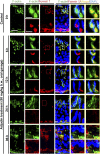
Adjudin treatment induces considerable changes in the spatiotemporal expression of Arp3 and its colocalization with F-actin at the ES prior to germ cell release from the seminiferous epithelium that mimics spermiation. Adult rats (∼300 g b.w.) were treated with a single dose of adjudin at 50 mg/kg b.w. by oral gavage at time 0 (controls; n = 3 rats). Thereafter, rats (n = 5) were terminated at specified time points for IF microscopy to visualize changes in the spatiotemporal expression of Arp3 (red fluorescence) and its colocalization with F-actin (green fluorescence) using frozen sections of testes. Cell nuclei were visualized by DAPI. Micrographs were enlarged in corresponding insets as indicated by the green, red, or yellow boxed areas. Relative location of the basement membrane was annotated by the dashed white line. F-actin organization was considerably disrupted within 6 hours after adjudin treatment, which continued to worsen. It appeared that F-actin disruption was mediated by considerable changes in spatial expression of Arp3, the barbed-end actin nucleation protein that effectively induced branched actin polymerization, causing the linear and bundled actin microfilaments at the ES to become disassembled and branched, destabilizing the ES. In control testes, Arp3 appeared as bulb-like structures at the concave (ventral) side of spermatid heads, which work together with Eps8 (see Fig. 3) to support endocytic vesicle-based protein trafficking for endocytosis and recycling in stage VII tubules. By 6 hours following adjudin treatment, however, Arp3 no longer appeared as bulb-like structures at the apical ES but considerably diminished (see also Supplemental Fig. 4); and by 24 and 96 hours (4 days), most of the elongated spermatids were not even supported by F-actin and Arp3, destabilizing spermatid adhesion that led to their eventual release from the epithelium that mimicked spermiation. Scale bars = 80 and 30 µm in the first micrograph and the corresponding enlarged images shown in insets.
Figure 6.
Figure 6.
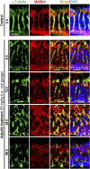
Adjudin treatment perturbs the organization of MTs through changes in the spatial expression of EB1. Adult rats (∼300 g b.w.) were treated with a single dose of adjudin at 50 mg/kg b.w. by oral gavage at time 0 (controls; n = 5 rats). Thereafter, rats (n = 5) were terminated at specified time points for IF microscopy to visualize changes in the organization of MTs by staining α-tubulin (green fluorescence; the building blocks of MTs). This disruptive organization of MTs following adjudin treatment appeared to be mediated by changes in the spatial expression of EB1 (red fluorescence), a +TIP protein known to stabilize MTs in Sertoli cells. It was noted that the track-like structures conferred by MTs, known to support the transport of spermatids and other organelles (e.g., residual bodies, phagosomes, endocytic vesicles) and to support actin microfilaments at the ES, across the seminiferous epithelium that laid perpendicular to the basement membrane as found in control testes were considerably disrupted. For instance, within 6 hours after adjudin treatment, these MT-based tracks and the colocalized EB1 were truncated, no longer stretched across the entire epithelium; and by 12 hours, virtually no identifiable track-like structures were found but were considerably truncated. For the residual MTs and the colocalized EB1 that were detected, some were laid in parallel, instead of perpendicular, to the basement membrane. Cell nuclei were visualized by DAPI. Scale bars = 180 and 80 µm in the first micrograph and the corresponding enlarged images shown in insets.
Figure 7.
Figure 7.
Adjudin treatment perturbs the organization of MTs through changes in the spatial expression of MARK4. Adults rats (∼300 g b.w.) were treated with a single dose of adjudin at 50 mg/kg b.w. by oral gavage at time 0 (controls; n = 5 rats). Thereafter, rats (n = 5) were terminated at specified time points for IF microscopy to visualize changes in the organization of MTs by staining α-tubulin (green fluorescence) vs MARK4 (red fluorescence). This disruptive organization of MTs following adjudin treatment appeared to be mediated by changes in the spatial expression of MARK4, a Ser/Thr protein kinase known to induce MT catastrophe by phosphorylating MAPs, causing their detachment from MTs, thereby rendering MTs to become less stable in Sertoli cells. The MT-conferred track-like structures necessary to support actin microfilaments at the ES as found in control testes were considerably disrupted following adjudin treatment. For instance, within 6 hours after adjudin treatment, these MT-based tracks that laid across the epithelium and aligned perpendicular to the basement membrane had became less prominent and the colocalized MARK4 were less concentrated to the MT-based tracks but diffusely localized, no longer stretched across the entire epithelium. By 12 hours, most of the tracts were truncated, and by 24 hours, there were virtually no identifiable track-like structures. For the residual MTs and the colocalized MARK4 that were detected, some were laid in parallel, instead of perpendicular, to the basement membrane. Cell nuclei were visualized by DAPI. Scale bar = 80 µm, which applies to all other images.
Figure 8.
Figure 8.
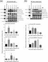
A study by Co-IP to assess changes in the interactions between actin- and MT-based binding and regulatory proteins with the corresponding cytoskeleton during the release of germ cells from the testis in the adjudin model. Lysates of testes (1 mg protein in each Co-IP reaction) from rats were obtained and used for Co-IP (as described in Materials and Methods) following treatment with adjudin (50 mg/kg b.w., oral gavage) at 6, 12, 24, and 96 hours vs 0 hours (control) with n = 3 rats per time point including control. (a) Actin regulatory proteins: Eps8 (an actin barbed-end capping and bundling protein causing actin filaments to assume a bundled configuration as those found at the ES), formin 1 (an actin nucleation protein capable of generating long stretches of actin microfilaments), and Arp3 (a branched actin polymerization protein causing linear actin filaments to become a branched network) were used for this study. Also shown is immunoprecipitated β-actin, which served as the positive control. Uncropped gel images shown herein can be found in Supplemental Fig. 5. (b) MT regulatory proteins: EB1 (a +TIP protein known to stabilize MTs) and MARK4 (a Ser/Thr protein kinase known to induce MT catastrophe) were also used for this study. Also shown is the immunoprecipitated α-tubulin, which was confirmed by using an anti–β-tubulin antibody for immunoblotting (IB) (see Table 1), which served as the positive control. Changes in protein-protein interaction of these selected regulatory proteins with the corresponding β-actin and α-tubulin, the building blocks of actin- and MT-based cytoskeletons, were assessed by Co-IP. In short, anti-actin IgG or anti–α-tubulin IgG, serving as the immunoprecipitating antibody, was incubated with testis lysates obtained from rats treated with adjudin for 6, 12, 24, and 96 hours vs 0 hours (control). Thereafter, immunocomplexes were pulled out by Protein A/G Plus-agarose beads, the interacting proteins with either actin or α-tubulin were resolved by sodium dodecyl sulfate-polyacrylamide gel electrophoresis, and the interacting proteins (and their changes following adjudin treatment) with either actin or MT were visualized by immunoblottings using corresponding specific antibodies (Table 1). Uncropped gel images shown herein can be found in Supplemental Fig. 6. The steady state of the regulatory proteins (i.e., Eps8, formin 1, Arp3, EB1, and MARK4) in testis lysates following adjudin treatment (without Co-IP) was also assessed and annotated as “Lysate.” Representative immunoblot data were shown in the upper panel in either (a) or (b), and the histograms below summarize results of these findings with n = 3 independent experiments using testes from different rats. *P < 0.05 by ANOVA.

Similar articles

Cited by

References

    1. Cheng CY, Mruk DD. Biochemistry of Sertoli cell/germ cell junctions, germ cell transport, and spermiation in the seminiferous epithelium In: Griswold MD, ed. Sertoli Cell Biology. 2nd ed. Amsterdam: Elsevier; 2015:333–383.
    1. Xiao X, Mruk DD, Wong CKC, Cheng CY. Germ cell transport across the seminiferous epithelium during spermatogenesis. Physiology (Bethesda). 2014;29(4):286–298. - PMC - PubMed
    1. Amann RP. The cycle of the seminiferous epithelium in humans: a need to revisit? J Androl. 2008;29(5):469–487. - PubMed
    1. Hess RA, Renato de Franca L. Spermatogenesis and cycle of the seminiferous epithelium. Adv Exp Med Biol. 2008;636:1–15. - PubMed
    1. Vogl AW, Vaid KS, Guttman JA. The Sertoli cell cytoskeleton. Adv Exp Med Biol. 2008;636:186–211. - PubMed

MeSH terms