. 2018 Feb 1;21(2):187-200.
doi: 10.1093/ijnp/pyx083.

Interferon-Alpha Reduces Human Hippocampal Neurogenesis and Increases Apoptosis via Activation of Distinct STAT1-Dependent Mechanisms

Affiliations

Interferon-Alpha Reduces Human Hippocampal Neurogenesis and Increases Apoptosis via Activation of Distinct STAT1-Dependent Mechanisms

Alessandra Borsini et al. Int J Neuropsychopharmacol. .

Abstract

Background: In humans, interferon-α treatment for chronic viral hepatitis is a well-recognized clinical model for inflammation-induced depression, but the molecular mechanisms underlying these effects are not clear. Following peripheral administration in rodents, interferon-α induces signal transducer and activator of transcription-1 (STAT1) within the hippocampus and disrupts hippocampal neurogenesis.

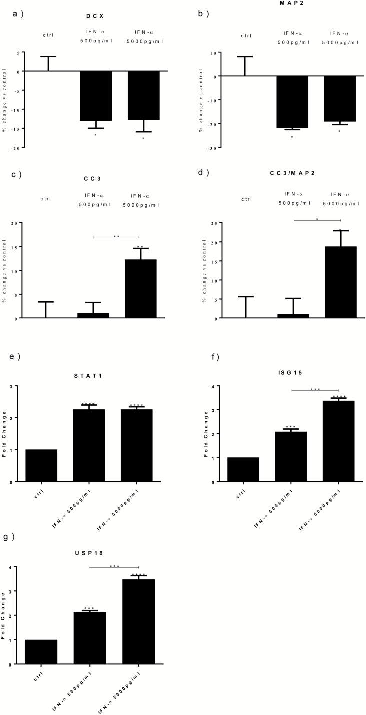
Methods: We used the human hippocampal progenitor cell line HPC0A07/03C to evaluate the effects of 2 concentrations of interferon-α, similar to those observed in human serum during its therapeutic use (500 pg/mL and 5000 pg/mL), on neurogenesis and apoptosis.

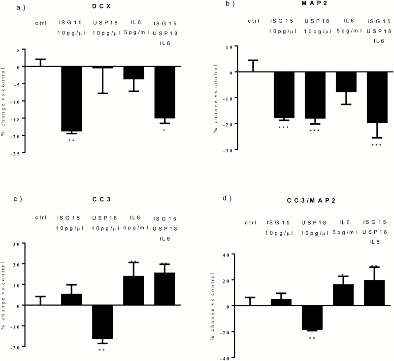
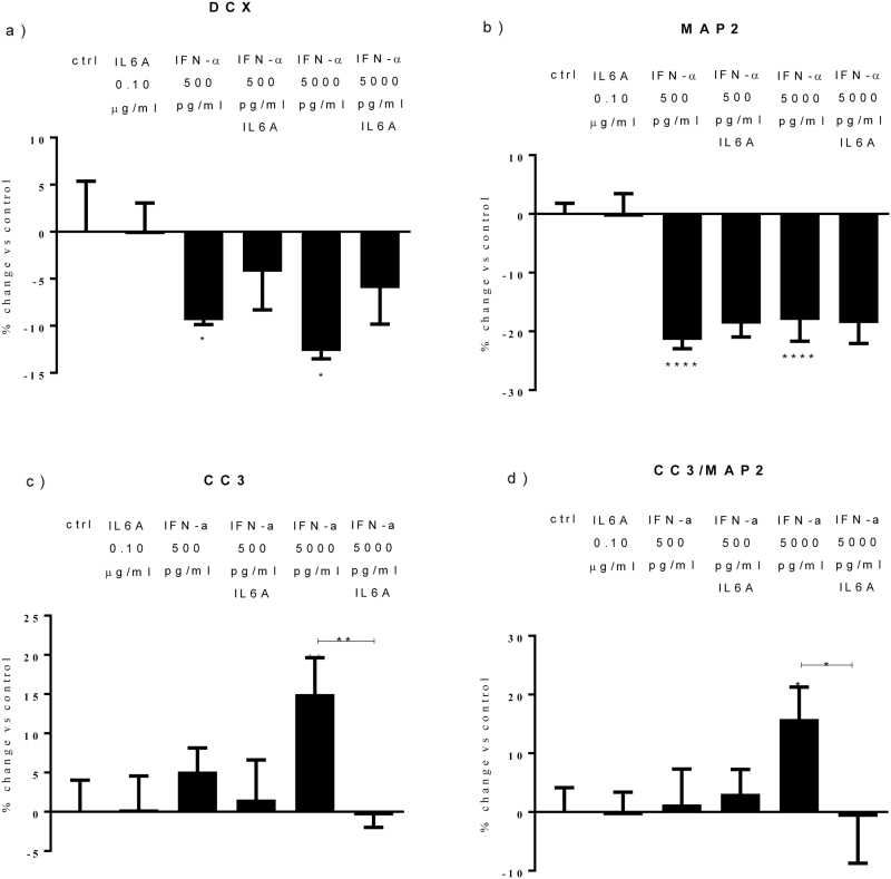
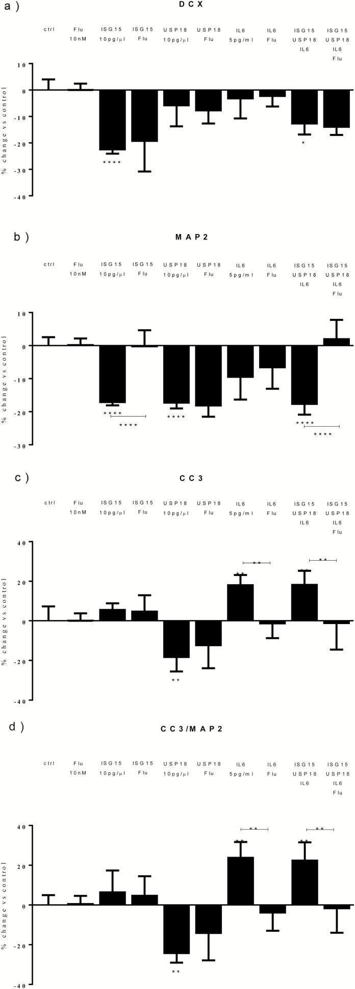
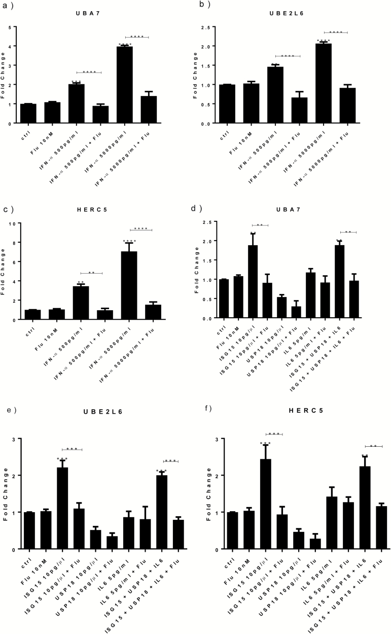
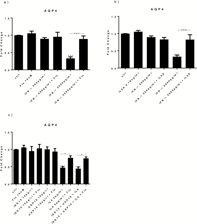
Results: Both concentrations of interferon-α decreased hippocampal neurogenesis, with the high concentration also increasing apoptosis. Moreover, interferon-α increased the expression of interferon-stimulated gene 15 (ISG15), ubiquitin-specific peptidase 18 (USP18), and interleukin-6 (IL-6) via activation of STAT1. Like interferon-α, co-treatment with a combination of ISG15, USP18, and IL-6 was able to reduce neurogenesis and enhance apoptosis via further downstream activation of STAT1. Further experiments showed that ISG15 and USP18 mediated the interferon-α-induced reduction in neurogenesis (potentially through upregulation of the ISGylation-related proteins UBA7, UBE2L6, and HERC5), while IL-6 mediated the interferon-α-induced increase in apoptosis (potentially through downregulation of aquaporin 4). Using transcriptomic analyses, we showed that interferon-α regulated pathways involved in oxidative stress and immune response (e.g., Nuclear Factor (erythroid-derived 2)-like 2 [Nrf2] and interferon regulatory factor [IRF] signaling pathway), neuronal formation (e.g., CAMP response element-binding protein [CREB] signaling), and cell death regulation (e.g., tumor protein(p)53 signaling).

Conclusions: We identify novel molecular mechanisms mediating the effects of interferon-α on the human hippocampus potentially involved in inflammation-induced neuropsychiatric symptoms.

Keywords: apoptosis; depression; inflammation; interferon-alpha; neurogenesis.


Figures

Figure 1.
Figure 1.
Interferon (IFN)-α reduces differentiation, increases apoptosis, and upregulates signal transducer and activator of transcription-1 (STAT1), interferon-stimulated gene 15 (ISG15), and ubiquitin-specific peptidase 18 (USP18) gene expression. IFN-α (500 and 5000 pg/mL) decreased doublecortin (DCX)+ (a) and microtubule-associated protein 2 (MAP2)+ cells (b). IFN-α 5000 pg/mL increased both caspase 3 (CC3)+ cells (c) and CC3+/MAP2+ cells (d). IFN-α (500 and 5000 pg/mL) increased STAT1 (e), ISG15 (f), and USP18 (g) gene expression. Three independent experiments were conducted on independent cultures (n=3). Data are shown as mean±SEM. 1-way ANOVA, Newman–Keuls’ posthoc test *P<.05, **P<.01, ***P<.001, ****P<.0001, compared with vehicle treatment or as indicated.
Figure 2.
Figure 2.
Cotreatment with interferon-stimulated gene 15 (ISG15), ubiquitin-specific peptidase 18 (USP18), and interleukin (IL)-6 mimics the effect of interferon (IFN)-α on neurogenesis and apoptosis. ISG15 and all proteins reduced doublecortin (DCX)+ cells (a). ISG15, USP18, and all proteins reduced microtubule-associated protein 2 (MAP2)+ cells (b). USP18 also decreased caspase 3 (CC3)+ cells, whereas IL-6 and all proteins significantly increased both CC3+ and CC3/MAP2+ cells (c-d). Three independent experiments were conducted on independent cultures (n=3). Data are shown as mean±SEM. 1-way ANOVA, Newman–Keuls’ posthoc test *P<.05, **P<.01, ***P<.001, compared with vehicle treatment or as indicated.
Figure 3.
Figure 3.
Interleukin (IL)-6A prevents the effects of interferon (IFN)-α on apoptosis. Treatment with IL-6A prevented the increase in caspase 3 (CC3)+ and CC3/microtubule-associated protein 2 (MAP2)+ cells detected upon IFN-α 5000 pg/mL (c-d). Three independent experiments were conducted on independent cultures (n=3). Data are shown as mean±SEM. 1-way ANOVA, Newman–Keuls’ posthoc test *P<.05, **P<.01, ****P<.0001, compared with vehicle treatment or as indicated.
Figure 4.
Figure 4.
Treatment with a signal transducer and activator of transcription-1 (STAT1) inhibitor reverts the effects of interferon (IFN)-α on neurogenesis and apoptosis by preventing the upregulation of interferon-stimulated gene 15 (ISG15), ubiquitin-specific peptidase 18 (USP18), and interleukin (IL)-6. Fludarabine prevented the decrease in doublecortin (DCX)+ and microtubule-associated protein 2 (MAP2)+ cells caused by IFN-α (500 and 5000 pg/mL) (a-b), as well as the decrease in caspase 3 (CC3)+ and CC3+/MAP2+ cells detected upon IFN-α 5000 pg/mL (c-d). Fludarabine prevented the increase in ISG15 (e) and USP18 (f) genes and IL-6 protein expression (g) observed upon IFN-α (500 and 5000 pg/mL). Three independent experiments were conducted on independent cultures (n=3). Data are shown as mean±SEM. 1-way ANOVA, Newman–Keuls’ posthoc test **P<.01, ***P<.001, ****P<.0001, compared with vehicle treatment or as indicated.
Figure 5.
Figure 5.
Treatment with a signal transducer and activator of transcription-1 (STAT1) inhibitor prevents the effect of interferon-stimulated gene 15 (ISG15) on neurogenesis and of interleukin (IL)-6 on apoptosis. Fludarabine prevented the decrease in microtubule-associated protein 2 (MAP2)+ cells caused by ISG15 and all proteins (b), as well as the increase in caspase 3 (CC3)+ and CC3+/MAP2+ cells detected upon treatment with IL-6 (c) and all proteins (d). Three independent experiments were conducted on independent cultures (n=3). Data are shown as mean±SEM. One-way ANOVA, Newman–Keuls’ posthoc test *P<.05, **P<.01, ****P<.0001, compared with vehicle treatment or as indicated.
Figure 6.
Figure 6.
Treatment with interferon (IFN)-α, interferon-stimulated gene 15 (ISG15), and all proteins increase UBA7, UBE2L6, and HERC5 gene expression via activation of signal transducer and activator of transcription-1 (STAT1) protein. IFN-α (500 and 5000 pg/mL) increased UBA7 (a), UBE2L6 (b), and HERC5 (c) gene expression, whereas cotreatment with fludarabine prevented such upregulation (a-c). ISG15 and all proteins induced UBA7 (d), UBE2L6 (e), and HERC5 (f). Cotreatment with either ISG15 or all proteins and fludarabine prevented such increase (d-f). Three independent experiments were conducted on independent cultures (n=3). Data are shown as mean±SEM. One-way ANOVA, Newman–Keuls’ posthoc test **P<.01, ***P<.001, ****P<.0001, compared with vehicle treatment or as indicated.
Figure 7.
Figure 7.
Treatment with interferon (IFN)-α, interleukin (IL)-6, and all proteins decrease AQP4 gene expression via activation of signal transducer and activator of transcription-1 (STAT1) protein. IFN-α 5000 pg/mL downregulated AQP4 gene expression, whereas cotreatment with IFN-α 5000 pg/mL and fludarabine prevented such downregulation (a). IL-6A blocked the reduction in AQP4 seen upon IFN-α 5000 pg/mL (b). Treatment with IL-6 and all proteins decreased AQP4 gene expression, whereas cotreatment of IL-6 or all proteins with fludarabine prevented the reduction in AQP4 gene expression (c). Three independent experiments were conducted on independent cultures (n=3). Data are shown as mean±SEM. 1-way ANOVA, Newman–Keuls’ posthoc test *P<.05, ***P<.001, compared with vehicle treatment or as indicated.

Similar articles

Cited by

References

    1. Alboni S, Gibellini L, Montanari C, Benatti C, Benatti S, Tascedda F, Brunello N, Cossarizza A, Pariante CM(2013)N-acetyl-cysteine prevents toxic oxidative effects induced by IFN-alpha in human neurons. Int J Neuropsychopharmacol 16:1849–1865. - PubMed
    1. Alexander K, Yang HS, Hinds PW(2004)Cellular senescence requires CDK5 repression of Rac1 activity. Mol Cell Biol 24:2808–2819. - PMC - PubMed
    1. Anacker C, Zunszain PA, Cattaneo A, Carvalho LA, Garabedian MJ, Thuret S, Price J, Pariante CM(2011)Antidepressants increase human hippocampal neurogenesis by activating the glucocorticoid receptor. Mol Psychiatry 16:738–750. - PMC - PubMed
    1. Anacker C, Cattaneo A, Luoni A, Musaelyan K, Zunszain PA, Milanesi E, Rybka J, Berry A, Cirulli F, Thuret S, Price J, Riva MA, Gennarelli M, Pariante CM (2013a) Glucocorticoid-related molecular signaling pathways regulating hippocampal neurogenesis. Neuropsychopharmacology 38:872–883. - PMC - PubMed
    1. Anacker C, Cattaneo A, Musaelyan K, Zunszain PA, Horowitz M, Molteni R, Luoni A, Calabrese F, Tansey K, Gennarelli M, Thuret S, Price J, Uher R, Riva MA, Pariante CM (2013b) Role for the kinase SGK1 in stress, depression, and glucocorticoid effects on hippocampal neurogenesis. Proc Natl Acad Sci U S A 110:8708–8713. - PMC - PubMed

Publication types

MeSH terms