. 2018 Aug 1;15(2):184-193.
doi: 10.1093/ons/opx205.

Navigation-Linked Heads-Up Display in Intracranial Surgery: Early Experience

Affiliations

Navigation-Linked Heads-Up Display in Intracranial Surgery: Early Experience

Justin R Mascitelli et al. Oper Neurosurg. .

Abstract

Background: The use of intraoperative navigation during microscope cases can be limited when attention needs to be divided between the operative field and the navigation screens. Heads-up display (HUD), also referred to as augmented reality, permits visualization of navigation information during surgery workflow.

Objective: To detail our initial experience with HUD.

Methods: We retrospectively reviewed patients who underwent HUD-assisted surgery from April 2016 through April 2017. All lesions were assessed for accuracy and those from the latter half of the study were assessed for utility.

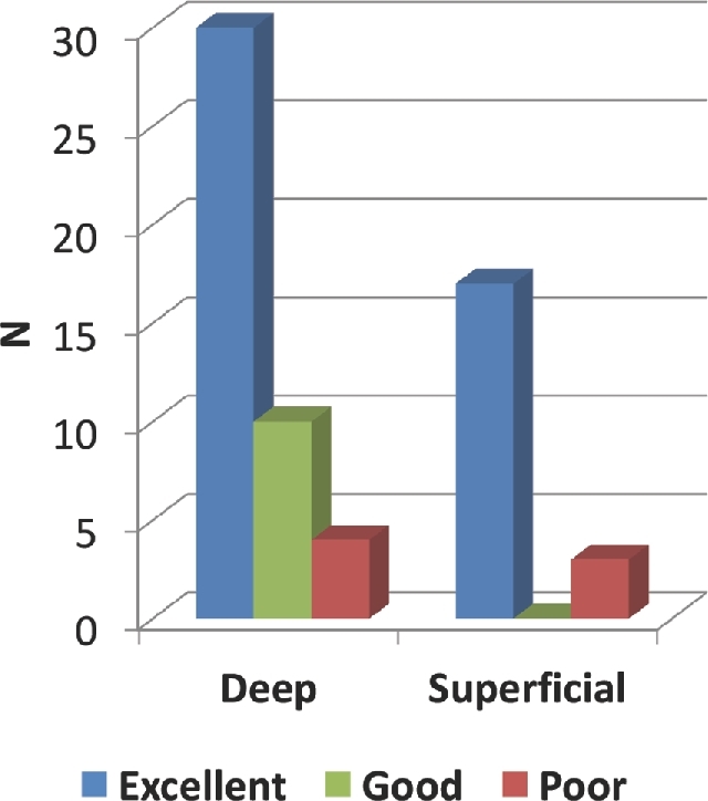
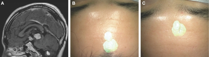
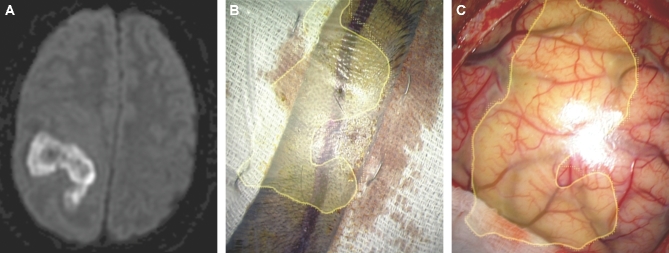
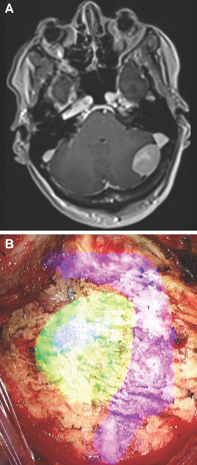
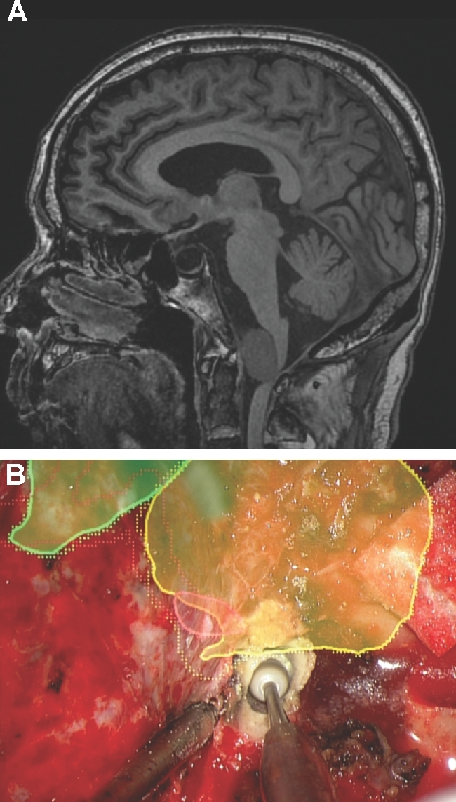
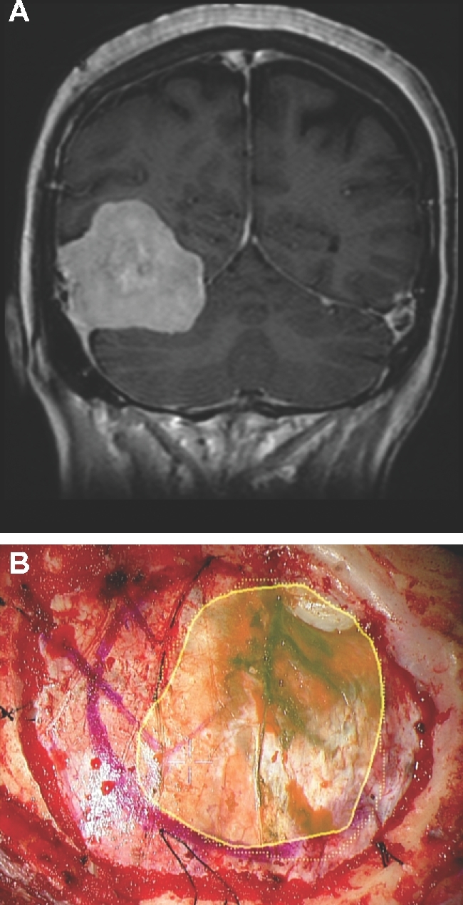
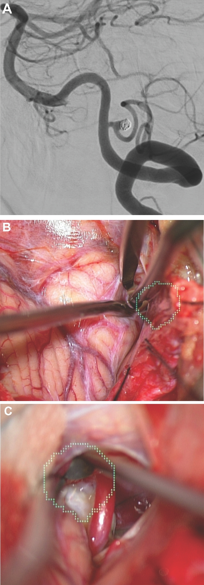
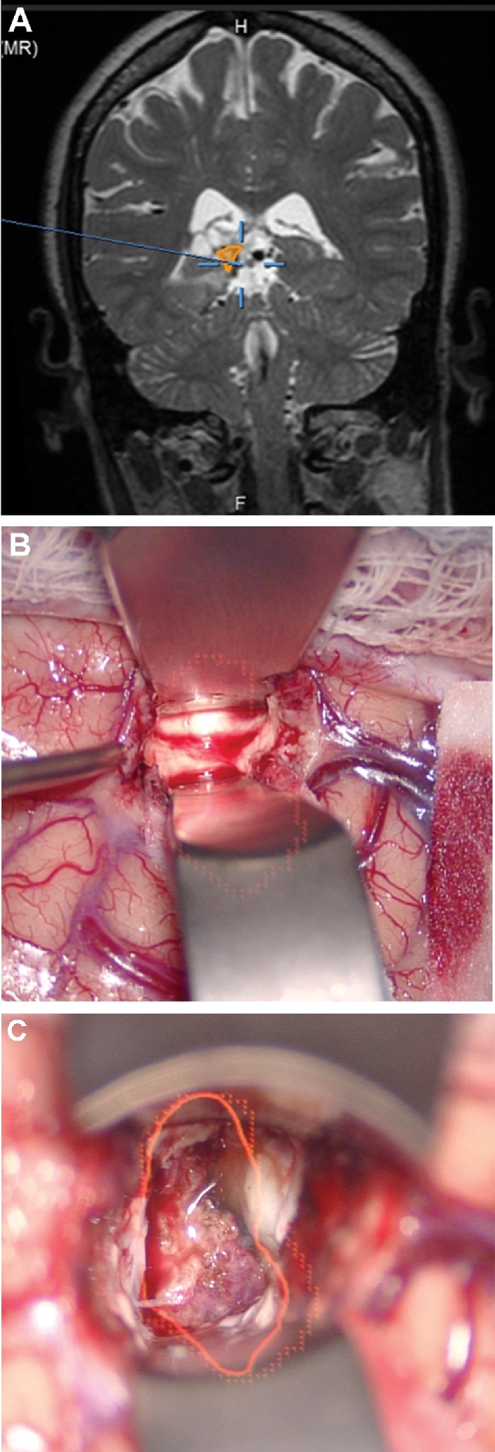
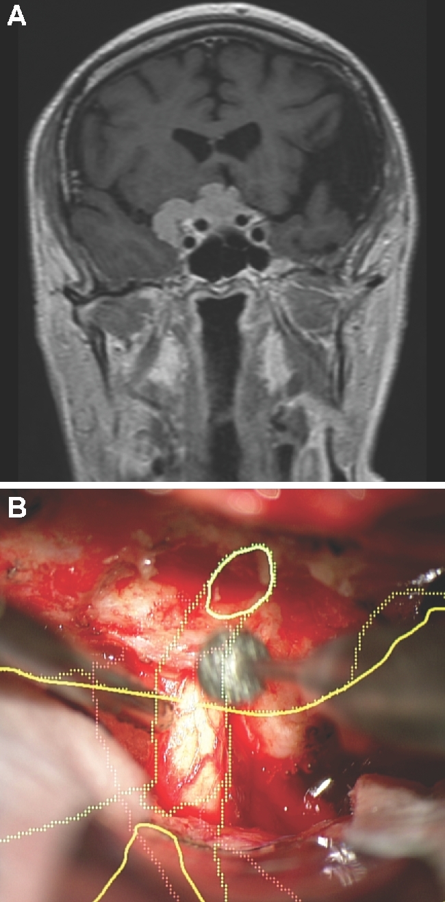
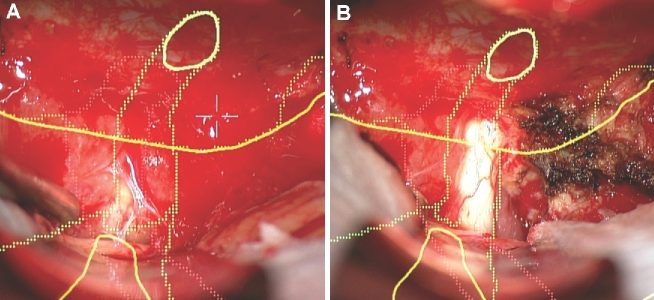
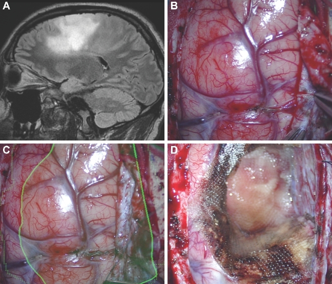
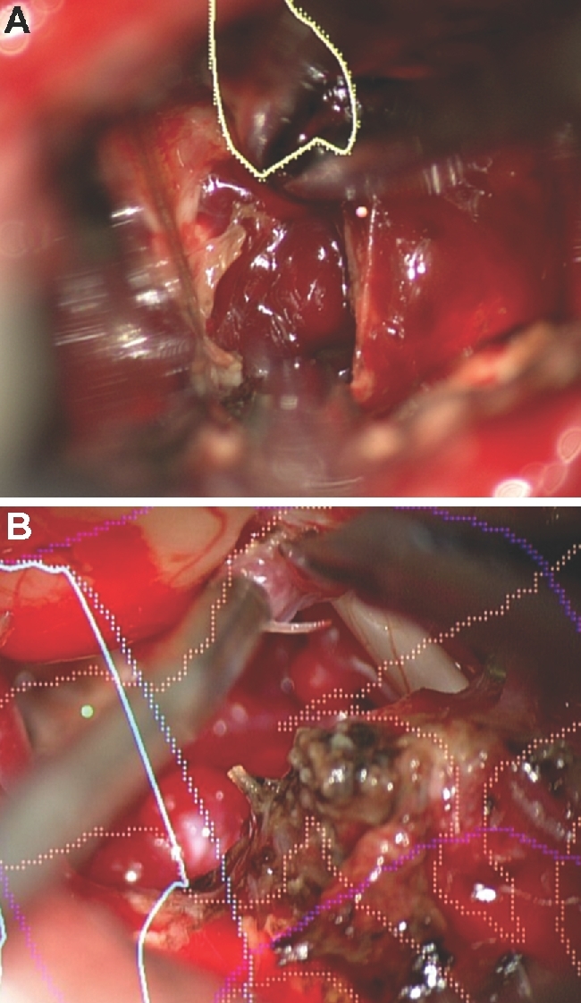
Results: Seventy-nine patients with 84 pathologies were included. Pathologies included aneurysms (14), arteriovenous malformations (6), cavernous malformations (5), intracranial stenosis (3), meningiomas (27), metastasis (4), craniopharygniomas (4), gliomas (4), schwannomas (3), epidermoid/dermoids (3), pituitary adenomas (2) hemangioblastoma (2), choroid plexus papilloma (1), lymphoma (1), osteoblastoma (1), clival chordoma (1), cerebrospinal fluid leak (1), abscess (1), and a cerebellopontine angle Teflon granuloma (1). Fifty-nine lesions were deep and 25 were superficial. Structures identified included the lesion (81), vessels (48), and nerves/brain tissue (31). Accuracy was deemed excellent (71.4%), good (20.2%), or poor (8.3%). Deep lesions were less likely to have excellent accuracy (P = .029). HUD was used during bed/head positioning (50.0%), skin incision (17.3%), craniotomy (23.1%), dural opening (26.9%), corticectomy (13.5%), arachnoid opening (36.5%), and intracranial drilling (13.5%). HUD was deactivated at some point during the surgery in 59.6% of cases. There were no complications related to HUD use.

Conclusion: HUD can be safely used for a wide variety of vascular and oncologic intracranial pathologies and can be utilized during multiple stages of surgery.


Figures

FIGURE 1.
FIGURE 1.
HUD convention. Dashed lines represent the maximum dimension of the painted lesion. Solid lines represent the dimension of the lesion that is in focus at any given time. Therefore, dashed lines will not change in size, but solid lines will change depending on focal length. In addition, the object can be displayed as outline-only or filled in with different degrees of transparency.
FIGURE 2.
FIGURE 2.
Accuracy analysis by depth. Deep lesions were less likely have excellent accuracy compared to superficial lesions (64.4% vs 88.0%, P = .029).
FIGURE 3.
FIGURE 3.
Bed/head positioning. A, A patient with a third ventricular hemangioblastoma. An interhemispheric and subfrontal approach was planned. HUD was activated prior to surgery. B, The patient's head was positioned with too much flexion, at first, and the HUD demonstrated that the approach to the lesion would transgress the frontal lobe. C, By extending the patient's head, the lesion was translated to a corridor along the skull base.
Figure 4.
Figure 4.
Skin incision. A, A patient with diabetes presenting with hemiplegia had a MRI with diffusion weighted imaging sequences performed, which demonstrated an extensive frontoparietal abscess. B, The HUD was used to outline the abscess (yellow) and was activated early to help tailor the correct skin incision. C, The HUD was then projected onto the cortical surface to understand the abscess location beneath the cortical surface.
FIGURE 5.
FIGURE 5.
Craniotomy/craniectomy. A, A patient with a cerebellar meningioma at the junction of the transverse and sigmoid sinuses. B, The HUD was activated prior to performing the craniectomy and was used to demonstrate the tumor (green), dural sinuses (purple), and guide the craniectomy.
FIGURE 6.
FIGURE 6.
Extradural drilling. A, A patient with ataxia and hemiparesis was found to have large anterior foramen magnum meningioma, as seen here in this noncontrast sagittal MRI. The patient underwent a left far lateral craniectomy. B, The HUD was used to outline the tumor (yellow), vertebral artery (red), and brainstem (green). A C1 laminectomy was performed to reach the bottom of the tumor and drilling of the occipital condyle was tailored to reach the lateral aspect of the tumor as well as the intracranial vertebral artery.
FIGURE 7.
FIGURE 7.
Dural opening. A, A patient with headaches was found to have a large right-sided tentorial meningioma, as seen here in a contrast enhanced coronal MRI. B, The patient underwent a temporal craniotomy. The HUD was used to outline the tumor (yellow), and was used to help guide the dural opening.
FIGURE 8.
FIGURE 8.
Arachnoid opening. A, A patient with previous subarachnoid hemorrhage from a ruptured posterior inferior cerebellar artery aneurysm with aneurysm recurrence following coiling, as seen here on a lateral digital subtraction angiography. B and C, In this case, the aneurysm was painted (green). The HUD was used to tailor a focused arachnoid opening directly over the aneurysm.
FIGURE 9.
FIGURE 9.
Corticectomy. A, A patient with a lateral ventricular AVM that had previously undergone radiation, but was not obliterated, had undergone cystic change as seen in this contrast-enhanced coronal MRI with a planned temporal trajectory. In this case, the AVM was painted (red). The HUD was activated after dural opening and was used to choose a precise temporal cortisectomy (B) to reach this deep lesion (C). The HUD allowed for visualization of an accurate, narrow, and safe trajectory to a deep location.
FIGURE 10.
FIGURE 10.
Intradural drilling. A, A patient with a tuberculum meningioma with a lateral extent, as seen on contrast enhanced coronal MRI. Given the lateral extent, the patient was selected for a transcranial approach, specifically a bifrontal craniectomy and subfrontal approach to the tumor. In this case, the tumor (yellow), optic nerves (green), and carotid arteries (red) were painted. The optic nerve can be seen entering the optic canal and then taking a normal slightly lateral trajectory. B, The HUD is used here to understand the course of the optic nerve within the optic canal while drilling the orbital roof.
FIGURE 11.
FIGURE 11.
Identification of nerves/vessels within tumor. A, In the same patient shown in Figure 10, the optic nerve is first embedded within the tumor and difficult to visualize. B, The HUD provides guidance as to its location, and once a portion of the tumor has been removed, the optic nerve is better visualized.
FIGURE 12.
FIGURE 12.
Identification of normal appearing pathological tissue. A, A patient with a first-time grand mal seizure was found to have a nonenhancing, flair positive left frontal lesion as seen in this sagittal flair MRI. In this case, the flair positive area was painted (green). B, After dural opening, normal appearing cortex was encountered. C, The HUD was turned on and outlined the flair positive lesion. D, The resection of normal-appearing tissue was guided by the HUD.
Figure 13.
Figure 13.
HUD limitations. The main limitations of HUD are inaccuracy as seen here with the HUD not accurately overlaying a cerebellar cavernous malformation (A) and distractibility as seen here during resection of a cerebellopontine angle AVM (B).

Comment in

References

    1. Yoon JW, Chen RE, Han PK, Si P, Freeman WD, Pirris SM. Technical feasibility and safety of an intraoperative head-up display device during spine instrumentation. Int J Med Robot. 2016. doi:10.1002/rcs.1770. - PubMed
    1. Ehlers JP, Srivastava SK, Feiler Det al. Integrative advances for OCT-guided ophthalmic surgery and intraoperative OCT: microscope integration, surgical instrumentation, and heads-Up display surgeon feedback PLoS One. 2014;9(8):e105224. - PMC - PubMed
    1. Eckardt C, Paulo EB. Heads-Up surgery for vitreoretinal procedures Retina. 2016;36(1):137-147. - PubMed
    1. Armstrong DG, Rankin TM, Giovinco NA, Mills JL, Matsuoka Y. A heads-up display for diabetic limb salvage surgery J Diabetes Sci Technol. 2014;8(5):951-956. - PMC - PubMed
    1. Chimenti PC, Mitten DJ. Google glass as an alternative to standard fluoroscopic visualization for percutaneous fixation of hand fractures Plast Reconstr Surg. 2015;136(2):328-330. - PubMed