

A case report of choledocholithiasis 33 years after cholecystectomy
- PMID: 29040906
- PMCID: PMC5645002
- DOI: 10.1016/j.ijscr.2017.09.029
A case report of choledocholithiasis 33 years after cholecystectomy
Abstract
Introduction: Choledocholithiasis after cholecystectomy is rare and often attributed to surgical clip migration and subsequent nidus formation.
Presentation of case: This case demonstrates choledocholithiasis following cholecystectomy with a latency period of 33 years.
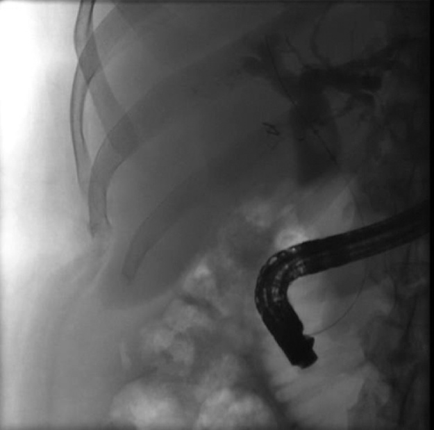
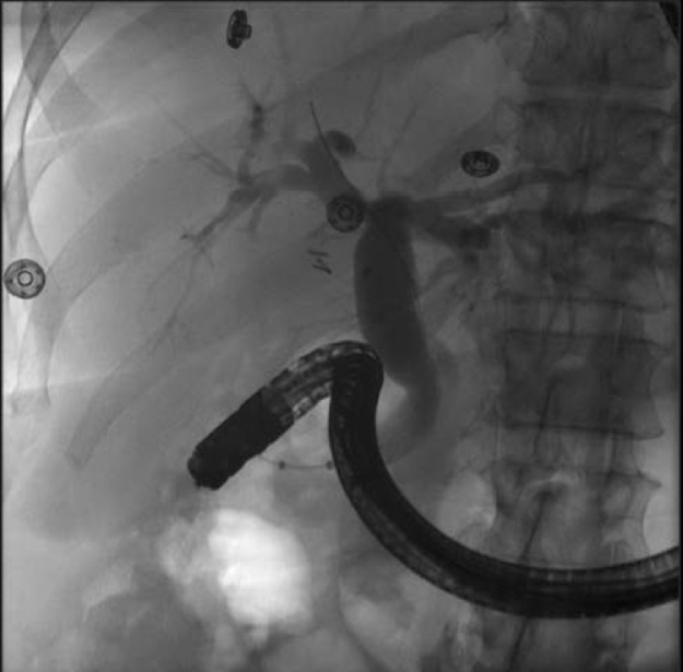
Discussion: The patient presented with pain of the right upper quadrant (RUQ). Subsequent abdominal-pelvic CT imaging revealed dilation of the common bile duct. Further Endoscopic Retrograde Cholangiopancreatography was indicative of choledocholithiasis. Additional findings included a long cystic duct remnant and surgical clips in the RUQ.
Conclusion: The patient underwent biliary sphincterotomy and sludge and stone fragments were swept from the biliary tree. To our knowledge, a latency of 33 years between cholecystectomy and choledocholithiasis has never been reported before, at least not in a patient without coexisting duodenal diverticulum, a condition associated with lithiasis of the common bile duct. Our case raises discussion of potential etiologies for such long latency, including surgical clip migration, remnant cystic duct lithiasis, and primary choledocholithiasis; and further details the incidence of such long latency periods following cholecystectomy.
Keywords: Case report; Cholecystectomy; Choledocolithiasis; Surgical clip migration.
Published by Elsevier Ltd.
Figures
References
-
- Agha R.A., Fowler A.J., Saetta A., Barai I., Rajmohan S., Orgill D.P., SCARE Group The SCARE Statement: Consensus-based surgical case report guidelines. Int. J. Surg. 2016 - PubMed
-
- Pernas Canadell J.C. Choledochal lithiasis and stenosis secondary to the migration of a surgical clip. Radiologia. 2014;56(6):e46–e49. 2011. - PubMed
-
- Copelan A., Baljendra K.S. Choledocholithiasis: diagnosis and management. Tech. Vasc. Interv. Radiol. 2015;18(4):244–255. - PubMed
-
- Gonzalez F.J., Dominguez E., Lede A., Portela J., Miguel P. Migration of vessel clip into the common bile duct and late formation of choledocholithiasis after laparoscopic cholecystectomy. Am. J. Surg. 2011;202(4):e41–e43. - PubMed
LinkOut - more resources
Full Text Sources
Other Literature Sources
Research Materials
