. 2018 Apr:14:354-360.
doi: 10.1016/j.redox.2017.10.007. Epub 2017 Oct 11.

A novel S-sulfhydrated human serum albumin preparation suppresses melanin synthesis

Affiliations

A novel S-sulfhydrated human serum albumin preparation suppresses melanin synthesis

Mayumi Ikeda et al. Redox Biol. 2018 Apr.

Abstract

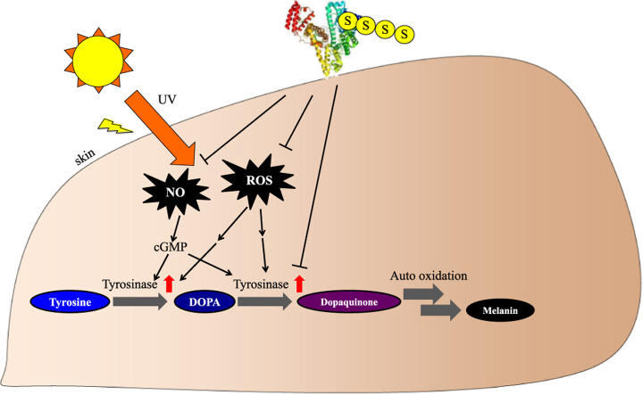
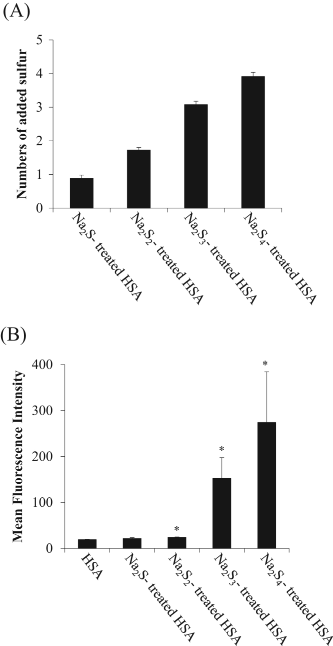
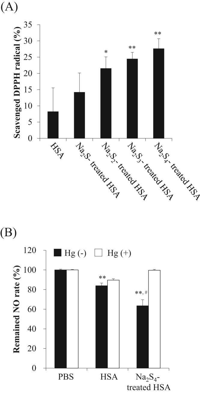
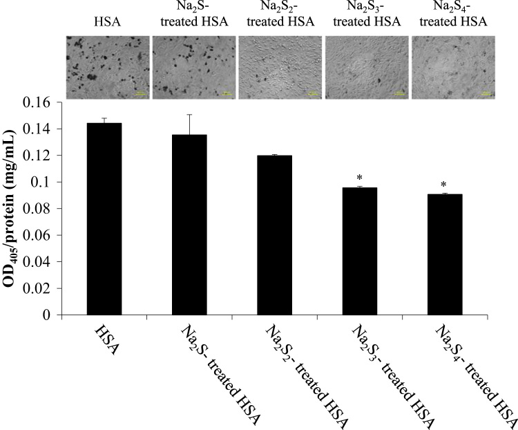
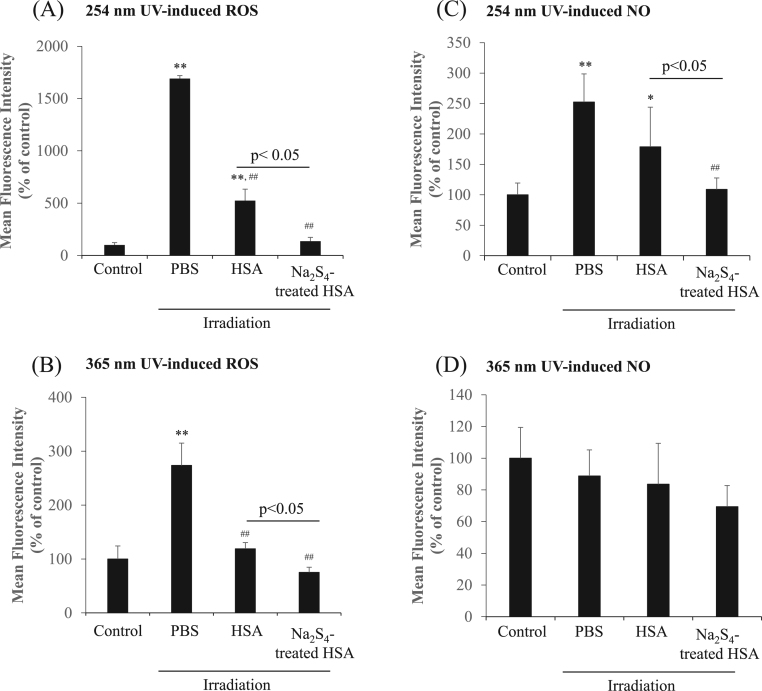
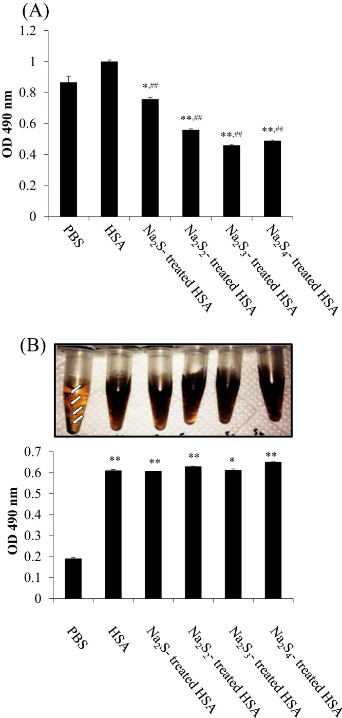
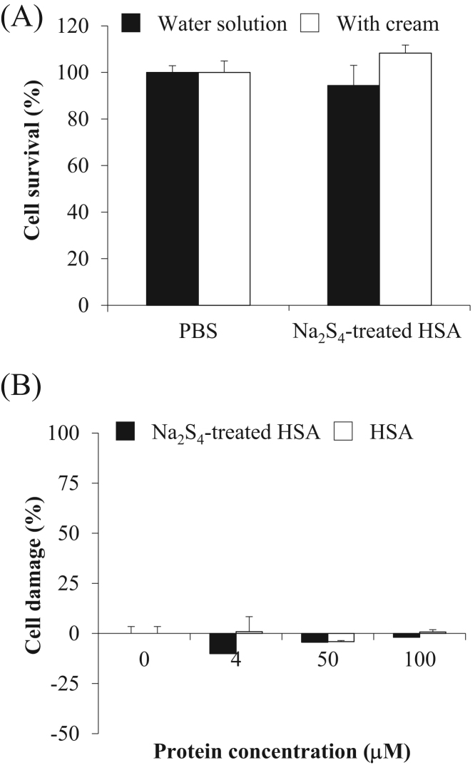
Products of ultraviolet (UV) irradiation such as reactive oxygen species (ROS) and nitric oxide (NO) stimulate melanin synthesis. Reactive sulfur species (RSS) have been shown to have strong ROS and NO scavenging effects. However, the instability and low retention of RSS limit their use as inhibitors of melanin synthesis. The free thiol at Cys34 on human serum albumin (HSA) is highly stable, has a long retention and possess a high reactivity for RSS. We report herein on the development of an HSA based RSS delivery system. Sulfane sulfur derivatives released from sodium polysulfides (Na2Sn) react readily with HSA. An assay for estimating the elimination of sulfide from polysulfide showed that almost all of the sulfur released from Na2Sn bound to HSA. The Na2Sn-treated HSA was found to efficiently scavenge ROS and NO produced from chemical reagents. The Na2Sn-treated HSA was also found to inhibit melanin synthesis in B16 melanoma cells and this inhibition was independent of the number of added sulfur atoms. In B16 melanoma cells, the Na2Sn-treated HSA also inhibited the levels of ROS and NO induced by UV radiation. Finally, the Na2Sn-treated HSA inhibited melanin synthesis from L-DOPA and mushroom tyrosinase and suppressed the extent of aggregation of melanin pigments. These data suggest that Na2Sn-treated HSA inhibits tyrosinase activity for melanin synthesis via two pathways; by directly inhibiting ROS signaling and by scavenging NO. These findings indicate that Na2Sn-treated HSA has potential to be an attractive and effective candidate for use as a skin whitening agent.

Keywords: Human serum albumin; Oxidative stress; Reactive sulfur species; Ultraviolet irradiation; Whitening agent.


Figures

fx1
Graphical abstract
Fig. 1
Fig. 1
Polysulfur binding to HSA by incubation with sodium polysulfide. (A) Valance dependency for adding sulfur to HSA with Na2Sn. Sulfane sulfur in Na2Sn-treated HSA samples were measured by EMSP. The values were subtracted from the untreated HSA. (B) SSP2 fluorescence intensity of Na2Sn-treated HSA samples was analyzed by SSP2. Each value represents the mean ± S.E. (n = 3). *p < 0.05 as compared with HSA.
Fig. 2
Fig. 2
Anti-oxidant properties of Na2Sn-treated HSA. (A) DPPH radical scavenging activity of HSA and Na2Sn-treated HSA. The concentration of DPPH radicals was measured by the oxidation of linoleic acid in the presence of HSA and Na2S4-treated HSA samples. (B) Scavenging of NO by Na2Sn-treated HSA. NO concentration was measured by a Griess assay after the reaction with Na2S4-treated HSA (50 μM) and NOC7 (200 μM). Each value represents the mean ± S.E. n = 3. *p < 0.05, **p < 0.01 as compared with control. #p < 0.05 as compared with HSA.
Fig. 3
Fig. 3
Effect of Na2Sn-treated HSA on melanin synthesis in B16 melanoma cells. Melanin content was measured by the absorption of at 405 nm after incubating Na2Sn-treated HSA with 0.4 mM tyrosine and 10 mM NH4Cl for 72 h. Protein contents were analyzed by BCA protein Assay. Each value represents the mean ± S.E. n = 3. *p < 0.05 as compared with HSA. Cell image after the treatment with Na2Sn-treated HSA in B16 melanoma cells. The photos were taken after a 72 h treatment with Na2Sn-treated HSA in the presence of 0.4 mM tyrosine and 10 mM NH4Cl.
Fig. 4
Fig. 4
ROS and NO scavenging effects of Na2S4-treated HSA under UV irradiation. ROS in B16 melanoma cells was detected by CM-H2DCF-DA in the presence of HSA and Na2S4-treated HSA with 15 min irradiation of 2 different UV, (A) 254 nm, (B) 365 nm. Each value represents the mean ± S.E. n = 3. **p < 0.01 as compared with control. ##p < 0.01 as compared with HSA. NO in B16 melanoma cells was detected by DAF-FM-DA in the presence of HSA and Na2S4-treated HSA after irradiation for 15 min with 2 different wavelengths, (C) 254 nm, (D) 365 nm. NO synthesis was measured by DAF-FM-DA. Each value represents the mean ± S.E. n = 3. *p < 0.05, **p < 0.01 as compared with control. ##p < 0.01 as compared with PBS.
Fig. 5
Fig. 5
Na2Sn-treated HSA inhibits the oxidation of L-DOPA and melanin aggregation. (A) Melanin synthesis from tyrosinase and L-DOPA in the presence of HSA and Na2Sn–treated HSA. Tyrosinase and samples were mixed and incubated at room temperature for 10 min. After the reaction, L-DOPA was added and the incubation continued for an additional 30 min (B) After 3 h, the mixture was centrifuged for 20,000 g, 15 min. Non-aggregated melanin in supernatant was measured using OD 490 nm. Tyrosinase and L-DOPA were co-incubated for 10 min and added HSA samples. White arrow showed the aggregation. Each value represents the mean ± S.E. n = 3. *p < 0.05, **p < 0.01 as compared with PBS. ##p < 0.01 as compared with HSA.
Fig. 6
Fig. 6
Safety testing of Na2S4-treated HSA for 3D cultured human epidermis. (A) Cell survival tests were performed using Autologous Cultured Epidermis kit with in PBS solution (white column) and in cream (black). (B) Cell damage tests were performed using an LDH Cytotoxicity Detection Kit. Each value represents the mean ± S.E. n = 3.

Similar articles

Cited by

References

    1. Simon H.U., Haj-Yehia A., Levi-Schaffer F. Role of reactive oxygen species (ROS) in apoptosis induction. Apoptosis. 2000;5:415–418. - PubMed
    1. Brenner M., Hearing V.J. The protective role of melanin against UV damage in human skin. Photochem. PhotoBiol. 2008;84 - PMC - PubMed
    1. Samson N., Fink B., Matts P. Interaction of skin color distribution and skin surface topography cues in the perception of female facial age and health. J. Cosmet. Dermatol. 2011;10:78–84. - PubMed
    1. Korner A., Pawelek J. Mammalian tyrosinase catalyzes three reactions in the biosynthesis of melanin. Science. 1982;217:1163–1165. - PubMed
    1. Kim D.S., Kim S.Y., Chung J.H., Kim K.H., Eun H.C., Park K.C. Delayed ERK activation by ceramide reduces melanin synthesis in human melanocytes. Cell. Signal. 2002;14:779–785. - PubMed

Publication types