

The effects of melatonin on colonization of neonate spermatogonial mouse stem cells in a three-dimensional soft agar culture system
- PMID: 29041987
- PMCID: PMC5646105
- DOI: 10.1186/s13287-017-0687-y
The effects of melatonin on colonization of neonate spermatogonial mouse stem cells in a three-dimensional soft agar culture system
Abstract
Background: Melatonin is a pleiotropic hormone with powerful antioxidant activity both in vivo and in vitro. The present study aimed to investigate the effects of melatonin on the proliferation efficiency of neonatal mouse spermatogonial stem cells (SSCs) using a three-dimensional soft agar culture system (SACS) which has the capacity to induce development of SSCs similar to in vivo conditions.
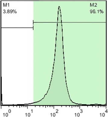
Methods: SSCs were isolated from testes of neonate mice and their purities were assessed by flow cytometry using PLZF antibody. Isolated testicular cells were cultured in the upper layer of the SACS in αMEM medium in the absence or presence of melatonin extract for 4 weeks.
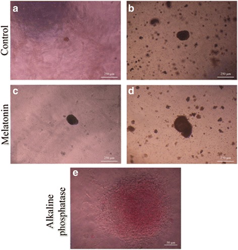
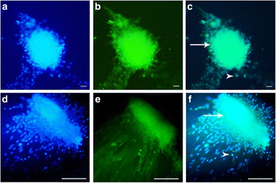
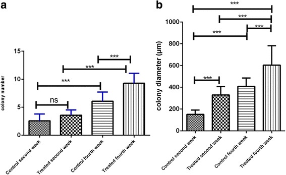
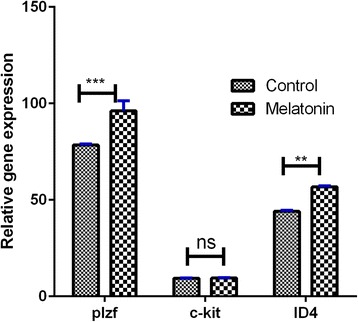
Results: The identity of colonies was confirmed by alkaline phosphatase staining and immunocytochemistry using PLZF and α6 integrin antibodies. The number and diameter of colonies of SSCs in the upper layer were evaluated at days 14 and 28 of culture. The number and diameter of colonies of SSCs were significantly higher in the melatonin group compared with the control group. The levels of expression of ID-4 and Plzf, unlike c-kit, were significantly higher in the melatonin group than in the control group.
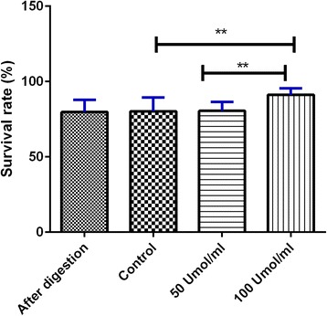
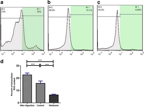
Conclusions: Results of the present study show that supplementation of the culture medium (SACS) with 100 μM melatonin significantly decreased reactive oxygen species (ROS) production in the treated group compared with the control group, and increased SSC proliferation.
Keywords: Colonization; Melatonin; Proliferation; Spermatogonial stem cell; Three-dimensional soft agar culture system.
Conflict of interest statement
Authors’ information
Not applicable.
Ethics approval
Animal experiments were approved by the Ethics Committee of Tehran University of Medical Sciences and all procedures were performed in accordance with the university’s guidelines. In the present work, we used an animal model considering all the rights based on the Ethical Committee of the Medical Faculty of Tehran University.
Consent for publication
Not applicable.
Competing interests
The authors declare that they have no competing interests.
Publisher’s note
Springer Nature remains neutral with regard to jurisdictional claims in published maps and institutional affiliations.
Figures
Similar articles
-
In vitro effects of melatonin on colonization of neonate mouse spermatogonial stem cells.Syst Biol Reprod Med. 2017 Dec;63(6):370-381. doi: 10.1080/19396368.2017.1358774. Epub 2017 Aug 28. Syst Biol Reprod Med. 2017. PMID: 28846448
-
Three-dimensional co-culture of human spermatogonial stem cells with Sertoli cells in soft agar culture system supplemented by growth factors and Laminin.Acta Histochem. 2020 Jul;122(5):151572. doi: 10.1016/j.acthis.2020.151572. Epub 2020 Jun 13. Acta Histochem. 2020. PMID: 32622422
-
Melatonin Promotes Differentiation of Human Spermatogonial Stem Cells Cultured on Three-Dimensional Decellularized Human Testis Matrix.Urol J. 2024 Jun 9;21(4):250-264. doi: 10.22037/uj.v20i.7846. Urol J. 2024. PMID: 38087969
-
The effect of biological mechanisms of melatonin on the proliferation of spermatogonial stem cells: a systematic review.Anat Cell Biol. 2024 Jun 30;57(2):163-171. doi: 10.5115/acb.23.256. Epub 2024 Apr 9. Anat Cell Biol. 2024. PMID: 38590095 Free PMC article. Review.
-
Spermatogonial stem cells.Biol Reprod. 2018 Jul 1;99(1):52-74. doi: 10.1093/biolre/ioy077. Biol Reprod. 2018. PMID: 29617903 Free PMC article. Review.
Cited by
-
Melatonin Protects Goat Spermatogonial Stem Cells against Oxidative Damage during Cryopreservation by Improving Antioxidant Capacity and Inhibiting Mitochondrial Apoptosis Pathway.Oxid Med Cell Longev. 2020 Dec 31;2020:5954635. doi: 10.1155/2020/5954635. eCollection 2020. Oxid Med Cell Longev. 2020. PMID: 33488926 Free PMC article.
-
MT1/cAMP/PKA Pathway in Melatonin-Regulated Sperm Capacitation.Reprod Sci. 2025 Mar;32(3):792-803. doi: 10.1007/s43032-024-01782-7. Epub 2025 Jan 21. Reprod Sci. 2025. PMID: 39838260
-
Protective effects of melatonin against physical injuries to testicular tissue: A systematic review and meta-analysis of animal models.Front Endocrinol (Lausanne). 2023 Jan 31;14:1123999. doi: 10.3389/fendo.2023.1123999. eCollection 2023. Front Endocrinol (Lausanne). 2023. PMID: 36798664 Free PMC article.
-
The Effect of Chitosan/Alginate/Graphene Oxide Nanocomposites on Proliferation of Mouse Spermatogonial Stem Cells.J Funct Biomater. 2023 Nov 22;14(12):556. doi: 10.3390/jfb14120556. J Funct Biomater. 2023. PMID: 38132810 Free PMC article.
-
The Role of Promyelocytic Leukemia Zinc Finger (PLZF) and Glial-Derived Neurotrophic Factor Family Receptor Alpha 1 (GFRα1) in the Cryopreservation of Spermatogonia Stem Cells.Int J Mol Sci. 2023 Jan 18;24(3):1945. doi: 10.3390/ijms24031945. Int J Mol Sci. 2023. PMID: 36768269 Free PMC article. Review.
References
-
- Morena AR, et al. Isolation of highly purified type A spermatogonia from prepubertal rat testis. J Androl. 1996;17(6):708–17. - PubMed
Publication types
MeSH terms
Substances
LinkOut - more resources
Full Text Sources
Other Literature Sources
Medical
