. 2018 Feb;93(2):403-415.
doi: 10.1016/j.kint.2017.08.005. Epub 2017 Oct 14.

Adenylyl cyclase 5 deficiency reduces renal cyclic AMP and cyst growth in an orthologous mouse model of polycystic kidney disease

Affiliations

Adenylyl cyclase 5 deficiency reduces renal cyclic AMP and cyst growth in an orthologous mouse model of polycystic kidney disease

Qian Wang et al. Kidney Int. 2018 Feb.

Abstract

Cyclic AMP promotes cyst growth in polycystic kidney disease (PKD) by stimulating cell proliferation and fluid secretion. Previously, we showed that the primary cilium of renal epithelial cells contains a cAMP regulatory complex comprising adenylyl cyclases 5 and 6 (AC5/6), polycystin-2, A-kinase anchoring protein 150, protein kinase A, and phosphodiesterase 4C. In Kif3a mutant cells that lack primary cilia, the formation of this regulatory complex is disrupted and cAMP levels are increased. Inhibition of AC5 reduces cAMP levels in Kif3a mutant cells, suggesting that AC5 may mediate the increase in cAMP in PKD. Here, we examined the role of AC5 in an orthologous mouse model of PKD caused by kidney-specific ablation of Pkd2. Knockdown of AC5 with siRNA attenuated the increase in cAMP levels in Pkd2-deficient renal epithelial cells. Levels of cAMP and AC5 mRNA transcripts were elevated in the kidneys of mice with collecting duct-specific ablation of Pkd2. Compared with Pkd2 single mutant mice, AC5/Pkd2 double mutant mice had less kidney enlargement, lower cyst index, reduced kidney injury, and improved kidney function. Importantly, cAMP levels and cAMP-dependent signaling were reduced in the kidneys of AC5/Pkd2 double mutant compared to the kidneys of Pkd2 single mutant mice. Additionally, we localized endogenous AC5 in the primary cilium of renal epithelial cells and showed that ablation of AC5 reduced ciliary elongation in the kidneys of Pkd2 mutant mice. Thus, AC5 contributes importantly to increased renal cAMP levels and cyst growth in Pkd2 mutant mice, and inhibition of AC5 may be beneficial in the treatment of PKD.

Keywords: adenylyl cyclase; cilia; cyclic AMP; knockout mice; polycystic kidney disease; polycystin-2.


Figures

Figure 1
Figure 1. Adenylyl cyclase 5 (AC5) contributes to the elevation in cAMP caused by mutation of Pkd2
(A) cAMP levels are elevated in homozygous Pkd2 null renal epithelial cells (Pkd2−/−) compared with heterozygous Pkd2+/− cells (left panel). cAMP was measured in confluent cells and normalized to protein content. Results from six independent experiments are shown. Right panel shows that cAMP levels are elevated in kidneys from Pkhd1/Cre;Pkd2F/F mice compared with control Pkhd1/Cre;Pkd2+/+ littermates (n=9). cAMP and protein concentration were measured at P14. Error bars represent SEM. (B) Expression of AC5 is increased in kidneys from Pkhd1/Cre;Pkd2F/F mice (open bar) compared with wild-type mice (filled bar). mRNA was extracted from kidneys at P14 (n=4), and levels of AC5 mRNA transcripts were measured by qRT-PCR and normalized to 18S rRNA. Expression is shown relative to wild-type kidneys (AC5+/+). Middle bar shows absence of expression in kidneys from homozygous null AC5−/− mice. Error bars represent SEM. (C) Immunoblot analysis with an AC5-specific antibody (501AP) and an antibody that recognizes both AC5 and AC6 (C17) shows increased expression in Pkd2−/− cells compared to Pkd2+/− cells. GAPDH was used as a loading control, and an antibody against polycystin-2 (PKD2) confirmed the absence of the protein in Pkd2−/− cells. (D) qRT-PCR shows reduction of AC5 mRNA levels in Pkd2+/− cells (left) and Pkd2−/− cells (right) transfected with AC5 siRNA (open bar, stippled bar) compared with cells treated with control siRNA (filled bar, hatched bar). Results from three independent experiments are shown. Error bars represent SEM. (E) Knockdown of AC5 reduces cAMP level in Pkd2 null cells. cAMP levels are elevated in Pkd2−/− cells (hatched bar) compared to Pkd2+/− cells (filled bar). Knockdown of AC5 normalizes cAMP levels in Pkd2−/− cells (stippled bar) and has no significant effect in Pkd2+/− cells (open bar). Error bars represent SEM.
Figure 2
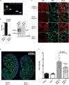
Figure 2. Knockout of AC5 in Pkd2 mutant mice
(A) Genotyping of AC5−/−, wild-type, Pkd2F/F, and Pkhd1/Cre mice. Sequences of PCR primers are provided in the Materials and Methods. PCR amplification of tail DNA shows a 650-bp product that is specific for the AC5 deletion (lane 2) and is absent in wild-type mice (lane 3). Lane 4 shows the 450-bp Pkd2F/F product, and lane 5 shows the 235-bp Pkhd1/Cre product. (B) Left panel shows qRT-PCR analysis confirming the absence of AC5 mRNA transcripts in kidneys from AC5−/−;Pkd2F/F;Pkhd1/Cre mice (open bar) compared to AC5+/+;Pkd2F/F;Pkhd1/Cre controls (filled bar). Error bars represent SEM (n=5). Right panel shows immunoblot analysis of kidney lysates from AC5−/−;Pkd2F/F;Pkhd1/Cre mice (right lanes) and AC5+/+;Pkd2F/F;Pkhd1/Cre controls (left lanes). Arrow indicates the predicted 140-kDa AC5 protein and * indicates higher molecular weight glycoforms that are present in controls and absent in AC5−/−;Pkd2F/F;Pkhd1/Cre mice. Lower panel shows GAPDH as a loading control. (C) Indirect immunofluorescence of kidneys from P14 mice with the indicated genotypes (all are Pkhd1/Cre-positive). Staining with anti-AC5 (501AP, 1:200) shows expression of AC5 (red) in renal tubules in AC5+/+ mice and only background staining in AC5−/− mice. Co-staining with FITC-DBA (1:1,000, green) labels collecting ducts in Pkd2+/+ mice and kidney cysts in Pkd2F/F mice. (D) Low magnification sagittal sections of P14 kidneys from AC5+/+;Pkd2F/F;Pkhd1/Cre mice (left) and AC5−/−;Pkd2F/F;Pkhd1/Cre mice (right) stained with FITC-DBA (green, 1:1,000) shows that renal cysts are derived from DBA-positive collecting ducts. Nuclei are counterstained blue with DAPI. Images were acquired using AxioScan Z1 microscope (Carl Zeiss) at 20× magnification and processed using Zenlift and ImageJ software. (E) Knockout of AC5 inhibits kidney enlargement caused by collecting-duct-specific deletion of Pkd2. Kidneys were collected from P14 mice with the indicated genotypes (all are Pkhd1/Cre-positive) and weighed. The ratio of kidney weight to body weight is shown. Deletion of Pkd2 (hatched bar) causes kidney enlargement compared to wild-type mice (filled bar). Concomitant deletion of AC5 in Pkd2 mutant mice reduces kidney enlargement (stippled bar) and has no effect in wild-type mice (open bar). Error bars represent SEM (n=9–17).
Figure 3
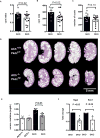
Figure 3. Ablation of AC5 slows cyst progression caused by kidney-specific deletion of Pkd2
(A) Cyst index of kidneys from Pkd2/AC5 double knockout mice (DKO: AC5−/−;Pkd2F/F;Pkhd1/Cre) is lower than Pkd2 single knockout mice (SKO: AC5+/+;Pkd2F/F;Pkhd1/Cre) at P14. (B) Average size of cysts in kidneys from DKO mice is lower than SKO mice at P14. (C) Average number of cysts per kidney sagittal section in DKO mice is not significantly different from SKO mice at P14. (D) H&E staining of kidneys at P14 shows less severe cystic disease and smaller kidneys in five representative DKO mice (lower panel) compared to SKO mice (upper panel). (E) Serum creatinine is elevated in Pkd2 mutant mice (filled bar) compared to wild-type littermates (open bar) at P14. Concomitant ablation of AC5 normalizes serum creatinine (hatched bar). Deficiency of AC5 by itself does not affect serum creatinine compared to wild-type mice (vertical bars). Error bars represent SEM (n=7–8). (F) qRT-PCR analysis shows that the expression of the kidney injury markers Ngal and Kim-1 is reduced in kidneys from DKO mice compared to SKO mice at P14. Error bars represent SEM (n=4).
Figure 4
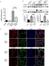
Figure 4. Knockout of AC5 reduces cAMP levels and attenuates cAMP-dependent signaling in Pkd2 mutant kidneys
(A) cAMP levels are significantly elevated in Pkd2 mutant kidneys at P14 (hatched bar) compared with wild-type kidneys (filled bar). Concomitant ablation of AC5 significantly reduces cAMP levels in Pkd2 mutant kidneys (stippled bar). (B) Immunoblot analysis shows that pCREB and pPKA substrate1 are increased in Pkd2 mutant kidneys (AC5+/+;Pkd2F/F) compared to wild-type controls (AC5+/+). Concomitant ablation of AC5 reduces CREB phosphorylation (pCREB) and phosphorylated PKA substrates (pPKA-S1) in Pkd2 mutant kidneys (AC5−/−;Pkd2F/F). Mice also carried the Pkhd1/Cre transgene. 20 µg protein was used for the analysis, and GAPDH was used as a loading control. (C) Quantification by band densitometry shows that phosphorylated PKA substrates are significantly increased in SKO kidneys (hatched bars) and reduced in DKO kidneys (open bars). pCREB is elevated in SKO kidneys and shows a trend towards reduction in DKO kidneys that was not statistically significant. GAPDH was used for normalization. Error bars represent SEM (n=3). (D) Indirect immunofluorescence on kidneys from P14 mice shows reduced nuclear staining of pCREB and pPKA-S1 in collecting ducts of DKO mice (AC5−/−;Pkd2F/F) compared to SKO mice (AC5+/+;Pkd2F/F). Red: pCREB (1:400), pPKA-S1 (1:400). Green: FITC-DBA (1:1,000). Blue: DAPI.
Figure 5
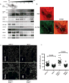
Figure 5. AC5 is located in renal cilia and AC5 deficiency reduces ciliary elongation in Pkd2 mutant kidneys
(A) Sucrose density gradient centrifugation showing co-fractionation of AC5 with ciliary proteins in Pkd2+/− renal epithelial cells transfected with PDE4C-Flag. Fractions were analyzed by SDS-PAGE followed by immunoblotting with antibodies against AC5 (501AP) and markers of Golgi (GM130), endoplasmic reticulum (calnexin), plasma membrane (CD44), and primary cilia (acetylated α-tubulin, Ac- Tub; intraflagellar transport protein 140, IFT140). AC5 is enriched in fraction 4 and to a lesser extent fraction 6, which contain acetylated α-tubulin, IFT140, polycystin-2 (PKD2), PDE4C (anti-Flag), AKAP150, and PKA regulatory subunit II (PKA-RII). Immunoblotting with an antibody that recognizes both AC5 and AC6 (C17) also shows expression in cilia as well as other organelles. (B) Indirect immunofluorescence of wild-type mouse kidney sections stained with anti-AC5 antibody (PAC-501AP). Endogenous AC5 is located in the primary cilia of renal tubular epithelial cells in addition to the cell body. Cilia were co-stained with an antibody against acetylated α-tubulin. (C) Indirect immunofluorescence showing primary cilia in renal collecting ducts from wild-type mice (AC5+/+), AC5 knockout mice (AC5−/−), Pkd2 single knockout mice (AC5+/+;Pkd2F/F), and Pkd2/AC5 double knockout mice (AC5−/−;Pkd2F/F). Mice were euthanized at P14, and primary cilia were stained with anti-ARL13B antibody (1:400). Insets show that ablation of AC5 reduces ciliary length compared to wild-type and Pkd2 mutant mice. (D) Quantification shows elongation of primary cilia in Pkd2 mutant mice. Concomitant ablation of AC5 normalizes cilia length. Cilia length was measured using ImageJ after 3D reconstruction of Z-stack images. Statistical analysis was performed with GraphPad Prism 6 using ANOVA for multiple comparisons. Error bars represent SEM.

Similar articles

Cited by

References

    1. Igarashi P, Somlo S. Genetics and pathogenesis of polycystic kidney disease. J Am Soc Nephrol. 2002;13:2384–2398. - PubMed
    1. Pazour GJ, San Agustin JT, Follit JA, et al. Polycystin-2 localizes to kidney cilia and the ciliary level is elevated in orpk mice with polycystic kidney disease. Curr Biol. 2002;12:R378–R380. - PubMed
    1. Yoder BK, Hou X, Guay-Woodford LM. The polycystic kidney disease proteins, polycystin-1, polycystin-2, polaris, and cystin, are co-localized in renal cilia. J Am Soc Nephrol. 2002;13:2508–2516. - PubMed
    1. Nauli SM, Alenghat FJ, Luo Y, et al. Polycystins 1 and 2 mediate mechanosensation in the primary cilium of kidney cells. Nat Genet. 2003;33:129–137. - PubMed
    1. Delling M, Indzhykulian AA, Liu X, et al. Primary cilia are not calcium-responsive mechanosensors. Nature. 2016;531:656–660. - PMC - PubMed

Publication types

MeSH terms